

CALP1 TFA |
Catalog No.GC65081
 One-Click Copy Product Info
|

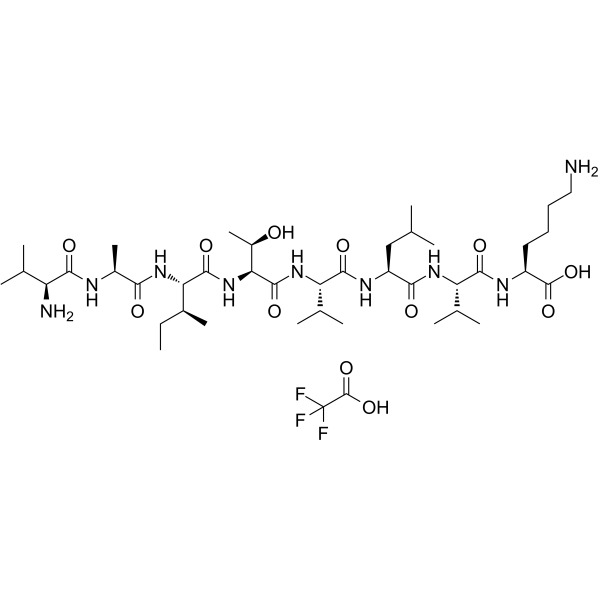
CALP1 TFA is an eight-residue complementary peptide that binds to the calcium-binding sites (the EF hand) of calmodulin.
Products are for research use only. Not for human use. We do not sell to patients.
Sample solution is provided at 25 µL, 10mM.
_1-3.$$$39978408@51.jpg');)
_1-9.$$$38538795@65.jpg');)
_1-9.$$$39112701@51.jpg');)
_1-7.$$$37316672@70.jpg');)
_366-372.$$$36198801@70.jpg');)
.$$$38565142@65.jpg');)
_1867-1883.$$$33248023@67.jpg');)
_eads6055.$$$39977546@45.jpg');)
_eadm9805.$$$40080571@45.jpg');)
_eadj3020.$$$39666821@45.jpg');)
_1-20.$$$39757301@28.jpg');)
_1-24.$$$38898113@28.jpg');)
_273-287.$$$36806353@46.jpg');)
_1-16.$$$37156877@44.jpg');)
_1-19.$$$37460805@44.jpg');)
_1291-1307.$$$34518654@46.jpg');)
CALP1 TFA is an eight-residue complementary peptide that binds to the calcium-binding sites (the EF hand) of calmodulin [1]. CALP1 TFA interacts with the two carboxyl-terminal Ca2+-binding sites of calmodulin as well as the EF hands of troponin C, which results in a conformational change and activates phosphodiesterase in the absence of Ca2+[2]. CALP1 TFA has been widely used in allergic asthma models to mitigate the development of airway hyperresponsiveness [3].
In vitro, CALP1 TFA treatment (20µM) for 2 hours can increase the expression of the VPAC1 protein on the surface of monocytes infected by Salmonella[4]. 100µM of CALP1 TFA pretreatment for 15 minutes can reduce the excessive cytoplasmic Ca2+ signal and necrosis in mouse pancreatic acinar cells (PACs) induced by BH3I-2′[5]. Treatment with 100µM CALP1 TFA for 60 minutes can inhibit the adhesion of murine bone marrow-derived mast cells (BMMCs) to fibronectin induced by FcεRI[6]. Treatment with 10µM CALP1 TFA for 48 hours significantly inhibited the expression of TGF-β2 and IL-10 in C2C12 cells and promoted the upregulation of p-CaMKII, p-CaMKIV, and NFATc2 levels after IFN-γ stimulation[7].
In vivo, thirty minutes before ovalbumin challenge, a single dose of CALP1 TFA (1mg/ml; 0.1ml) was injected intratracheally, significantly reducing the infiltration of alveolar macrophages in the lungs of guinea pigs stimulated by ovalbumin and inhibiting the generation of free radicals[8].
References:
[1] Folkerts G, Nijkamp F P. Airway nitrergic pathways: is there therapeutic potential in asthma and COPD?[J]. Current Opinion in Pharmacology, 2004, 4(3): 202-206.
[2] Villain M, Jackson P L, Manion M K, et al. De novo design of peptides targeted to the EF hands of calmodulin[J]. Journal of Biological Chemistry, 2000, 275(4): 2676-2685.
[3] Ten Broeke R, Leusink-Muis T, Hilberdink R, et al. Specific modulation of calmodulin activity induces a dramatic production of superoxide by alveolar macrophages[J]. Laboratory investigation, 2004, 84(1): 29-40.
[4] Askar B, Higgins J, Barrow P, et al. Immune evasion by Salmonella: exploiting the VPAC 1/VIP axis in human monocytes[J]. Immunology, 2019, 158(3): 230-239.
[5] Ferdek P E, Jakubowska M A, Nicolaou P, et al. BH3 mimetic-elicited Ca2+ signals in pancreatic acinar cells are dependent on Bax and can be reduced by Ca2+-like peptides[J]. Cell Death & Disease, 2017, 8(3): e2640-e2640.
[6] Houtman R, Ten Broeke R, Blalock J E, et al. Attenuation of very late antigen-5-mediated adhesion of bone marrow-derived mast cells to fibronectin by peptides with inverted hydropathy to EF-hands[J]. The Journal of Immunology, 2001, 166(2): 861-867.
[7] Hu J, Shi D, Ding M, et al. Calmodulin‐dependent signalling pathways are activated and mediate the acute inflammatory response of injured skeletal muscle[J]. The Journal of physiology, 2019, 597(21): 5161-5177.
[8] Ten Broeke R, Brandhorst M C, Leusink-Muis T, et al. Ca2+ sensors modulate asthmatic symptoms in an allergic model for asthma[J]. European journal of pharmacology, 2003, 476(1-2): 151-157.
| Cell experiment [1]: | |
Cell lines | Bone marrow-derived mast cells (BMMCs) |
Preparation Method | BMMCs from BALB/c mice were cultured in RPMI 1640 medium supplemented with 4mM L-glutamine, 0.05µM 2-ME, 1mM sodium pyruvate, 100U/ml penicillin, 100mg/ml streptomycin, and 0.1mM nonessential amino acids in 5% CO2 at 37°C. Cells were placed in a 96 well-plate at a concentration of 5×10⁵ cells per well and were treated with different concentrations of CALP1 TFA (0, 1, 10, 100µM) and 30ng/ml DNP-HAS (FcεRI clustering) for 30min, and then the Effect of CALP1 TFA on the degranulation of BMMCs was analyzed. |
Reaction Conditions | 0, 1, 10, 100µM; 30min |
Applications | CALP1 TFA treatment significantly reduced FcεRI-induced degranulation in BMMCs. |
| Animal experiment [2]: | |
Animal models | Guinea pigs |
Preparation Method | Specified-pathogen-free guinea pigs (300-400g) were housed in a specialized animal care facility, in a room with constant temperature (25°C), humidity control, and a 12/12h light/dark cycle with free access to food and water. Thirty minutes before ovalbumin challenge, guinea pigs received the control solution (0.1ml; positive control), CALP1 TFA (1mg/ml; 0.1ml), or CALP2 (3mg/ml; 0.1ml) intratracheally. Therefore, the guinea pigs were anaesthetized with halothane and placed in a supine position on a table. The jaws were kept apart by two elastic bands, and a needle with a bulbous tip was inserted just behind the glottis. Thereafter, 0.1ml of the solution was gently injected into the trachea, the bands were removed, and the chest was gently massaged. Broncho-alveolar lavage cells were obtained from pigs for analysis. |
Dosage form | 1mg/ml (1ml) for once; intratracheal injection |
Applications | CALP1 TFA treatment significantly reduced the influx of alveolar macrophages into the lungs of ovalbumin-challenged guinea pigs 6h after challenge. |
References: | |
| Cas No. | SDF | ||
| Formula | C42H76F3N9O12 | M.Wt | 956.1 |
| Solubility | Water : 16.67 mg/mL (17.44 mM; Need ultrasonic) | Storage | -20°C, away from moisture |
| General tips | Please select the appropriate solvent to prepare the stock solution according to the
solubility of the product in different solvents; once the solution is prepared, please store it in
separate packages to avoid product failure caused by repeated freezing and thawing.Storage method
and period of the stock solution: When stored at -80°C, please use it within 6 months; when stored
at -20°C, please use it within 1 month. To increase solubility, heat the tube to 37°C and then oscillate in an ultrasonic bath for some time. |
||
| Shipping Condition | Evaluation sample solution: shipped with blue ice. All other sizes available: with RT, or with Blue Ice upon request. | ||
| Prepare stock solution | |||

|
1 mg | 5 mg | 10 mg |
| 1 mM | 1.0459 mL | 5.2296 mL | 10.4592 mL |
| 5 mM | 209.2 μL | 1.0459 mL | 2.0918 mL |
| 10 mM | 104.6 μL | 523 μL | 1.0459 mL |
Step 1: Enter information below (Recommended: An additional animal making an allowance for loss during the experiment)
 g
μL
g
μL
Step 2: Enter the in vivo formulation (This is only the calculator, not formulation. Please contact us first if there is no in vivo formulation at the solubility Section.)
Calculation results:
Working concentration: mg/ml;
Method for preparing DMSO master liquid: mg drug pre-dissolved in μL DMSO ( Master liquid concentration mg/mL, Please contact us first if the concentration exceeds the DMSO solubility of the batch of drug. )
Method for preparing in vivo formulation: Take μL DMSO master liquid, next addμL PEG300, mix and clarify, next addμL Tween 80, mix and clarify, next add μL saline, mix and clarify.
Method for preparing in vivo formulation: Take μL DMSO master liquid, next add μL Corn oil, mix and clarify.
Note: 1. Please make sure the liquid is clear before adding the next solvent.
2. Be sure to add the solvent(s) in order. You must ensure that the solution obtained, in the previous addition, is a clear solution before proceeding to add the next solvent. Physical methods such as vortex, ultrasound or hot water bath can be used to aid dissolving.
3. All of the above co-solvents are available for purchase on the GlpBio website.
Quality Control & SDS
- View current batch:
- Purity: >98.00% Appearance: A solid
- COA (Certificate of Analysis)
- SDS (Safety Data Sheet)
- Datasheet
Average Rating: 5 (Based on Reviews and 30 reference(s) in Google Scholar.)