

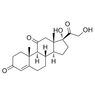
Cortisone (17-Hydroxy-11-dehydrocorticosterone) |
Catalog No.GC33839
 One-Click Copy Product Info
|

Cortisone (17-Hydroxy-11-dehydrocorticosterone) is an oxidized metabolite of Cortisol with anti-inflammatory properties.
Products are for research use only. Not for human use. We do not sell to patients.
Cas No.: 53-06-5
Sample solution is provided at 25 µL, 10mM.
_1-3.$$$39978408@51.jpg');)
_1-9.$$$38538795@65.jpg');)
_1-9.$$$39112701@51.jpg');)
_1-7.$$$37316672@70.jpg');)
_366-372.$$$36198801@70.jpg');)
.$$$38565142@65.jpg');)
_1867-1883.$$$33248023@67.jpg');)
_eads6055.$$$39977546@45.jpg');)
_eadm9805.$$$40080571@45.jpg');)
_eadj3020.$$$39666821@45.jpg');)
_1-20.$$$39757301@28.jpg');)
_1-24.$$$38898113@28.jpg');)
_273-287.$$$36806353@46.jpg');)
_1-16.$$$37156877@44.jpg');)
_1-19.$$$37460805@44.jpg');)
_1291-1307.$$$34518654@46.jpg');)
Cortisone (17-Hydroxy-11-dehydrocorticosterone) is an oxidized metabolite of Cortisol with anti-inflammatory properties[1]. Cortisone suppresses intraocular inflammation by reducing inflammatory exudation and inhibiting proliferation of fibroblasts and formation of granulation tissue[2]. Cortisone in saliva is a useful surrogate for circulating free cortisol in research and preclinical assessment and is a stress biomarker with high discriminatory power and significant correlations with subjective and autonomic stress measures[3-4]. Cortisone has been widely used as an internal standard to optimize the parameters of chromatography and mass spectrometry analysis, facilitating the precise identification of Cortisone and related compounds in saliva and hair samples[5].
In vitro, Cortisone (10nM) treatment for 6 days promoted cell growth and prostate-specific antigen (PSA) secretion in MDA PCa 2b cells[6]. Treatment with 1nM Cortisone for 24 hours abolished the platelet-derived growth factor (PDGF)-induced expression of the VEGF gene and VEGF secretion in human vascular smooth muscle cells (hVSMCs)[7].
In vivo, Cortisone treatment via subcutaneous injection at a dose of 40mg/kg/day for 4 weeks aggravated the Mycobacteroides abscessus infection in C3HeB/FeJ mice, and increased the bacterial load in the lungs of mice[8]. Subcutaneous injection of Cortisone (5mg/day) for 8 days inhibited the growth of rats and caused weight loss[9].
References:[1] Hillier S G. Diamonds are forever: the cortisone legacy[J]. Journal of Endocrinology, 2007, 195(1): 1-6.
[2] Jonas J B, Hayler J K, Söfker A, et al. Intravitreal injection of crystalline cortisone as adjunctive treatment of proliferative diabetic retinopathy[J]. American journal of ophthalmology, 2001, 131(4): 468-471.
[3] Perogamvros I, Keevil B G, Ray D W, et al. Salivary cortisone is a potential biomarker for serum free cortisol[J]. The Journal of Clinical Endocrinology & Metabolism, 2010, 95(11): 4951-4958.
[4] Bae Y J, Reinelt J, Netto J, et al. Salivary cortisone, as a biomarker for psychosocial stress, is associated with state anxiety and heart rate[J]. Psychoneuroendocrinology, 2019, 101: 35-41.
[5] Jones R L, Owen L J, Adaway J E, et al. Simultaneous analysis of cortisol and cortisone in saliva using XLC–MS/MS for fully automated online solid phase extraction[J]. Journal of Chromatography B, 2012, 881: 42-48.
[6] Zhao X Y, Malloy P J, Krishnan A V, et al. Glucocorticoids can promote androgen-independent growth of prostate cancer cells through a mutated androgen receptor[J]. Nature medicine, 2000, 6(6): 703-706.
[7] Nauck M, Karakiulakis G, Perruchoud A P, et al. Corticosteroids inhibit the expression of the vascular endothelial growth factor gene in human vascular smooth muscle cells[J]. European journal of pharmacology, 1998, 341(2-3): 309-315.
[8] Maggioncalda E C, Story-Roller E, Mylius J, et al. A mouse model of pulmonary Mycobacteroides abscessus infection[J]. Scientific reports, 2020, 10(1): 3690.
[9] Krieg Jr R J, Niimi K, Chan J C M, et al. Cortisone effects on growth, food efficiency, and in vitro growth hormone release[J]. Kidney international, 1991, 39(6): 1135-1139.
| Cell experiment [1]: | |
|
Cell lines |
MDA PCa 2b cells |
|
Preparation Method |
MDA PCa 2b cells were cultured in RPMI-1640 medium, supplemented with 5% charcoal-stripped fetal bovine serum at 37°C in an incubator with 5% CO2. Cells (5×10⁴/well) were seeded in 96-well plates, allowed to adhere overnight, and treated for 6 days at 37°C with different concentrations of Cortisone (0, 1, 10, and 100nM), the cell proliferation was tested. |
|
Reaction Conditions |
0, 1, 10, and 100nM; 6 days |
|
Applications |
Cortisone treatment stimulated MDA PCa 2b cell proliferation in a dose-dependent manner. |
| Animal experiment [2]: | |
|
Animal models |
Female C3HeB/FeJ mice |
|
Preparation Method |
Female C3HeB/FeJ mice (4 weeks old; 19-23g) were housed in SPF conditions with an automatic 12h/12h light-dark cycle at a constant temperature (21±1°C). To achieve an implantation of 3.0-3.5 log10 CFU in the lungs of a mouse, a primary M. abscessus culture at exponential phase, A600nm of 1.00-1.20, was used to prepare a suspension by diluting to a calculated A600nm of 0.1 in Middlebrook 7H9 broth prewarmed to 37°C. Infections were performed by aerosolizing 10ml of this suspension. The infection cycle comprised 15 minutes of pre-heat, 30 minutes of nebulization, 30 minutes of cloud decay, and 15 minutes of surface decontamination. A 200μl Cortisone (40mg/kg/day) was administered by subcutaneous injection in the dorsal abdominal flank using a 26-gauge syringe. Cortisone treatment began one week prior to infection and lasted for 4 weeks. M. abscessus lung burden in mice was measured. |
|
Dosage form |
40mg/kg/day; 4 weeks; s.c. |
|
Applications |
Cortisone treatment aggravated M. abscessus lung burden in mice. |
| References: [1] Zhao X Y, Malloy P J, Krishnan A V, et al. Glucocorticoids can promote androgen-independent growth of prostate cancer cells through a mutated androgen receptor[J]. Nature medicine, 2000, 6(6): 703-706. [2] Maggioncalda E C, Story-Roller E, Mylius J, et al. A mouse model of pulmonary Mycobacteroides abscessus infection[J]. Scientific reports, 2020, 10(1): 3690. | |
| Cas No. | 53-06-5 | SDF | |
| Canonical SMILES | C[C@@]1(C2)[C@](C(CO)=O)(O)CC[C@@]1([H])[C@]3([H])CCC4=CC(CC[C@]4(C)[C@@]3([H])C2=O)=O | ||
| Formula | C21H28O5 | M.Wt | 360.44 |
| Solubility | DMSO : 100 mg/mL (277.44 mM) | Storage | Store at RT |
| Shipping Condition | Evaluation sample solution: shipped with blue ice. All other sizes available: with RT, or with Blue Ice upon request. | ||
| Prepare stock solution | |||

|
1 mg | 5 mg | 10 mg |
| 1 mM | 2.7744 mL | 13.8719 mL | 27.7439 mL |
| 5 mM | 554.9 μL | 2.7744 mL | 5.5488 mL |
| 10 mM | 277.4 μL | 1.3872 mL | 2.7744 mL |
Step 1: Enter information below (Recommended: An additional animal making an allowance for loss during the experiment)
 g
μL
g
μL
Step 2: Enter the in vivo formulation (This is only the calculator, not formulation. Please contact us first if there is no in vivo formulation at the solubility Section.)
Calculation results:
Working concentration: mg/ml;
Method for preparing DMSO master liquid: mg drug pre-dissolved in μL DMSO ( Master liquid concentration mg/mL, Please contact us first if the concentration exceeds the DMSO solubility of the batch of drug. )
Method for preparing in vivo formulation: Take μL DMSO master liquid, next addμL PEG300, mix and clarify, next addμL Tween 80, mix and clarify, next add μL saline, mix and clarify.
Method for preparing in vivo formulation: Take μL DMSO master liquid, next add μL Corn oil, mix and clarify.
Note: 1. Please make sure the liquid is clear before adding the next solvent.
2. Be sure to add the solvent(s) in order. You must ensure that the solution obtained, in the previous addition, is a clear solution before proceeding to add the next solvent. Physical methods such as vortex, ultrasound or hot water bath can be used to aid dissolving.
3. All of the above co-solvents are available for purchase on the GlpBio website.
Quality Control & SDS
- View current batch:
- Purity: >97.00% Appearance: A solid
- COA (Certificate of Analysis)
- SDS (Safety Data Sheet)
- Datasheet
Average Rating: 5 (Based on Reviews and 5 reference(s) in Google Scholar.)