

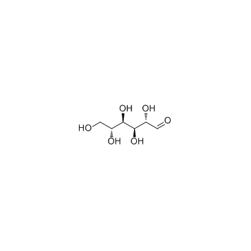
D-Mannose (Synonyms: Carubinose, (+)-Mannose, NSC 26247) |
Catalog No.GC33460
 One-Click Copy Product Info
|

D-Mannose is a natural hexose monosaccharide with anti-inflammatory and antitumor activities.
Products are for research use only. Not for human use. We do not sell to patients.
Cas No.: 3458-28-4
Sample solution is provided at 25 µL, 10mM.
_1-3.$$$39978408@51.jpg');)
_1-9.$$$38538795@65.jpg');)
_1-9.$$$39112701@51.jpg');)
_1-7.$$$37316672@70.jpg');)
_366-372.$$$36198801@70.jpg');)
.$$$38565142@65.jpg');)
_1867-1883.$$$33248023@67.jpg');)
_eads6055.$$$39977546@45.jpg');)
_eadm9805.$$$40080571@45.jpg');)
_eadj3020.$$$39666821@45.jpg');)
_1-20.$$$39757301@28.jpg');)
_1-24.$$$38898113@28.jpg');)
_273-287.$$$36806353@46.jpg');)
_1-16.$$$37156877@44.jpg');)
_1-19.$$$37460805@44.jpg');)
_1291-1307.$$$34518654@46.jpg');)
D-Mannose is a natural hexose monosaccharide with anti-inflammatory and antitumor activities. D-Mannose is rapidly absorbed in the upper intestine. D-Mannose inhibits bacterial adhesion to urothelium by competitively binding to adhesins of uropathogenic Escherichia coli. D-Mannose accumulates as mannose-6-phosphate to regulate phosphomannose isomerase and inhibits the succinate-HIF-1α axis in macrophages to reduce the production of inflammatory factors such as IL-1β. D-Mannose can be used in studies of recurrent urinary tract infection, cystitis, and inflammation-related diseases[1-4].
In vitro, 0-20 mM D-Mannose was applied to INS-1 cells and islet cells from 0-16-week-old mice. D-Mannose increased IRS-2 expression and phosphorylation[5]. 25 mM D-Mannose was applied to MDA-MB-231 and BT-549 cells for 0-72 hours. D-Mannose decreased PD-L1 protein levels and induced aberrant PD-L1 glycosylation[6]. 25 mM D-Mannose was applied to HUVEC cells for 24 hours. D-Mannose inhibited VEGF-enhanced cell proliferation, migration, and capillary tube formation, and decreased VEGFR2 protein levels to promote lysosomal degradation[7].
In vivo, ethanol-induced gastric injury C57BL/6J mice received 0.2 mL 20%-40% (w/v) D-Mannose by gavage once daily for 7 days. D-Mannose significantly reduced ulcer index and pathological scores, increased SOD, GSH-Px, and CAT activities, decreased ROS and MDA production, and activated HSP90 and downstream Nrf2/HO-1 pathway-related genes and protein expression[8]. Experimental autoimmune encephalomyelitis C57BL/6 mouse models received 450 mg/kg D-Mannose by intraperitoneal injection once daily for 19 days. D-Mannose improved encephalomyelitis symptoms and improved survival rate, reduced lesion area, decreased oxidative stress, decreased infiltrating M1-like macrophages and microglia[9]. Diabetic back wound BALB/c mouse models received 100 μL 3% (w/v) D-Mannose in PBS topically on the wound once daily for 13 days. D-Mannose accelerated skin wound closure and shortened complete healing time, reduced AGEs formation in skin lesions, upregulated AMPK/Nrf2/HO-1 signaling molecules, decreased CD86+ macrophage counts, and increased dermal collagen deposition[10].
References:[1] Ala-Jaakkola R, Laitila A, Ouwehand AC, et al. Role of D-mannose in urinary tract infections - a narrative review. Nutr J. 2022 Mar 22;21(1):18.
[2] Kranjčec B, Papeš D, Altarac S. D-mannose powder for prophylaxis of recurrent urinary tract infections in women: a randomized clinical trial. World J Urol. 2014 Feb;32(1):79-84.
[3] Wu H, Zhang W, Mu W. Recent studies on the biological production of D-mannose. Appl Microbiol Biotechnol. 2019 Nov;103(21-22):8753-8761.
[4] Joshi CS, Salazar AM, Wang C, et al. D-mannose reduces cellular senescence and NLRP3/GasderminD/IL-1β-driven pyroptotic uroepithelial cell shedding in the murine bladder. Dev Cell. 2024 Jan 8;59(1):33-47.e5.
[5] Amacker-Francoys I, Mohanty S, Niessen M, et al. The metabolisable hexoses D-glucose and D-mannose enhance the expression of IRS-2 but not of IRS-1 in pancreatic β-cells. Exp Clin Endocrinol Diabetes. 2005;113(7):423-429.
[6] Zhang R, Yang Y, Dong W, et al. D-mannose facilitates immunotherapy and radiotherapy of triple-negative breast cancer via degradation of PD-L1. Proc Natl Acad Sci U S A. 2022 Feb 22;119(8):e2114851119.
[7] Du Y, Zhang X, Xu Y, et al. D-mannose suppresses the angiogenesis and progression of colorectal cancer. Acta Biochim Biophys Sin. 2025;57(8):1270-1280.
[8] Yan F, Yan X, Li X, et al. D-Mannose Attenuates Ethanol-Induced Gastric Ulcers via Antioxidant Activity Through the HSP90/Nrf2/HO-I Pathway. Drug Des Devel Ther. 2025;19:10967-10989.
[9] Wang J, Jalali Motlagh N, Wang C, et al. D-mannose suppresses oxidative response and blocks phagocytosis in experimental neuroinflammation. Proc Natl Acad Sci U S A. 2021 Oct 26;118(43):e2107663118.
[10] Luo J, Wu T, Zhang J, et al. D-mannose promotes diabetic wound healing through inhibiting advanced glycation end products formation in keratinocytes. Mol Med. 2025;31:15.
| Cell experiment [1]: | |
|
Cell lines |
HUVEC (human umbilical vein endothelial cells) |
|
Preparation Method |
HUVEC were cultured in DMEM supplemented with 10% FBS and 100μg/mL penicillin and streptomycin at 37°C, 5% CO2. HUVEC were treated with D-Mannose at 10mM for 24h. After treatment, cell proliferation was evaluated by colony formation assay, cell migration was assessed by wound healing and transwell migration assays, capillary tube formation was examined by Matrigel tube formation assay, and protein levels were analyzed by western blot. |
|
Reaction Conditions |
10mM; 24h |
|
Applications |
D-Mannose significantly inhibited VEGF-enhanced cell proliferation, migration and capillary tube formation, reduced VEGF-induced p-AKT and p-ERK1/2 levels without affecting their total protein levels. D-Mannose decreased VEGFR2 protein level in HUVEC. |
| Animal experiment [2]: | |
|
Animal models |
C57BL/6 mice |
|
Preparation Method |
Mice were intraperitoneally administered 450mg/kg D-Mannose once a day from the day of experimental autoimmune encephalomyelitis induction (day 0) until day 19. Clinical scores and weights were recorded daily. On day 15, multiagent molecular MRI was performed using MPO-Gd and CLIO to assess oxidative stress and phagocytosis in vivo. On day 19, mice were transcardially perfused and spinal cords were collected for flow cytometric analysis and immunohistochemical staining. |
|
Dosage form |
450mg/kg; i.p.; daily for 19 days |
|
Applications |
D-Mannose improved survival rate and clinical symptoms, and reduced weight loss, decreased total MPO-Gd+ lesion areas, indicating attenuated MPO-mediated oxidative stress, reduced CLIO+ lesion numbers and increased mean T2 values of CLIO+ lesions, revealing partial blockade of phagocytosis, decreased the percentage of infiltrating macrophages/activated microglia and M1-like proinflammatory macrophages/microglia, increasing M2-like antiinflammatory macrophages/microglia in spinal cords. |
| References: [1] Du Y, Zhang X, Xu Y, et al. D-mannose suppresses the angiogenesis and progression of colorectal cancer. Acta Biochim Biophys Sin. 2025;57(8):1270-1280. [2] Wang J, Jalali Motlagh N, Wang C, et al. D-mannose suppresses oxidative response and blocks phagocytosis in experimental neuroinflammation. Proc Natl Acad Sci U S A. 2021 Oct 26;118(43):e2107663118. | |
| Cas No. | 3458-28-4 | SDF | |
| Synonyms | Carubinose, (+)-Mannose, NSC 26247 | ||
| Canonical SMILES | OC[C@@H](O)[C@@H](O)[C@H](O)[C@H](O)C=O | ||
| Formula | C6H12O6 | M.Wt | 180.16 |
| Solubility | Water : ≥ 50 mg/mL (277.53 mM);DMSO : 50 mg/mL (277.53 mM) | Storage | Store at -20°C |
| Shipping Condition | Evaluation sample solution: shipped with blue ice. All other sizes available: with RT, or with Blue Ice upon request. | ||
| Prepare stock solution | |||

|
1 mg | 5 mg | 10 mg |
| 1 mM | 5.5506 mL | 27.7531 mL | 55.5062 mL |
| 5 mM | 1.1101 mL | 5.5506 mL | 11.1012 mL |
| 10 mM | 555.1 μL | 2.7753 mL | 5.5506 mL |
Step 1: Enter information below (Recommended: An additional animal making an allowance for loss during the experiment)
 g
μL
g
μL
Step 2: Enter the in vivo formulation (This is only the calculator, not formulation. Please contact us first if there is no in vivo formulation at the solubility Section.)
Calculation results:
Working concentration: mg/ml;
Method for preparing DMSO master liquid: mg drug pre-dissolved in μL DMSO ( Master liquid concentration mg/mL, Please contact us first if the concentration exceeds the DMSO solubility of the batch of drug. )
Method for preparing in vivo formulation: Take μL DMSO master liquid, next addμL PEG300, mix and clarify, next addμL Tween 80, mix and clarify, next add μL saline, mix and clarify.
Method for preparing in vivo formulation: Take μL DMSO master liquid, next add μL Corn oil, mix and clarify.
Note: 1. Please make sure the liquid is clear before adding the next solvent.
2. Be sure to add the solvent(s) in order. You must ensure that the solution obtained, in the previous addition, is a clear solution before proceeding to add the next solvent. Physical methods such as vortex, ultrasound or hot water bath can be used to aid dissolving.
3. All of the above co-solvents are available for purchase on the GlpBio website.
Quality Control & SDS
- View current batch:
- Purity: >98.00% Appearance: A solid
- COA (Certificate of Analysis)
- SDS (Safety Data Sheet)
- Datasheet
Average Rating: 5 (Based on Reviews and 36 reference(s) in Google Scholar.)