

Ruxolitinib sulfate |
Catalog No.GC37575
 One-Click Copy Product Info
|

Ruxolitinib sulfate (INCB018424 sulfate) is the first potent, selective JAK1/2 inhibitor to enter the clinic with IC50s of 3.3 nM/2.8 nM, and has > 130-fold selectivity for JAK1/2 versus JAK3.
Products are for research use only. Not for human use. We do not sell to patients.
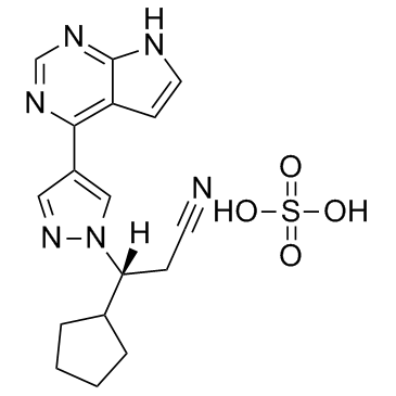
Cas No.: 1092939-16-6
Sample solution is provided at 25 µL, 10mM.
_1-3.$$$39978408@51.jpg');)
_1-9.$$$38538795@65.jpg');)
_1-9.$$$39112701@51.jpg');)
_1-7.$$$37316672@70.jpg');)
_366-372.$$$36198801@70.jpg');)
.$$$38565142@65.jpg');)
_1867-1883.$$$33248023@67.jpg');)
_eads6055.$$$39977546@45.jpg');)
_eadm9805.$$$40080571@45.jpg');)
_eadj3020.$$$39666821@45.jpg');)
_1-20.$$$39757301@28.jpg');)
_1-24.$$$38898113@28.jpg');)
_273-287.$$$36806353@46.jpg');)
_1-16.$$$37156877@44.jpg');)
_1-19.$$$37460805@44.jpg');)
_1291-1307.$$$34518654@46.jpg');)
Ruxolitinib sulfate is the first potent, selective JAK1/2 inhibitor to enter the clinic with IC50s of 3.3 nM/2.8 nM, and has > 130-fold selectivity for JAK1/2 versus JAK3. JAK2|2.8 nM (IC50)|JAK1|3.3 nM (IC50)|Tyk2|19 nM (IC50)|JAK3|428 nM (IC50)
Ruxolitinib sulfate is the first potent, selective JAK1/2 inhibitor to enter the clinic with IC50s of 3.3 nM/2.8 nM, and has > 130-fold selectivity for JAK1/2 versus JAK3. Ruxolitinib potently and selectively inhibits JAK2V617F-mediated signaling and proliferation, markedly increases apoptosis in a dose dependent manner, and at 64 nM results in a doubling of cells with depolarized mitochondria in Ba/F3 cells. Ruxolitinib demonstrates remarkable potency against erythroid colony formation with IC50 of 67 nM, and inhibits proliferating of erythroid progenitors from normal donors and polycythemia vera patients with IC50 values of 407 nM and 223 nM, respectively[1].
Ruxolitinib (180 mg/kg, orally, twice a day) results in survive rate of greater than 90% by day 22 and markedly reduces splenomegaly and circulating levels of inflammatory cytokines, and preferentially eliminated neoplastic cells, resulting in significantly prolonged survival without myelosuppressive or immunosuppressive effects in a JAK2V617F-driven mouse model[1]. In the Ruxolitinib group, the primary end point is reached in 41.9% of patients, as compared with 0.7% in the placebo group in the double-blind trial of myelofibrosis. Ruxolitinib results in maintaining of reduction in spleen volume and improvement of 50% or more in the total symptom score[2]. Ruxolitinib (15 mg twice daily) treatment leads a total of 28% of the patients to have at least a 35% reduction in spleen volume at week 48 in patients with myelofibrosis, as compared with 0% in the group receiving the best available therapy. The mean palpable spleen length has decreased by 56% with Ruxolitinib but has increased by 4% with the best available therapy at week 48. Patients in the ruxolitinib group has an improvement in overall quality-of-life measures and a reduction in symptoms associated with myelofibrosis[3].
[1]. Quintas-Cardama A, et al. Preclinical characterization of the selective JAK1/2 inhibitor INCB018424: therapeutic implications for the treatment of myeloproliferative neoplasms. Blood, 2010, 115(15), 3109-3117. [2]. Verstovsek S, et al. A double-blind, placebo-controlled trial of ruxolitinib for myelofibrosis. N Engl J Med, 2012, 366(9), 799-807. [3]. Harrison C, et al. JAK inhibition with ruxolitinib versus best available therapy for myelofibrosis. N Engl J Med. 2012 Mar 1;366(9):787-98.
Kinase experiment: | Recombinant proteins are expressed using Sf21 cells and baculovirus vectors and purified with affinity chromatography. JAK kinase assays use a homogeneous time-resolved fluorescence assay with the peptide substrate (-EQEDEPEGDYFEWLE). Each enzyme reaction is carried out with Ruxolitinib or control, JAK enzyme, 500 nM peptide, adenosine triphosphate (ATP; 1mM), and 2% dimethyl sulfoxide (DMSO) for 1 hour. The 50% inhibitory concentration (IC50) is calculated as Ruxolitinib concentration required for inhibition of 50% of the fluorescent signal[1]. |
Cell experiment: | Cells are seeded at 2 × 10³/well of white bottom 96-well plates, treated with Ruxolitinib from DMSO stocks (0.2% final DMSO concentration), and incubated for 48 hours at 37°C with 5% CO2. Viability is measured by cellular ATP determination using the Cell-Titer Glo luciferase reagent or viable cell counting. Values are transformed to percent inhibition relative to vehicle control, and IC50 curves are fitted according to nonlinear regression analysis of the data using PRISM GraphPad[1]. |
Animal experiment: | Mice[1]Mice are fed standard rodent chow and provided with water ad libitum. Ba/F3-JAK2V617F cells (105 per mouse) are inoculated intravenously into 6- to 8-week-old female BALB/c mice. Survival is monitored daily, and moribund mice are humanely killed and considered deceased at time of death. Treatment with vehicle (5% dimethyl acetamide, 0.5% methocellulose) or Ruxolitinib begin within 24 hours of cell inoculation, twice daily by oral gavage. Hematologic parameters are measured using a Bayer Advia120 analyzed, and statistical significance is determined using Dunnett testing[1]. |
References: [1]. Quintas-Cardama A, et al. Preclinical characterization of the selective JAK1/2 inhibitor INCB018424: therapeutic implications for the treatment of myeloproliferative neoplasms. Blood, 2010, 115(15), 3109-3117. | |
| Cas No. | 1092939-16-6 | SDF | |
| Canonical SMILES | [H][C@@](C1CCCC1)(N2N=CC(C3=C4C=CNC4=NC=N3)=C2)CC#N.O=S(O)(O)=O | ||
| Formula | C17H20N6O4S | M.Wt | 404.44 |
| Solubility | Soluble in DMSO | Storage | Store at -20°C |
| General tips | Please select the appropriate solvent to prepare the stock solution according to the
solubility of the product in different solvents; once the solution is prepared, please store it in
separate packages to avoid product failure caused by repeated freezing and thawing.Storage method
and period of the stock solution: When stored at -80°C, please use it within 6 months; when stored
at -20°C, please use it within 1 month. To increase solubility, heat the tube to 37°C and then oscillate in an ultrasonic bath for some time. |
||
| Shipping Condition | Evaluation sample solution: shipped with blue ice. All other sizes available: with RT, or with Blue Ice upon request. | ||
| Prepare stock solution | |||

|
1 mg | 5 mg | 10 mg |
| 1 mM | 2.4726 mL | 12.3628 mL | 24.7255 mL |
| 5 mM | 494.5 μL | 2.4726 mL | 4.9451 mL |
| 10 mM | 247.3 μL | 1.2363 mL | 2.4726 mL |
Step 1: Enter information below (Recommended: An additional animal making an allowance for loss during the experiment)
 g
μL
g
μL
Step 2: Enter the in vivo formulation (This is only the calculator, not formulation. Please contact us first if there is no in vivo formulation at the solubility Section.)
Calculation results:
Working concentration: mg/ml;
Method for preparing DMSO master liquid: mg drug pre-dissolved in μL DMSO ( Master liquid concentration mg/mL, Please contact us first if the concentration exceeds the DMSO solubility of the batch of drug. )
Method for preparing in vivo formulation: Take μL DMSO master liquid, next addμL PEG300, mix and clarify, next addμL Tween 80, mix and clarify, next add μL saline, mix and clarify.
Method for preparing in vivo formulation: Take μL DMSO master liquid, next add μL Corn oil, mix and clarify.
Note: 1. Please make sure the liquid is clear before adding the next solvent.
2. Be sure to add the solvent(s) in order. You must ensure that the solution obtained, in the previous addition, is a clear solution before proceeding to add the next solvent. Physical methods such as vortex, ultrasound or hot water bath can be used to aid dissolving.
3. All of the above co-solvents are available for purchase on the GlpBio website.
Quality Control & SDS
- View current batch:
- Purity: >98.00% Appearance: A solid
- COA (Certificate of Analysis)
- SDS (Safety Data Sheet)
- Datasheet
Average Rating: 5 (Based on Reviews and 9 reference(s) in Google Scholar.)