

Secalciferol |
Catalog No.GC37614
 One-Click Copy Product Info
|

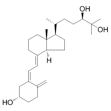
Secalciferol is a fat-soluble vitamin derived from plants and fungi, also known as (24R)-24,25-Dihydroxyvitamin D3. Secalciferol activates the vitamin D receptor (VDR) with an EC50 of 150nM.
Products are for research use only. Not for human use. We do not sell to patients.
Cas No.: 55721-11-4
Sample solution is provided at 25 µL, 10mM.
_1-3.$$$39978408@51.jpg');)
_1-9.$$$38538795@65.jpg');)
_1-9.$$$39112701@51.jpg');)
_1-7.$$$37316672@70.jpg');)
_366-372.$$$36198801@70.jpg');)
.$$$38565142@65.jpg');)
_1867-1883.$$$33248023@67.jpg');)
_eads6055.$$$39977546@45.jpg');)
_eadm9805.$$$40080571@45.jpg');)
_eadj3020.$$$39666821@45.jpg');)
_1-20.$$$39757301@28.jpg');)
_1-24.$$$38898113@28.jpg');)
_273-287.$$$36806353@46.jpg');)
_1-16.$$$37156877@44.jpg');)
_1-19.$$$37460805@44.jpg');)
_1291-1307.$$$34518654@46.jpg');)
Secalciferol is a fat-soluble vitamin derived from plants and fungi, also known as (24R)-24,25-Dihydroxyvitamin D3. Secalciferol activates the vitamin D receptor (VDR) with an EC50 of 150nM[1-2]. Secalciferol is involved in various biological functions, including calcium homeostasis, cell differentiation, proliferation processes, and other functions related to the immune system. Secalciferol can be used in research on rickets, osteomalacia, hypercalcemia, and autoimmune diseases[3-4].
In vitro, Secalciferol (100pM - 10μM) was used to treat the IZ-VDRE and IZ-CYP24 stably transfected reporter gene cell lines for 24 hours. Secalciferol demonstrated a dose-dependent activation of the vitamin D receptor (VDR). The half-maximal effective concentration (EC50) of Secalciferol in the IZ-VDRE and IZ-CYP24 cell lines was 140.8nM and 166.1nM[5]. In the chondrogenic ATDC5 cell differentiation model, treatment with Secalciferol (0.1μM) for 7-14 days enhanced the gene expression of type 2 collagen (Col2a1) in Tlcd3b2-overexpressing cells, promoted proteoglycan synthesis, and facilitated the early differentiation of chondrocytes[6].
In vivo, Cyp24a1 knockout (Cyp24a1-/-) mice in a fracture repair model received subcutaneous injections of Secalciferol (6.7μg/kg/day) for 15 days. Secalciferol significantly corrected the defects in callus formation and callus stiffness during fracture repair in Cyp24a1-/- mice, restored callus volume, and improved the mechanical properties of the callus[7]. In hypophosphatemic (Hyp) mice, daily intraperitoneal injections of Secalciferol (1-1000μg/kg/day) for 28 days. Secalciferol significantly promoted bone formation in the lumbar vertebrae and improved bone formation parameters such as bone volume/tissue volume ratio and mineral apposition rate in a dose-dependent manner[8].
References:
[1] Henderson CM, Fink SL, Bassyouni H, et al. Vitamin D-Binding Protein Deficiency and Homozygous Deletion of the GC Gene. The New England Journal of Medicine. 2019 Mar;380(12):1150-1157.
[2] Wilson RT, Roff AN, Dai PJ, et al. Genetic Ancestry, Skin Reflectance and Pigmentation Genotypes in Association with Serum Vitamin D Metabolite Balance. Hormone Molecular Biology and Clinical Investigation. 2011 Sep;7(1):279-293.
[3] Makris K, Bhattoa HP, Cavalier E, et al. Recommendations on the measurement and the clinical use of vitamin D metabolites and vitamin D binding protein - A position paper from the IFCC Committee on bone metabolism. Clinica Chimica Acta; International Journal of Clinical Chemistry. 2021 Jun;517:171-197.
[4] Ikezaki S, Nishikawa A, Furukawa F, et al. Influences of long-term administration of 24R, 25-dihydroxyvitamin D3, a vitamin D3 derivative, in rats. J Toxicol Sci. 1999 May;24(2):133-9.
[5] Bartonkova I, Grycova A, Dvorak Z. Profiling of Vitamin D Metabolic Intermediates toward VDR Using Novel Stable Gene Reporter Cell Lines IZ-VDRE and IZ-CYP24. Chem Res Toxicol. 2016 Jul 18;29(7):1211-22.
[6] Antonyan L, Martineau C, St-Arnaud R. The ER protein TLC domain 3B2 and its enzymatic product lactosylceramide enhance chondrocyte maturation. Connect Tissue Res. 2021 Mar;62(2):176-182.
[7] Martineau C, Naja RP, Husseini A, et al. Optimal bone fracture repair requires 24R,25-dihydroxyvitamin D3 and its effector molecule FAM57B2. J Clin Invest. 2018 Aug 1;128(8):3546-3557.
[8] Ono T, Tanaka H, Yamate T, et al. 24R,25-dihydroxyvitamin D3 promotes bone formation without causing excessive resorption in hypophosphatemic mice. Endocrinology. 1996 Jun;137(6):2633-7.
| Cell experiment [1]: | |
Cell lines | ATDC5 cells (mouse chondrogenic teratocarcinoma cell line) |
Preparation Method | The ATDC5 prechondrogenic cells were cultured in DMEM/F12 medium supplemented with 5% FBS. For differentiation, ATDC5 cells were cultured in DMEM/F12 medium supplemented with Insulin-Transferrin-Sodium Selenite (ITS). Where indicated, cells were treated with Secalciferol (0.1μM) in charcoal-stripped FBS. |
Reaction Conditions | 0.1μM; 14 day. |
Applications | In ATDC5 cells overexpressing Tlcd3b2, treatment with 0.1μM Secalciferol significantly increased the gene expression of the early chondrogenic differentiation marker Col2a1 compared to untreated cells. |
| Animal experiment [2]: | |
Animal models | Hypophosphatemic (Hyp) mice (a model for X-linked hypophosphatemic rickets) and normal control mice. |
Preparation Method | The mice were divided into groups and received daily intraperitoneal injections for 28 days. Hyp mice received either Secalciferol at doses of 1-1000μg/kg/day. Control mice received no treatment or vehicle. |
Dosage form | 1-1000μg/kg/day; i.p.; Daily for 28 days. |
Applications | Treatment of Hyp mice with Secalciferol dose-dependently promoted bone formation in the fifth lumbar (L5) vertebrae, as evidenced by increased parameters such as the length of the L5 spine, mineral apposition rate (MAR), and mineralized bone volume/bone volume (Md.BV/BV). |
References: | |
| Cas No. | 55721-11-4 | SDF | |
| Canonical SMILES | C=C(CC1)/C(C[C@H]1O)=C\C=C2[C@@]3([H])[C@@](CCC\2)(C)[C@@H]([C@H](C)CC[C@@H](O)C(C)(C)O)CC3 | ||
| Formula | C27H44O3 | M.Wt | 416.64 |
| Solubility | DMSO : 100 mg/mL (240.02 mM; Need ultrasonic); Ethanol : 50 mg/mL (120.01 mM; Need ultrasonic) | Storage | Store at -20°C, protect from light, stored under nitrogen,unstable in solution, ready to use. |
| General tips | Please select the appropriate solvent to prepare the stock solution according to the
solubility of the product in different solvents; once the solution is prepared, please store it in
separate packages to avoid product failure caused by repeated freezing and thawing.Storage method
and period of the stock solution: When stored at -80°C, please use it within 6 months; when stored
at -20°C, please use it within 1 month. To increase solubility, heat the tube to 37°C and then oscillate in an ultrasonic bath for some time. |
||
| Shipping Condition | Evaluation sample solution: shipped with blue ice. All other sizes available: with RT, or with Blue Ice upon request. | ||
| Prepare stock solution | |||

|
1 mg | 5 mg | 10 mg |
| 1 mM | 2.4002 mL | 12.0008 mL | 24.0015 mL |
| 5 mM | 480 μL | 2.4002 mL | 4.8003 mL |
| 10 mM | 240 μL | 1.2001 mL | 2.4002 mL |
Step 1: Enter information below (Recommended: An additional animal making an allowance for loss during the experiment)
 g
μL
g
μL
Step 2: Enter the in vivo formulation (This is only the calculator, not formulation. Please contact us first if there is no in vivo formulation at the solubility Section.)
Calculation results:
Working concentration: mg/ml;
Method for preparing DMSO master liquid: mg drug pre-dissolved in μL DMSO ( Master liquid concentration mg/mL, Please contact us first if the concentration exceeds the DMSO solubility of the batch of drug. )
Method for preparing in vivo formulation: Take μL DMSO master liquid, next addμL PEG300, mix and clarify, next addμL Tween 80, mix and clarify, next add μL saline, mix and clarify.
Method for preparing in vivo formulation: Take μL DMSO master liquid, next add μL Corn oil, mix and clarify.
Note: 1. Please make sure the liquid is clear before adding the next solvent.
2. Be sure to add the solvent(s) in order. You must ensure that the solution obtained, in the previous addition, is a clear solution before proceeding to add the next solvent. Physical methods such as vortex, ultrasound or hot water bath can be used to aid dissolving.
3. All of the above co-solvents are available for purchase on the GlpBio website.
Quality Control & SDS
- View current batch:
- Purity: >99.00% Appearance: A solid
- COA (Certificate of Analysis)
- SDS (Safety Data Sheet)
- Datasheet
Average Rating: 5 (Based on Reviews and 9 reference(s) in Google Scholar.)