

HBC599 |
Catalog No.GC63549
 One-Click Copy Product Info
|

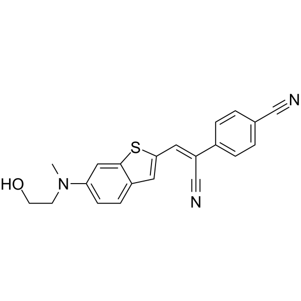
HBC599 is a non-fluorescent HBC mimetic that does not emit light in solution but activates and emits bright fluorescence upon forming a complex with the Pepper RNA aptamer.
Products are for research use only. Not for human use. We do not sell to patients.
Cas No.: 2530162-00-4
Sample solution is provided at 25 µL, 10mM.
_1-3.$$$39978408@51.jpg');)
_1-9.$$$38538795@65.jpg');)
_1-9.$$$39112701@51.jpg');)
_1-7.$$$37316672@70.jpg');)
_366-372.$$$36198801@70.jpg');)
.$$$38565142@65.jpg');)
_1867-1883.$$$33248023@67.jpg');)
_eads6055.$$$39977546@45.jpg');)
_eadm9805.$$$40080571@45.jpg');)
_eadj3020.$$$39666821@45.jpg');)
_1-20.$$$39757301@28.jpg');)
_1-24.$$$38898113@28.jpg');)
_273-287.$$$36806353@46.jpg');)
_1-16.$$$37156877@44.jpg');)
_1-19.$$$37460805@44.jpg');)
_1291-1307.$$$34518654@46.jpg');)
HBC599 is a non-fluorescent HBC mimetic that does not emit light in solution but activates and emits bright fluorescence upon forming a complex with the Pepper RNA aptamer. The complex can be detected using an excitation wavelength of 515nm and an emission wavelength of 599nm[1].
References:
[1] Chen X, Zhang D, Su N, et al. Visualizing RNA dynamics in live cells with bright and stable fluorescent RNAs. Nat Biotechnol. 2019 Nov;37(11):1287-1293.
Staining with HBC599 for Live-Cell RNA Imaging
This protocol is adapted from research data and provided for reference only. Adjustments may be necessary based on specific experimental requirements.
(1) Cell Preparation and Transfection: Seed appropriate cells (e.g., HeLa, U2OS) onto glass-bottom dishes or coverslips and culture until they reach 50-70% confluency. Transfect the cells with a plasmid or RNA expressing the RNA of interest tagged with the Pepper RNA aptamer sequence. Incubate for 12-48 hours to allow for sufficient RNA expression.
(2) HBC599 Solution Preparation: Prepare a 1-5mM stock solution of HBC599 using anhydrous DMSO. Aliquot and store at -20°C or -80°C protected from light. Dilute the HBC599 stock solution in pre-warmed, serum-free cell culture medium (e.g., DMEM or Opti-MEM) to achieve a final working concentration typically between 0.5 to 5µM. Mix thoroughly.
(3) Cell Staining: Gently wash the transfected cells 1-2 times with warm, serum-free medium or PBS. Completely remove the wash buffer and add the prepared HBC599 staining medium to the cells. Incubate the cells in the staining medium at 37°C, 5% CO₂ for 15 to 30 minutes. Carefully aspirate the staining solution. Wash the cells 2-3 times with warm, serum-free medium or 1X PBS.
(4) Live-Cell Imaging: After the final wash, add a small volume of pre-warmed, fresh culture medium to the cells. Proceed to image the cells immediately on a confocal or epifluorescence microscope. HBC599 fluorescence is typically excited with a 515nm laser and detected using a 599nm emission filter.
Precautions:
(1) The incubation time can range from 30 to 90 minutes with minimal impact on result accuracy; choosing 1 hour is most convenient.
(2) For your safety and health, please wear a lab coat and disposable gloves during operation.
References:
[1] Rees HC, Gogacz W, Li NS, et al. Structural Basis for Fluorescence Activation by Pepper RNA. ACS Chem Biol. 2022 Jul 15;17(7):1866-1875.
| Cas No. | 2530162-00-4 | SDF | |
| Formula | C21H17N3OS | M.Wt | 359.44 |
| Solubility | DMSO : ≥ 25 mg/mL (69.55 mM) | Storage | Store at 2-8°C,protect from light |
| General tips | Please select the appropriate solvent to prepare the stock solution according to the
solubility of the product in different solvents; once the solution is prepared, please store it in
separate packages to avoid product failure caused by repeated freezing and thawing.Storage method
and period of the stock solution: When stored at -80°C, please use it within 6 months; when stored
at -20°C, please use it within 1 month. To increase solubility, heat the tube to 37°C and then oscillate in an ultrasonic bath for some time. |
||
| Shipping Condition | Evaluation sample solution: shipped with blue ice. All other sizes available: with RT, or with Blue Ice upon request. | ||
| Prepare stock solution | |||

|
1 mg | 5 mg | 10 mg |
| 1 mM | 2.7821 mL | 13.9105 mL | 27.8211 mL |
| 5 mM | 556.4 μL | 2.7821 mL | 5.5642 mL |
| 10 mM | 278.2 μL | 1.3911 mL | 2.7821 mL |
Step 1: Enter information below (Recommended: An additional animal making an allowance for loss during the experiment)
 g
μL
g
μL
Step 2: Enter the in vivo formulation (This is only the calculator, not formulation. Please contact us first if there is no in vivo formulation at the solubility Section.)
Calculation results:
Working concentration: mg/ml;
Method for preparing DMSO master liquid: mg drug pre-dissolved in μL DMSO ( Master liquid concentration mg/mL, Please contact us first if the concentration exceeds the DMSO solubility of the batch of drug. )
Method for preparing in vivo formulation: Take μL DMSO master liquid, next addμL PEG300, mix and clarify, next addμL Tween 80, mix and clarify, next add μL saline, mix and clarify.
Method for preparing in vivo formulation: Take μL DMSO master liquid, next add μL Corn oil, mix and clarify.
Note: 1. Please make sure the liquid is clear before adding the next solvent.
2. Be sure to add the solvent(s) in order. You must ensure that the solution obtained, in the previous addition, is a clear solution before proceeding to add the next solvent. Physical methods such as vortex, ultrasound or hot water bath can be used to aid dissolving.
3. All of the above co-solvents are available for purchase on the GlpBio website.
Quality Control & SDS
- View current batch:
- Purity: >98.00% Appearance: A solid
- COA (Certificate of Analysis)
- SDS (Safety Data Sheet)
- Datasheet
Average Rating: 5 (Based on Reviews and 39 reference(s) in Google Scholar.)