

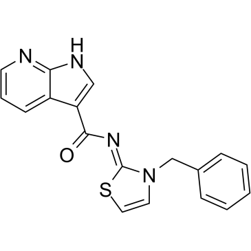
Lats-IN-1 (Synonyms: LATS Inhibitor 1, TRULI) |
Catalog No.GC61527
 One-Click Copy Product Info
|

LATS-IN-1 is an inhibitor of LATS1 and LATS2 (IC50 = 2nM for both). Lats-IN-1 inhibits core Hippo pathway kinases and activates downstream effector molecules YAP/TAZ, which translocate to the nucleus, driving expression of genes that promote cell proliferation and survival. Lats-IN-1 is primarily used to treat tissue regeneration and degenerative diseases.
Products are for research use only. Not for human use. We do not sell to patients.
Cas No.: 1424635-83-5
Sample solution is provided at 25 µL, 10mM.
_1-3.$$$39978408@51.jpg');)
_1-9.$$$38538795@65.jpg');)
_1-9.$$$39112701@51.jpg');)
_1-7.$$$37316672@70.jpg');)
_366-372.$$$36198801@70.jpg');)
.$$$38565142@65.jpg');)
_1867-1883.$$$33248023@67.jpg');)
_eads6055.$$$39977546@45.jpg');)
_eadm9805.$$$40080571@45.jpg');)
_eadj3020.$$$39666821@45.jpg');)
_1-20.$$$39757301@28.jpg');)
_1-24.$$$38898113@28.jpg');)
_273-287.$$$36806353@46.jpg');)
_1-16.$$$37156877@44.jpg');)
_1-19.$$$37460805@44.jpg');)
_1291-1307.$$$34518654@46.jpg');)
LATS-IN-1 is an inhibitor of LATS1 and LATS2 (IC50 = 2nM for both). Lats-IN-1 inhibits core Hippo pathway kinases and activates downstream effector molecules YAP/TAZ, which translocate to the nucleus, driving expression of genes that promote cell proliferation and survival. Lats-IN-1 is primarily used to treat tissue regeneration and degenerative diseases [1-4].
In HEK293A cells, LATS-IN-1 (10μM, 24h) inhibits phosphorylation of YAP1 in serum-starved HEK293A cells (EC50 = 510 nM) [1]. In K562 cells, LATS-IN-1 (30μM, 48h) significantly reduces the number of K562 colonies in the colony formation assay [5]. In human embryonic stem cells, LATS-IN-1 (30μM, 24h) inhibition of the Hippo Pathway Promotes Growth of ATM-Knockout human embryonic stem cells and of Their Derived NPCs [6]. In MCF-7 cells, the proliferation and migration ability of MCF-7 cells was significantly restored after LATS-IN-1 (30μM, 24h) inhibited the Hippo signaling [7]. In LX2 cells, Lats-IN-1 (5μM, 24h) was supplemented, expression of YAP and α-SMA was increased in Ctrl (scrambled)-siRNA-transfected LX2 cells [8].
In a subcutaneous xenograft tumor mouse model of 4T1 cells, LATS-IN-1 (10mg/kg, sc, 7d) increases xenograft tumor growth [7]. In myocardial infarction mice models, Lats-IN-1 (1mg/kg, ip, 14d) treated mice had higher ejection fraction (EF%) and fractional shortening (FS%) than those treated with the vehicle [9]. In lumbar spine instability surgery mice modes Lats-IN-1 (3mg/kg, ip, 56d) treated mice showed less pores or remodeling related signs in the cartilage endplate, with abundant Collagen II expression and rare Collagen X expression [10]. In vascular smooth muscle cell-specific Ddr1 knockout mice, Lats-IN-1 (0.5mg/kg, ip, 7d) abolishes the effect of DDR1-IN-1 on the inhibition of YAP activation [11].
References:
[1]. Kastan N, Gnedeva K, Alisch T, et al. Small-molecule inhibition of Lats kinases may promote Yap-dependent proliferation in postmitotic mammalian tissues. Nature communications. 2021 May 25; 12(1): 3100.
[2]. Cai X, Warburton C, Perez OF, et al. Hippo signaling modulates the inflammatory response of chondrocytes to mechanical compressive loading. bioRxiv. 2023 Jun 11.
[3]. Boopathy GT, Hong W. Role of hippo pathway-YAP/TAZ signaling in angiogenesis. Frontiers in cell and developmental biology. 2019 Apr 10; 7: 49.
[4] Ohnishi Y, Masui A, Suezawa T, et al. Screening of factors inducing alveolar type 1 epithelial cells using human pluripotent stem cells. Stem cell reports. 2024 Apr 9; 19(4): 529-544.
[5]. Klaihmon P, Lorthongpanich C, Kheolamai P, et al. Inhibition of LATS kinases reduces tumorigenicity and increases the sensitivity of human chronic myelogenous leukemia cells to imatinib. Scientific Reports. 2024 Feb 18; 14(1): 3993.
[6]. Viner-Breuer R, Golan-Lev T, Benvenisty N, et al. Genome-Wide Screening in Human Embryonic Stem Cells Highlights the Hippo Signaling Pathway as Granting Synthetic Viability in ATM Deficiency. Cells. 2023 May 29; 12(11): 1503.
[7]. Chen Y, Li J, Pu L, et al. DNAJB4 suppresses breast cancer progression and promotes tumor immunity by regulating the Hippo signaling pathway. Discover Oncology. 2023 Aug 7; 14(1): 144.
[8]. Xie L, Chen H, Zhang L, et al. JCAD deficiency attenuates activation of hepatic stellate cells and cholestatic fibrosis. Clinical and Molecular Hepatology. 2024 Jan 8; 30(2): 206.
[9]. Shen H, Wang Q, Liu B, et al. Lats-IN-1 protects cardiac function and promotes regeneration after myocardial infarction by targeting the hippo pathway. Frontiers in Pharmacology. 2024 Oct 3; 15: 1463465.
[10]. Li H, Tang Y, Liu Z, et al. Lumbar instability remodels cartilage endplate to induce intervertebral disc degeneration by recruiting osteoclasts via Hippo-CCL3 signaling. Bone Research. 2024 May 30; 12(1): 34.
[11]. Liu J, Wang J, Liu Y, et al. Liquid-liquid phase separation of DDR1 counteracts the hippo pathway to orchestrate arterial stiffening. Circulation Research. 2023 Jan 6; 132(1): 87-105.
| Kinase experiment [1]: | |
Preparation Method | The LATS-IN-1 kinase assay uses an in vitro kinase activity detection method, using recombinant Lats1 and Lats2 proteins containing the main kinase domain, and STK1 peptides known to be phosphorylated by them as substrates. Gradient concentrations of LATS-IN-1 are added under different ATP concentrations (10, 50, 250μM). After the reaction system is configured, incubate for 1h. The degree of substrate phosphorylation is detected by fluorescence or FRET and the IC₅₀ value is calculated. |
Reaction Conditions | 0.01nM-1μM; 1h |
Applications | The IC₅₀ of TRULI against Lats1/2 is 0.2nM at 10μM ATP, and the IC₅₀ increases accordingly with increasing ATP concentration, TRULI is an ATP-competitive Lats kinase inhibitor. |
| Cell experiment [1]: | |
Cell lines | HEK293A cells |
Preparation Method | HEK293A cells were plated overnight in 96-well culture plates at a density of 50,000 cells per well in the medium described above. To start the assay, new serum-free medium with 10μM LATS-IN-1 was added to the cells, which were then incubated for 24h at 37 °C. |
Reaction Conditions | 10μM; 24h |
Applications | Lats-IN-1 inhibits YAP phosphorylation and promotes YAP nuclear translocation, activating its downstream transcriptional activity. |
| Animal experiment [2]: | |
Animal models | Myocardial infarction (MI) mice models |
Preparation Method | The mice in the Sham + Lats-IN-1 group and the MI + Lats-IN-1 group were administered intraperitoneally with Lats-IN-1 (1mg/kg) dissolved in corn oil on the first day after myocardial infarction surgery, while the mice in the Sham + Vehicle group and the MI + Vehicle group were given corn oil. Following a period of 2 weeks post-MI, the mice were anesthetized and subsequently sacrificed to facilitate the collection of heart tissues for subsequent examinations and analyses. |
Dosage form | 1mg/kg; ip; 14d |
Applications | Lats-IN-1 treated mice had higher ejection fraction (EF%) and fractional shortening (FS%) than those treated with the vehicle. |
References: | |
| Cas No. | 1424635-83-5 | SDF | |
| Synonyms | LATS Inhibitor 1, TRULI | ||
| Canonical SMILES | O=C(C1=CNC2=NC=CC=C12)/N=C(SC=C3)/N3CC4=CC=CC=C4 | ||
| Formula | C18H14N4OS | M.Wt | 334.39 |
| Solubility | DMSO: 83.33 mg/mL (249.20 mM) | Storage | Store at -20°C |
| General tips | Please select the appropriate solvent to prepare the stock solution according to the
solubility of the product in different solvents; once the solution is prepared, please store it in
separate packages to avoid product failure caused by repeated freezing and thawing.Storage method
and period of the stock solution: When stored at -80°C, please use it within 6 months; when stored
at -20°C, please use it within 1 month. To increase solubility, heat the tube to 37°C and then oscillate in an ultrasonic bath for some time. |
||
| Shipping Condition | Evaluation sample solution: shipped with blue ice. All other sizes available: with RT, or with Blue Ice upon request. | ||
| Prepare stock solution | |||

|
1 mg | 5 mg | 10 mg |
| 1 mM | 2.9905 mL | 14.9526 mL | 29.9052 mL |
| 5 mM | 598.1 μL | 2.9905 mL | 5.981 mL |
| 10 mM | 299.1 μL | 1.4953 mL | 2.9905 mL |
Step 1: Enter information below (Recommended: An additional animal making an allowance for loss during the experiment)
 g
μL
g
μL
Step 2: Enter the in vivo formulation (This is only the calculator, not formulation. Please contact us first if there is no in vivo formulation at the solubility Section.)
Calculation results:
Working concentration: mg/ml;
Method for preparing DMSO master liquid: mg drug pre-dissolved in μL DMSO ( Master liquid concentration mg/mL, Please contact us first if the concentration exceeds the DMSO solubility of the batch of drug. )
Method for preparing in vivo formulation: Take μL DMSO master liquid, next addμL PEG300, mix and clarify, next addμL Tween 80, mix and clarify, next add μL saline, mix and clarify.
Method for preparing in vivo formulation: Take μL DMSO master liquid, next add μL Corn oil, mix and clarify.
Note: 1. Please make sure the liquid is clear before adding the next solvent.
2. Be sure to add the solvent(s) in order. You must ensure that the solution obtained, in the previous addition, is a clear solution before proceeding to add the next solvent. Physical methods such as vortex, ultrasound or hot water bath can be used to aid dissolving.
3. All of the above co-solvents are available for purchase on the GlpBio website.
Quality Control & SDS
- View current batch:
- Purity: >98.00% Appearance: A solid
- COA (Certificate of Analysis)
- SDS (Safety Data Sheet)
- Datasheet
Average Rating: 5 (Based on Reviews and 40 reference(s) in Google Scholar.)