

Lysing Enzymes from Arthrobacter luteus |
Catalog No.GC19846
 One-Click Copy Product Info
|

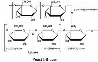
Lysing Enzymes from Arthrobacter luteus, also known as cell wall lysing enzymes or lysing enzymes in Chinese, are highly efficient hydrolases that can specifically degrade poly-β(1→3)-glucose (pol-β(1→3)-glucose) and destroy the integrity of yeast cell walls.
Products are for research use only. Not for human use. We do not sell to patients.
Cas No.: 37340-57-1
Sample solution is provided at 25 µL, 10mM.
Lysing Enzymes from Arthrobacter luteus, also known as cell wall lysing enzymes or lysing enzymes in Chinese, are highly efficient hydrolases that can specifically degrade poly-β(1→3)-glucose (pol-β(1→3)-glucose) and destroy the integrity of yeast cell walls.
Lysing Enzymes from Arthrobacter luteus specifically cut poly-β(1→3)-glucose chains, which are one of the main structural components of yeast cell walls. Yeast cell walls are mainly composed of glucan, chitin and mannoprotein, among which poly-β(1→3)-glucose is the main skeleton component of the cell wall. Yeast cell walls are usually difficult to destroy by conventional physical or chemical methods due to their complex multi-layered structure and the possible formation of capsules or resistant spores. Lysing Enzymes from Arthrobacter luteus significantly reduce the mechanical strength of the cell wall by targeted hydrolysis of key components in the cell wall, thereby inducing the formation of partial protoplasts, providing an efficient and gentle solution for subsequent cell lysis and DNA release. In practical applications, Lysing Enzymes from Arthrobacter luteus can be used to lyse the cell walls of a variety of yeasts and fungi, including Saccharomyces cerevisiae and Schizosaccharomyces pombe. Lysing Enzymes from Arthrobacter luteus has the advantages of efficient hydrolysis, broad-spectrum adaptability and high stability[1] [2].
References:
[1] KITAMURA K, KANEKO T, YAMAMOTO Y. Lysis of viable yeast cells by enzymes of Arthrobacter luteus II. Purification and properties of an enzyme, zymolyase, which lyses viable yeast cells[J]. The Journal of General and Applied Microbiology, 1974, 20(6): 323-344.
[2] Połomska X, Kierul M, Dąbrowska A, et al. The effectiveness comparison of linear dsDNA plasmids isolation from Debaryomyces hansenii yeasts depending on cell disintegration technique[J]. Acta Scientiarum Polonorum. Biotechnology, 2014.
This protocol is for guidance only. The actual application should be modified according to your needs.
F. oxysporum pathogen protoplasm preparation[1]:
1. Enzyme solution preparation: Dissolve 500mg Driselase, 200mg Lysing Enzymes from Arthrobacter luteus and 200mg Snailase in 20mL 1.2M KCl solution to prepare enzyme digestion solution. Mix 1mL 1M Tris-HCl (pH 7.5), 2mL 2.5M CaCl2, and 96mL ddH2O to prepare sucrose-tris-calcium (STC) buffer and store in a refrigerator at 4℃.
2. Cell treatment: After F. oxysporum is cultured in peptone dextrose (YPD) liquid medium and spores are collected, they are inoculated into YPD medium containing ampicillin for overnight culture. Then, the mycelium was washed with sterile distilled water and 1.2M KCl in turn to remove the culture medium and other impurities in preparation for subsequent enzymatic digestion.
3. Incubation reaction: The washed mycelium was incubated with the prepared enzyme solution for 4 hours at 28°C and a shaking speed of 90rpm to break the mycelial wall and release the protoplasts by the digestive action of the enzyme. After the incubation, the protoplasts were separated by filtration and centrifugation at 5000rpm for 90s at 4°C, and washed three times with STC buffer to remove residual enzymes and other impurities.
Notes:
1) For your safety and health, please wear lab coats and protective gloves when using.
2) The activity of Lysing Enzymes from Arthrobacter luteus is affected by factors such as ion concentration, reaction temperature and pH. It is recommended to find the optimal concentration when using for the first time.
References:
[1] Fan S, Zhou Y, Zhu N, et al. Exogenous Application of dsRNA—Inducing Silencing of the Fusarium oxysporum Tup1 Gene and Reducing Its Virulence[J]. International Journal of Molecular Sciences, 2024, 25(19): 10286.
| Cas No. | 37340-57-1 | SDF | |
| Formula | M.Wt | ||
| Solubility | Storage | Store at 2-8°C | |
| General tips | Please select the appropriate solvent to prepare the stock solution according to the
solubility of the product in different solvents; once the solution is prepared, please store it in
separate packages to avoid product failure caused by repeated freezing and thawing.Storage method
and period of the stock solution: When stored at -80°C, please use it within 6 months; when stored
at -20°C, please use it within 1 month. To increase solubility, heat the tube to 37°C and then oscillate in an ultrasonic bath for some time. |
||
| Shipping Condition | Evaluation sample solution: shipped with blue ice. All other sizes available: with RT, or with Blue Ice upon request. | ||
Step 1: Enter information below (Recommended: An additional animal making an allowance for loss during the experiment)
 g
μL
g
μL
Step 2: Enter the in vivo formulation (This is only the calculator, not formulation. Please contact us first if there is no in vivo formulation at the solubility Section.)
Calculation results:
Working concentration: mg/ml;
Method for preparing DMSO master liquid: mg drug pre-dissolved in μL DMSO ( Master liquid concentration mg/mL, Please contact us first if the concentration exceeds the DMSO solubility of the batch of drug. )
Method for preparing in vivo formulation: Take μL DMSO master liquid, next addμL PEG300, mix and clarify, next addμL Tween 80, mix and clarify, next add μL saline, mix and clarify.
Method for preparing in vivo formulation: Take μL DMSO master liquid, next add μL Corn oil, mix and clarify.
Note: 1. Please make sure the liquid is clear before adding the next solvent.
2. Be sure to add the solvent(s) in order. You must ensure that the solution obtained, in the previous addition, is a clear solution before proceeding to add the next solvent. Physical methods such as vortex, ultrasound or hot water bath can be used to aid dissolving.
3. All of the above co-solvents are available for purchase on the GlpBio website.
Quality Control & SDS
- View current batch:
- Biological Activity: 298u/mg Appearance: A solid
- COA (Certificate of Analysis)
- SDS (Safety Data Sheet)
- Datasheet
Average Rating: 5 (Based on Reviews and 10 reference(s) in Google Scholar.)