Home>>Signaling Pathways>> Others>>MitoMark Red I

MitoMark Red I

Catalog No.GC50454

Red fluorescent mitochondrial stain; cell permeable

Products are for research use only. Not for human use. We do not sell to patients.
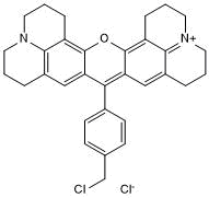
MitoMark Red I Chemical Structure

Cas No.: 167095-09-2

Size Price Stock Qty
50ug
$108.00
In stock
50ug
$108.00
In stock

Tel:(909) 407-4943 Email: sales@glpbio.com


Customer Reviews

Based on customer reviews.

  • GlpBio Citations

    GlpBio Citations
  • Bioactive Compounds Premium Provider

    Bioactive Compounds Premium Provider

Sample solution is provided at 25 µL, 10mM.

Product has been cited by 1 publications

Description of MitoMark Red I

Red fluorescent mitochondrial stain. Fluorescence intensity is dependent on mitochondrial membrane potential. Excitation/emission maxima λ ~ 578/599 nm.

References:
[1]. Poot et al (1996) Analysis of mitochondrial morphology and function with novel fixable fluorescent stains. J.Histochem.Cytochem. 44 1363 PMID:8985128
[2]. Buckman et al (2001) MitoTracker labeling in primary neuronal and astrocytic cultures: influence of mitochondrial membrane potential and oxidants. J.Neuroscience Methods. 104 165 PMID:11164242

Protocol of MitoMark Red I

This plan only provides a guide, please modify it to meet your specific needs.

1. Prepare dyeing solution

(1) Dye storage solution: Use DMSO to dissolve MitoMark Red I into a 1-5mM storage solution. The prepared storage solution was aliquoted and stored in the dark at -20°C.

(2) Dye working solution: Dilute the storage solution with a suitable buffer (such as serum-free medium, HBSS or PBS) to prepare a MitoMark Red I working solution with a concentration of 25-500 nM.

Notice:

① The commonly used working concentration range of MitoMark Red I is 25-500nM. It is recommended to use a relatively low concentration for staining. If you use a higher concentration, other cellular structures may be stained. In order to reduce false positives that may be caused by overloading the probe, it is recommended to use as low a concentration as possible without affecting the staining effect;

② For subsequent cell staining that requires fixation and permeabilization, it is recommended to use a working concentration range of 100-500nM;

③ Please adjust and optimize the concentration of the working fluid according to the actual situation, and prepare it now.


2. Cell suspension staining (taking a 6-well plate as an example)

(1) Centrifuge suspended cells at 1000g for 3-5 minutes. Discard the supernatant and wash twice with PBS for 5 minutes each time.

(2) Wash the adherent cells twice with PBS, add trypsin to digest the cells, and centrifuge at 1000g for 3-5 minutes after digestion is completed.

(3) Add 1 mL of dye working solution to resuspend the cells, and incubate at room temperature in the dark for 15-45 minutes. The optimal culture time for different cells is different.

(4) After the incubation, centrifuge at 1000g for 5 minutes, remove the supernatant, and add PBS to wash 2-3 times, 5 minutes each time.

(5) Use serum-free cell culture medium or PBS to resuspend the cells and observe them through fluorescence microscopy or flow cytometry.

3. Cell adhesion staining

(1) Culture adherent cells on sterile coverslips.

(2) Remove the coverslip from the culture medium, aspirate the excess culture medium, and place the coverslip in a humid environment.

(3) Add 100μL of dye working solution from one corner of the coverslip, shake gently to evenly cover all cells with the dye, and incubate at room temperature in the dark for 15-45 minutes.

(4) Aspirate away the dye working solution and use culture solution to wash the coverslip 2 to 3 times for 5 minutes each time.

4. Observe using a fluorescence microscope or flow cytometer. MitoMark Red I has a maximum excitation/emission wavelength of 578/599 nm.


Precautions:

① If subsequent steps such as ICC require permeabilization, it is recommended to use a buffer containing surfactant (such as Triton X-100) to permeabilize the cells. Cells can also be permeabilized with ice-cold acetone for 5 minutes and then washed with PBS (even if the cells are not subsequently labeled with other antibodies, this acetone permeabilization step may also improve the signal-to-noise ratio);

② If cells need to be fixed and stained, it is recommended to use freshly prepared preheated buffer or culture medium containing 2-4% paraformaldehyde for fixation;

③ Fluorescent dyes all have quenching problems. Please try to avoid light to slow down fluorescence quenching;

④ For your safety and health, please wear a lab coat and disposable gloves.


References:

[1]. K Gilmore, M Wilson. The use of chloromethyl-X-rosamine (Mitotracker red) to measure loss of mitochondrial membrane potential in apoptotic cells is incompatible with cell fixation. 1999 Aug 1;36(4):355-8. doi: 10.1002/(sici)1097-0320(19990801)36:4<355::aid-cyto11>3.0.co;2-9.

Chemical Properties of MitoMark Red I

Cas No. 167095-09-2 SDF
Canonical SMILES ClCC(C=C1)=CC=C1C(C(C2=C3CCC4)=CC5=C3N4CCC5)=C6C(O2)=C7C8=[N+](CCC7)CCCC8=C6.[Cl-]
Formula C32H32Cl2N2O M.Wt 531.52
Solubility Soluble in DMSO Storage Store at -20°C,protect from light
General tips Please select the appropriate solvent to prepare the stock solution according to the solubility of the product in different solvents; once the solution is prepared, please store it in separate packages to avoid product failure caused by repeated freezing and thawing.Storage method and period of the stock solution: When stored at -80°C, please use it within 6 months; when stored at -20°C, please use it within 1 month.
To increase solubility, heat the tube to 37°C and then oscillate in an ultrasonic bath for some time.
Shipping Condition Evaluation sample solution: shipped with blue ice. All other sizes available: with RT, or with Blue Ice upon request.

Complete Stock Solution Preparation Table of MitoMark Red I

Prepare stock solution
1 mg 5 mg 10 mg
1 mM 1.8814 mL 9.407 mL 18.814 mL
5 mM 0.3763 mL 1.8814 mL 3.7628 mL
10 mM 0.1881 mL 0.9407 mL 1.8814 mL
  • Molarity Calculator

  • Dilution Calculator

  • Molecular Weight Calculator

Mass
=
Concentration
x
Volume
x
MW*


**When preparing stock solutions always use the batch-specific molecular weight of the product found on the vial label and MSDS / CoA (available online).

Calculate

In vivo Formulation Calculator (Clear solution) of MitoMark Red I

Step 1: Enter information below (Recommended: An additional animal making an allowance for loss during the experiment)

mg/kg g μL

Step 2: Enter the in vivo formulation (This is only the calculator, not formulation. Please contact us first if there is no in vivo formulation at the solubility Section.)

% DMSO % % Tween 80 % ddH2O
%DMSO %

Calculation results:

Working concentration: mg/ml;

Method for preparing DMSO master liquid: mg drug pre-dissolved in μL DMSO ( Master liquid concentration mg/mL, Please contact us first if the concentration exceeds the DMSO solubility of the batch of drug. )

Method for preparing in vivo formulation: Take μL DMSO master liquid, next addμL PEG300, mix and clarify, next addμL Tween 80, mix and clarify, next add μL ddH2O, mix and clarify.

Method for preparing in vivo formulation: Take μL DMSO master liquid, next add μL Corn oil, mix and clarify.

Note: 1. Please make sure the liquid is clear before adding the next solvent.
2. Be sure to add the solvent(s) in order. You must ensure that the solution obtained, in the previous addition, is a clear solution before proceeding to add the next solvent. Physical methods such as vortex, ultrasound or hot water bath can be used to aid dissolving.
3. All of the above co-solvents are available for purchase on the GlpBio website.

Product Documents

Quality Control & SDS

View current batch:

Reviews

Review for MitoMark Red I

Average Rating: 5 ★★★★★ (Based on Reviews and 30 reference(s) in Google Scholar.)

5 Star
100%
4 Star
0%
3 Star
0%
2 Star
0%
1 Star
0%
Review for MitoMark Red I

GLPBIO products are for RESEARCH USE ONLY. Please make sure your review or question is research based.

Required fields are marked with *

You may receive emails regarding this submission. Any emails will include the ability to opt-out of future communications.