

PKH 26 |
Catalog No.GC19925
 One-Click Copy Product Info
|

PKH 26 is a lipophilic fluorescent dye that is widely used in cell labeling, cell proliferation, and cell tracing. PKH 26 has almost no toxic side effects, with an optimal excitation wavelength of 551 nm and an optimal emission wavelength of 567 nm.
Products are for research use only. Not for human use. We do not sell to patients.
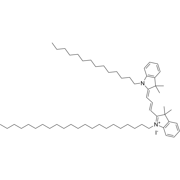
Cas No.: 154214-55-8
Sample solution is provided at 25 µL, 10mM.
PKH 26 is a lipophilic fluorescent dye that is widely used in cell labeling, cell proliferation, and cell tracing, and has almost no toxic side effects, with an optimal excitation wavelength of 551 nm and an optimal emission wavelength of 567 nm[1-4].
The proliferation of human colon epithelial cells can be studied by labeling them with PKH 26(1∶250 (v/v)) [5]. PKH 26 can also be used to trace the effector induced apoptosis of target cells[6].
In rat autologous mature adipocyte transplantation, PKH 26-stained adipocytes could still be detected after 14.5 months[7]. Intravitreal injection of fluorescent (PKH 26)-labeled or exosomes (106 exosomes/μL) were given to the wild-type mouse or laser-induced Choroidal Neovascularization(CNV) mouse model. Intravitreally delivered PKH 26-labeled exosomes reached inner and outer retinal layers at 1 and 7 days post-injection after the intravitreal injection of exosomes[8].
References:
[1]. Horan PK, Slezak SE. Stable cell membrane labelling. Nature. 1989 Jul 13;340(6229):167-8. doi: 10.1038/340167a0. PMID: 2662017.
[2]. Horan PK, Melnicoff MJ, et,al. Fluorescent cell labeling for in vivo and in vitro cell tracking. Methods Cell Biol. 1990;33:469-90. doi: 10.1016/s0091-679x(08)60547-6. PMID: 2084480.
[3]. Samlowski WE, Robertson BA, et,al. Effects of supravital fluorochromes used to analyze the in vivo homing of murine lymphocytes on cellular function. J Immunol Methods. 1991 Nov 5;144(1):101-15. doi: 10.1016/0022-1759(91)90236-9. PMID: 1960398.
[4]. Fischer K, Mackensen A. The flow cytometric PKH-26 assay for the determination of T-cell mediated cytotoxic activity. Methods. 2003 Oct;31(2):135-42. doi: 10.1016/s1046-2023(03)00123-3. PMID: 12957571.
[5]. Pastò A, Marchesi M, et,al. PKH26 staining defines distinct subsets of normal human colon epithelial cells at different maturation stages. PLoS One. 2012;7(8):e43379. doi: 10.1371/journal.pone.0043379. Epub 2012 Aug 22. PMID: 22927961; PMCID: PMC3425557.
[6]. Drvar V, Ćurko-Cofek B, et,al. Granulysin expression and granulysin-mediated apoptosis in the peripheral blood of osteoarthritis patients. Biomed Rep. 2022 May;16(5):44. doi: 10.3892/br.2022.1527. Epub 2022 Mar 24. PMID: 35478928; PMCID: PMC9016702.
[7]. Rieck B. Unexpected durability of PKH 26 staining on rat adipocytes. Cell Biol Int. 2003;27(5):445-7. doi: 10.1016/s1065-6995(03)00036-2. PMID: 12758093.
[8]. Pollalis D, Kim D, et,al. Intraocular RGD-Engineered Exosomes and Active Targeting of Choroidal Neovascularization (CNV). Cells. 2022 Aug 18;11(16):2573. doi: 10.3390/cells11162573. PMID: 36010651; PMCID: PMC9406786.
|
Protocol for cell fluorescence labelling using PKH-26 [1]: |
This protocol only provides a guideline, and should be modified according to your specific needs. |
|
Protocol for cell proliferation using PKH-26 [2]: |
This protocol only provides a guideline, and should be modified according to your specific needs. |
|
Protocol for intracellular uptake of exosomes in mice using PKH-26 [3]: |
This protocol only provides a guideline, and should be modified according to your specific needs. |
|
References: [1]. Fischer K, Mackensen A. The flow cytometric PKH-26 assay for the determination of T-cell mediated cytotoxic activity. Methods. 2003 Oct;31(2):135-42. doi: 10.1016/s1046-2023(03)00123-3. PMID: 12957571. [2].Pastò A, Marchesi M,et,al. PKH26 staining defines distinct subsets of normal human colon epithelial cells at different maturation stages. PLoS One. 2012;7(8):e43379. doi: 10.1371/journal.pone.0043379. Epub 2012 Aug 22. PMID: 22927961; PMCID: PMC3425557. [3]. Pollalis D, Kim D, et,al. Intraocular RGD-Engineered Exosomes and Active Targeting of Choroidal Neovascularization (CNV). Cells. 2022 Aug 18;11(16):2573. doi: 10.3390/cells11162573. PMID: 36010651; PMCID: PMC9406786. |
| Cas No. | 154214-55-8 | SDF | |
| Formula | C59H97IN2 | M.Wt | 961.32 |
| Solubility | DMSO:1.67 mg/mL (1.74 mM) | Storage | Store at -20°C,protect from light |
| General tips | Please select the appropriate solvent to prepare the stock solution according to the
solubility of the product in different solvents; once the solution is prepared, please store it in
separate packages to avoid product failure caused by repeated freezing and thawing.Storage method
and period of the stock solution: When stored at -80°C, please use it within 6 months; when stored
at -20°C, please use it within 1 month. To increase solubility, heat the tube to 37°C and then oscillate in an ultrasonic bath for some time. |
||
| Shipping Condition | Evaluation sample solution: shipped with blue ice. All other sizes available: with RT, or with Blue Ice upon request. | ||
| Prepare stock solution | |||

|
1 mg | 5 mg | 10 mg |
| 1 mM | 1.0402 mL | 5.2012 mL | 10.4024 mL |
| 5 mM | 208 μL | 1.0402 mL | 2.0805 mL |
| 10 mM | 104 μL | 520.1 μL | 1.0402 mL |
Step 1: Enter information below (Recommended: An additional animal making an allowance for loss during the experiment)
 g
μL
g
μL
Step 2: Enter the in vivo formulation (This is only the calculator, not formulation. Please contact us first if there is no in vivo formulation at the solubility Section.)
Calculation results:
Working concentration: mg/ml;
Method for preparing DMSO master liquid: mg drug pre-dissolved in μL DMSO ( Master liquid concentration mg/mL, Please contact us first if the concentration exceeds the DMSO solubility of the batch of drug. )
Method for preparing in vivo formulation: Take μL DMSO master liquid, next addμL PEG300, mix and clarify, next addμL Tween 80, mix and clarify, next add μL saline, mix and clarify.
Method for preparing in vivo formulation: Take μL DMSO master liquid, next add μL Corn oil, mix and clarify.
Note: 1. Please make sure the liquid is clear before adding the next solvent.
2. Be sure to add the solvent(s) in order. You must ensure that the solution obtained, in the previous addition, is a clear solution before proceeding to add the next solvent. Physical methods such as vortex, ultrasound or hot water bath can be used to aid dissolving.
3. All of the above co-solvents are available for purchase on the GlpBio website.
Quality Control & SDS
- View current batch:
- Purity: >98.00% Appearance: A solid
- COA (Certificate of Analysis)
- SDS (Safety Data Sheet)
- Datasheet
Average Rating: 5 (Based on Reviews and 29 reference(s) in Google Scholar.)