

PRIMA-1MET (Synonyms: APR-246) |
Catalog No.GC11247
 One-Click Copy Product Info
|

PRIMA-1MET is a small molecule organic compound that can restore tumor suppression function, mainly targeting mutant p53 and inducing cell death in various types of cancer cells.
Products are for research use only. Not for human use. We do not sell to patients.
Cas No.: 5291-32-7
Sample solution is provided at 25 µL, 10mM.
_1-3.$$$39978408@51.jpg');)
_1-9.$$$38538795@65.jpg');)
_1-9.$$$39112701@51.jpg');)
_1-7.$$$37316672@70.jpg');)
_366-372.$$$36198801@70.jpg');)
.$$$38565142@65.jpg');)
_1867-1883.$$$33248023@67.jpg');)
_eads6055.$$$39977546@45.jpg');)
_eadm9805.$$$40080571@45.jpg');)
_eadj3020.$$$39666821@45.jpg');)
_1-20.$$$39757301@28.jpg');)
_1-24.$$$38898113@28.jpg');)
_273-287.$$$36806353@46.jpg');)
_1-16.$$$37156877@44.jpg');)
_1-19.$$$37460805@44.jpg');)
_1291-1307.$$$34518654@46.jpg');)
PRIMA-1MET is a small molecule organic compound that can restore tumor suppression function, mainly targeting mutant p53 and inducing cell death in various types of cancer cells[1]. PRIMA-1MET is hydrolyzed into the active form of methylquinine cycloketone within cells and covalently binds to the thiol group of the core domain of the mutant p53 protein, thereby causing structural changes and restoring the active conformation of p53 protein [2]. PRIMA-1MET has been widely used in anti-cancer research[3].
In vitro, PRIMA-1MET inhibits the cell viability of BWM-1 cells (IC50 value=30μM) and MWCL-1 cells (IC50 value= 28μM) after treatment of 48 hours[4]. PRIMA-1MET treatment (60µM) for 24 hours significant induced autophagy and enhanced the mTOR/AMPK-ULK1-Vps34 autophagic signaling cascade in colorectal cancer (CRC) cells[5]. PRIMA-1 MET (100μM; 72H) effectively inhibited the growth of small cell lung cancer (SCLC) cell lines expressing mutant p53 induced apoptosis, accompanied by an increase in the proportion of DNA fragmented cells, caspase-3 activation, and downregulation of Bcl-2 expression[6].
In vivo, PRIMA-1MET treatment (100mg/kg/day; i.p.) for 15 consecutive days can inhibit the growth of multiple myeloma tumors containing mutant p53 in multiple myeloma xenograft SCID mouse models[7]. Intraperitoneal injection of PRIMA-1MET at the dosage of 100mg/kg/day for 5 consecutive days significantly reduced the tumor size and tumor volume in the mouse model of rectal cancer xenograft tumors, decreased ERK1/2 phosphorylation, and inhibited MEK activity[8].
References:
[1] Perdrix, A.; Najem, A.; Saussez, S.; Awada, A.; Journe, F.; Ghanem, G.; Krayem, M. PRIMA-1 and PRIMA-1Met (APR-246): From Mutant/Wild Type p53 Reactivation to Unexpected Mechanisms Underlying Their Potent Anti-Tumor Effect in Combinatorial Therapies. Cancers 2017, 9, 172.
[2] Xie X, Fan C, Luo B, et al. APR-246 enhances colorectal cancer sensitivity to radiotherapy[J]. Molecular cancer therapeutics, 2023, 22(8): 947-961.
[3] Menichini P, Monti P, Speciale A, et al. Antitumor effects of PRIMA-1 and PRIMA-1Met (APR246) in hematological malignancies: still a mutant p53-dependent affair?[J]. Cells, 2021, 10(1): 98.
[4] Sobhani M, Kwan K, Saha M N, et al. Small molecule PRIMA-1 met sensitizes Waldenstrom macroglobulinemia cells to apoptosis and displays synergistic cytotoxicity with bortezomib[J]. Blood, 2013, 122(21): 5143.
[5] Li X L, Zhou J, Xia C J, et al. PRIMA-1met induces autophagy in colorectal cancer cells through upregulation of the mTOR/AMPK-ULK1-Vps34 signaling cascade[J]. Oncology Reports, 2021, 45(5): 86.
[6] Zandi R, Selivanova G, Christensen C L, et al. PRIMA-1Met/APR-246 induces apoptosis and tumor growth delay in small cell lung cancer expressing mutant p53[J]. Clinical Cancer Research, 2011, 17(9): 2830-2841.
[7] Saha M N, Jiang H, Yang Y, et al. PRIMA-1Met/APR-246 displays high antitumor activity in multiple myeloma by induction of p73 and Noxa[J]. Molecular cancer therapeutics, 2013, 12(11): 2331-2341.
[8] Lu T, Zou Y, Xu G, et al. PRIMA-1Met suppresses colorectal cancer independent of p53 by targeting MEK[J]. Oncotarget, 2016, 7(50): 83017.
| Cell experiment [1]: | |
Cell lines | KKU-100 cell |
Preparation Method | KKU-100 cells were cultured in the modified Eagle medium (DMEM) supplemented with 10% heat-inactivated fetal bovine serum (FBS), 2mg/ml sodium bicarbonate and 1% antibiotic solution. All cells were cultured in a humidified incubator at 37℃ with a concentration of 5% CO2. KKU-100 cells (2×10³) suspended in 100μl of medium were inoculated into 96-well plates in triplicate. Cells were treated with PRIMA-1MET at different concentrations (0, 20, 40, 60, 80, and 100μM) for 48 hours. Cell viability was determined using sulfonylrhodamine B (SRB). The absorbance was measured at 540nm using a microplate reader. |
Reaction Conditions | 0, 20, 40, 60, 80, and 100μM; 48h |
Applications | PRIMA-1MET significantly inhibited the viability of KKU-100 cells in a dose-dependent manner. |
| Animal experiment [2]: | |
Animal models | Balb/c mice |
Preparation Method | Five Balb/ c mice in three groups were subcutaneously inoculated with 5×10⁵MCO4 cells. On the 7th day after inoculation, PBS or PRIMA-1MET at a dose of 100mg/kg was intravenously injected into the tail vein of mice once a day for 10 consecutive days. During the observation period, body weight and tumor size were monitored starting from vaccination. The tumor was immediately fixed in 4% formaldehyde for 12 hours after resection, stored in 70% ethanol at 4℃, and then embedded in paraffin. Hematoxylin and eosin staining were carried out according to the standard protocol. According to the manufacturer's instructions, apoptotic cells were detected in situ using TUNEL. |
Dosage form | 100mg/kg for 10 days; i.v. |
Applications | PRIMA-1MET treatment significantly suppressed tumor growth, and increased the survival rate of mice without obvious toxicity. |
References: | |
| Cas No. | 5291-32-7 | SDF | |
| Synonyms | APR-246 | ||
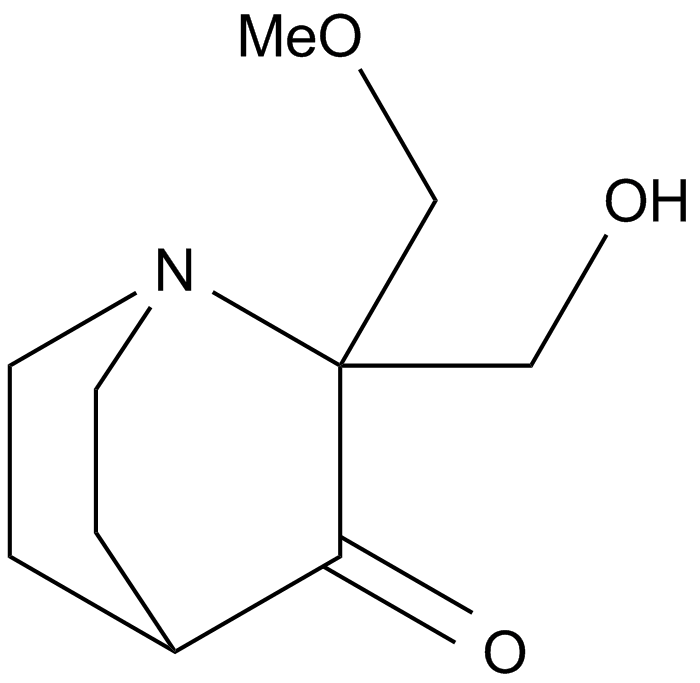
| Chemical Name | 2-(hydroxymethyl)-2-(methoxymethyl)-1-azabicyclo[2.2.2]octan-3-one | ||
| Canonical SMILES | COCC1(C(=O)C2CCN1CC2)CO | ||
| Formula | C10H17NO3 | M.Wt | 199.25 |
| Solubility | ≥ 19.9mg/mL in DMSO, ≥ 102 mg/mL in EtOH with ultrasonic, ≥ 104.2 mg/mL in Water | Storage | Store at 4°C |
| General tips | Please select the appropriate solvent to prepare the stock solution according to the
solubility of the product in different solvents; once the solution is prepared, please store it in
separate packages to avoid product failure caused by repeated freezing and thawing.Storage method
and period of the stock solution: When stored at -80°C, please use it within 6 months; when stored
at -20°C, please use it within 1 month. To increase solubility, heat the tube to 37°C and then oscillate in an ultrasonic bath for some time. |
||
| Shipping Condition | Evaluation sample solution: shipped with blue ice. All other sizes available: with RT, or with Blue Ice upon request. | ||
| Prepare stock solution | |||

|
1 mg | 5 mg | 10 mg |
| 1 mM | 5.0188 mL | 25.0941 mL | 50.1882 mL |
| 5 mM | 1.0038 mL | 5.0188 mL | 10.0376 mL |
| 10 mM | 501.9 μL | 2.5094 mL | 5.0188 mL |
Step 1: Enter information below (Recommended: An additional animal making an allowance for loss during the experiment)
 g
μL
g
μL
Step 2: Enter the in vivo formulation (This is only the calculator, not formulation. Please contact us first if there is no in vivo formulation at the solubility Section.)
Calculation results:
Working concentration: mg/ml;
Method for preparing DMSO master liquid: mg drug pre-dissolved in μL DMSO ( Master liquid concentration mg/mL, Please contact us first if the concentration exceeds the DMSO solubility of the batch of drug. )
Method for preparing in vivo formulation: Take μL DMSO master liquid, next addμL PEG300, mix and clarify, next addμL Tween 80, mix and clarify, next add μL saline, mix and clarify.
Method for preparing in vivo formulation: Take μL DMSO master liquid, next add μL Corn oil, mix and clarify.
Note: 1. Please make sure the liquid is clear before adding the next solvent.
2. Be sure to add the solvent(s) in order. You must ensure that the solution obtained, in the previous addition, is a clear solution before proceeding to add the next solvent. Physical methods such as vortex, ultrasound or hot water bath can be used to aid dissolving.
3. All of the above co-solvents are available for purchase on the GlpBio website.
Quality Control & SDS
- View current batch:
- Purity: >98.00% Appearance: A solid
- COA (Certificate of Analysis)
- SDS (Safety Data Sheet)
- Datasheet
Average Rating: 5 (Based on Reviews and 30 reference(s) in Google Scholar.)