

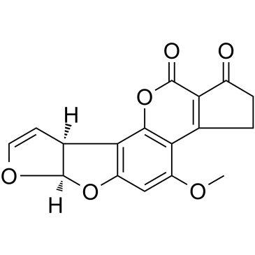
Aflatoxin B1 (Synonyms: AFB1, HSDB 3453, NSC 529592) |
Catalog No.GC35261
 One-Click Copy Product Info
|

Aflatoxin B1 is a mycotoxin produced by Aspergillus flavus and Aspergillus parasiticus with strong toxicity and hepatocarcinogenic activity.
Products are for research use only. Not for human use. We do not sell to patients.
Cas No.: 1162-65-8
Sample solution is provided at 25 µL, 10mM.
_1-3.$$$39978408@51.jpg');)
_1-9.$$$38538795@65.jpg');)
_1-9.$$$39112701@51.jpg');)
_1-7.$$$37316672@70.jpg');)
_366-372.$$$36198801@70.jpg');)
.$$$38565142@65.jpg');)
_1867-1883.$$$33248023@67.jpg');)
_eads6055.$$$39977546@45.jpg');)
_eadm9805.$$$40080571@45.jpg');)
_eadj3020.$$$39666821@45.jpg');)
_1-20.$$$39757301@28.jpg');)
_1-24.$$$38898113@28.jpg');)
_273-287.$$$36806353@46.jpg');)
_1-16.$$$37156877@44.jpg');)
_1-19.$$$37460805@44.jpg');)
_1291-1307.$$$34518654@46.jpg');)
Aflatoxin B1 is a mycotoxin produced by Aspergillus flavus and Aspergillus parasiticus with strong toxicity and hepatocarcinogenic activity. Aflatoxin B1 is metabolically activated by cytochrome P450 enzymes into the 8,9-epoxide, which covalently binds to DNA to form adducts and induces p53 gene G→T mutations. Aflatoxin B1 drives hepatocellular carcinogenesis through inducing oxidative stress and inflammatory responses. Aflatoxin B1 can be used in studies related to food safety, toxicology, and the pathogenesis of hepatocellular carcinoma[1-4].
In vitro, treatment of TM3 Leydig cells with 3.6µM Aflatoxin B1 for 24 hours upregulated Caspase 3, Caspase 8 and Caspase 9 expression, increased the apoptosis rate and DNA fragmentation, and reduced the proportion of cells in G1, S and G2 phases[5]. Treatment of MAC-T cells with 5µM Aflatoxin B1 for 48 hours decreased the proportion of cells in G1 phase, increased the proportion of cells in sub-G1, S and G2-M phases, elevated the proportion of late apoptotic cells, and increased DNA fragmentation[6]. Treatment of PLC/PRF/5 cells with 8µM Aflatoxin B1 for 12 days induced aryl hydrocarbon receptor nuclear translocation and upregulated AHR, CYP1A1, CYP1A2 and PD-L1 expression, promoted long-chain fatty acid accumulation and MLKL, RIPK1, RIPK3 expression, and reduced cell viability[7].
In vivo, a single intraperitoneal injection of 6µg/g Aflatoxin B1 in hepatitis C virus transgenic or wild-type C57BL/6J mice induced hepatocellular adenoma or carcinoma as well as hyperplastic or focal preneoplastic lesions, caused hepatic steatosis and increased liver triglyceride content, and upregulated immune cell chemotaxis-related gene expression and altered lipid metabolism gene expression such as fatty acid translocase and microsomal transport protein in hepatitis C virus transgenic mice[8]. Oral gavage of Swiss albino mice with 9mg/kg Aflatoxin B1 twice weekly for 2 weeks, or with 3-6mg/kg Aflatoxin B1 once daily for 10 days, caused destruction and damage of liver tissue architecture including hepatic sinusoidal congestion, hepatocyte hydropic degeneration, nuclear pyknosis, congestion, necrosis and giant cell transformation, and upregulated MMP1 and MMP7 immunohistochemical expression in liver tissue in a concentration-dependent manner[9]. Daily intraperitoneal injection of C57BL/6J mice with 100µg/kg Aflatoxin B1 for 3 weeks upregulated NLRP3, caspase-1, GSDMD-N, IL-1β and TNF-α levels in the hippocampus, reduced the numbers of Iba1+ microglia, DCX+ immature neurons and BrdU+, BrdU-DCX+, BrdU-NeuN+ cells in the hippocampal dentate gyrus, decreased sucrose preference and prolonged immobility time and shortened latency in the tail suspension test and forced swimming test, while increasing serum malondialdehyde content and decreasing superoxide dismutase activity[10].
References:
[1] Cao W, Yu P, Yang K, et al. Aflatoxin B1: metabolism, toxicology, and its involvement in oxidative stress and cancer development. Toxicol Mech Methods. 2022 Jul;32(6):395-419.
[2] Rushing BR, Selim MI. Aflatoxin B1: A review on metabolism, toxicity, occurrence in food, occupational exposure, and detoxification methods. Food Chem Toxicol. 2019 Feb;124:81-100.
[3] Dai Y, Huang K, Zhang B, et al. Aflatoxin B1-induced epigenetic alterations: An overview. Food Chem Toxicol. 2017 Nov;109(Pt 1):683-689.
[4] Engin AB, Engin A. DNA damage checkpoint response to aflatoxin B1. Environ Toxicol Pharmacol. 2019 Jan;65:90-96.
[5] Jalili C, Abbasi A, Rahmani-Kukia N, et al. The relationship between aflatoxin B1 with the induction of extrinsic/intrinsic pathways of apoptosis and the protective role of taraxasterol in TM3 leydig cell line. Ecotoxicology and Environmental Safety. 2024;276:116316.
[6] Park W, Park MY, Song G, et al. Exposure to aflatoxin B1 attenuates cell viability and induces endoplasmic reticulum-mediated cell death in a bovine mammary epithelial cell line(MAC-T). Toxicology in Vitro. 2019;104591.
[7] Zhu Q, Ma Y, Liang J, et al. AHR mediates the aflatoxin B1 toxicity associated with hepatocellular carcinoma. Signal Transduction and Targeted Therapy. 2021 Aug 9;6:299.
[8] Jeannot E, Boorman GA, Kosyk O, et al. Increased incidence of aflatoxin B1-induced liver tumors in hepatitis virus C transgenic mice. Int J Cancer. 2012 Mar 15;130(6):1347-56.
[9] Al-Mudallal NHA, et al. The Expression of MMP1 and MMP7 in Mice Liver after Exposure to Aflatoxin B1 Using Immunohistochemistry Technique. Archives of Razi Institute. 2023;78(1):63-72.
[10] Su D, Jiang W, Yuan Q, et al. Chronic exposure to aflatoxin B1 increases hippocampal microglial pyroptosis and vulnerability to stress in mice. Ecotoxicology and Environmental Safety. 2023;258:114991.
| Cell experiment [1]: | |
|
Cell lines |
TM3 Leydig cells (mouse Leydig cell line) |
|
Preparation Method |
TM3 Leydig cells were maintained in RPMI 1640 medium containing 1% glutamine, 10% fetal bovine serum, 100IU/mL penicillin and 100IU/mL streptomycin at 37°C, 5% CO2. TM3 Leydig cells were treated with Aflatoxin B1 at 3.6μM for 24 hours, followed by MTT assay, flow cytometry (annexin V/PI and PI staining), TUNEL assay, RT-qPCR and Western blot analysis. |
|
Reaction Conditions |
3.6μM; 24h |
|
Applications |
Aflatoxin B1 upregulated Caspase 3, Caspase 8 and Caspase 9 expression at mRNA and protein levels in TM3 Leydig cells. Aflatoxin B1 increased cellular apoptosis and DNA fragmentation, and reduced the proportion of cells in G1, S and G2 phases of the cell cycle. |
| Animal experiment [2]: | |
|
Animal models |
7-day-old C57BL/6J wild-type male mice and HCV transgenic (HCV-Tg) male mice on C57BL/6J background |
|
Preparation Method |
Neonatal (7-day-old) mice were intraperitoneally injected once with Aflatoxin B1 (6μg/g) dissolved in tricaprylin (15μL/g body weight vehicle), male offspring were maintained on regular chow until 12 months of age, then livers were examined macroscopically and histologically (H&E, Oil Red O, Sirius Red, immunohistochemistry for BrdU, 4-HNE, F4/80), with hepatic triglyceride, glutathione, 8-oxo-dG, mtDNA damage and gene expression (qPCR, microarray) assessments. |
|
Dosage form |
6μg/g; i.p.; single injection |
|
Applications |
Aflatoxin B1 induced liver tumors and preneoplastic lesions in 22.5% of wild-type and HCV-Tg mice at 12 months. Aflatoxin B1 caused hepatic steatosis, increased liver triglyceride content, elevated 4-HNE-adducted proteins, and produced small lobular inflammatory foci in both strains, with HCV-Tg mice showing higher incidence of inflammatory foci, steatosis and adenomas; Aflatoxin B1 increased BrdU-positive nuclei and F4/80-positive macrophage area in wild-type but not HCV-Tg livers, and altered expression of lipid metabolism genes without changing UPR, genomic/mtDNA oxidative DNA damage or glutathione levels. |
|
References: [1] Jalili C, Abbasi A, Rahmani-Kukia N, et al. The relationship between aflatoxin B1 with the induction of extrinsic/intrinsic pathways of apoptosis and the protective role of taraxasterol in TM3 leydig cell line. Ecotoxicology and Environmental Safety. 2024;276:116316. [2] Jeannot E, Boorman GA, Kosyk O, et al. Increased incidence of aflatoxin B1-induced liver tumors in hepatitis virus C transgenic mice. Int J Cancer. 2012 Mar 15;130(6):1347-56. |
|
| Cas No. | 1162-65-8 | SDF | |
| Synonyms | AFB1, HSDB 3453, NSC 529592 | ||
| Canonical SMILES | COC1=CC(O[C@]2([H])[C@@]3([H])C=CO2)=C3C(O4)=C1C(CCC5=O)=C5C4=O | ||
| Formula | C17H12O6 | M.Wt | 312.27 |
| Solubility | DMF: 20 mg/ml,DMF:PBS(pH 7.2)(1:1): 0.5 mg/ml,DMSO: 12 mg/ml | Storage | -20°C, protect from light |
| Shipping Condition | Evaluation sample solution: shipped with blue ice. All other sizes available: with RT, or with Blue Ice upon request. | ||
| Prepare stock solution | |||

|
1 mg | 5 mg | 10 mg |
| 1 mM | 3.2024 mL | 16.0118 mL | 32.0236 mL |
| 5 mM | 640.5 μL | 3.2024 mL | 6.4047 mL |
| 10 mM | 320.2 μL | 1.6012 mL | 3.2024 mL |
Step 1: Enter information below (Recommended: An additional animal making an allowance for loss during the experiment)
 g
μL
g
μL
Step 2: Enter the in vivo formulation (This is only the calculator, not formulation. Please contact us first if there is no in vivo formulation at the solubility Section.)
Calculation results:
Working concentration: mg/ml;
Method for preparing DMSO master liquid: mg drug pre-dissolved in μL DMSO ( Master liquid concentration mg/mL, Please contact us first if the concentration exceeds the DMSO solubility of the batch of drug. )
Method for preparing in vivo formulation: Take μL DMSO master liquid, next addμL PEG300, mix and clarify, next addμL Tween 80, mix and clarify, next add μL saline, mix and clarify.
Method for preparing in vivo formulation: Take μL DMSO master liquid, next add μL Corn oil, mix and clarify.
Note: 1. Please make sure the liquid is clear before adding the next solvent.
2. Be sure to add the solvent(s) in order. You must ensure that the solution obtained, in the previous addition, is a clear solution before proceeding to add the next solvent. Physical methods such as vortex, ultrasound or hot water bath can be used to aid dissolving.
3. All of the above co-solvents are available for purchase on the GlpBio website.
Quality Control & SDS
- View current batch:
- Purity: >99.50% Appearance: A solid
- COA (Certificate of Analysis)
- SDS (Safety Data Sheet)
- Datasheet
Average Rating: 5 (Based on Reviews and 36 reference(s) in Google Scholar.)