

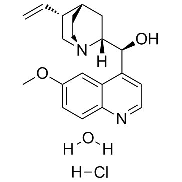
Quinidine hydrochloride monohydrate |
Catalog No.GC37062
 One-Click Copy Product Info
|

Quinidine hydrochloride monohydrate is an anti-arrythmic agent which is also a potent blocker of K+ channel with an IC50 of 19.9 μM.
Products are for research use only. Not for human use. We do not sell to patients.
Cas No.: 6151-40-2
Sample solution is provided at 25 µL, 10mM.
_1-3.$$$39978408@51.jpg');)
_1-9.$$$38538795@65.jpg');)
_1-9.$$$39112701@51.jpg');)
_1-7.$$$37316672@70.jpg');)
_366-372.$$$36198801@70.jpg');)
.$$$38565142@65.jpg');)
_1867-1883.$$$33248023@67.jpg');)
_eads6055.$$$39977546@45.jpg');)
_eadm9805.$$$40080571@45.jpg');)
_eadj3020.$$$39666821@45.jpg');)
_1-20.$$$39757301@28.jpg');)
_1-24.$$$38898113@28.jpg');)
_273-287.$$$36806353@46.jpg');)
_1-16.$$$37156877@44.jpg');)
_1-19.$$$37460805@44.jpg');)
_1291-1307.$$$34518654@46.jpg');)
Quinidine hydrochloride monohydrate is a clinical anti-arrythmic drug which is also a potent blocker of K+ channel with an IC50 of 19.9 μM. IC50: 19.9 μM (K+ channel)[1]
Quinidine hydrochloride monohydrate blocks WT mSlo3 (KCa5.1) channels with an IC50 of 19.9±1.41 μM and Hill slope of 1.15±0.15 (n=7). Again, the potency of inhibition by Quinidine hydrochloride monohydrate is higher for F304Y mSlo3 (IC50 of 2.42±0.60 μM, n=9, P<0.005; Hill slope of 0.98±0.12), but lower with R196Q mSlo3 (IC50 of 38.4±6.77 μM, n=5, P<0.001; Hill slope of 1.05±0.16). The inhibition of F304Y mSlo3 by Quinidine hydrochloride monohydrate is observed to have some time dependence[1].
Direct application of Quinidine hydrochloride monohydrate on the sciatic nerve produces a dose-related decrease in amplitude at ascending somato-sensory evoked potential (SSEP) and descending compound muscle action potentials (CMAP) when comparing baseline with other time points, or when comparing the experimental left limb to the right contra-lateral glucose-treated limb. The latencies of SSEPs and CMAP potentials after Quinidine hydrochloride monohydrate applications are increased compare to baseline and the contralateral side[2].
[1]. Wrighton DC, et al. Mechanism of inhibition of mouse Slo3 (KCa 5.1) potassium channels by quinine, quinidine and barium. Br J Pharmacol. 2015 Sep;172(17):4355-63. [2]. Cheng KI, et al. Application of quinidine on rat sciatic nerve decreases the amplitude and increases the latency of evoked responses. J Anesth. 2014 Aug;28(4):559-68.
Cell experiment: | Mouse (m) Slo3 (KCa5.1) channels or mutant forms are expressed in Xenopus oocytes and currents recorded with 2-electrode voltage-clamp. Gain-of-function mSlo3 mutations are used to explore the state-dependence of the inhibition. The interaction between Quinidine hydrochloride monohydrate and mSlo3 channels is modelled by in silico docking[1]. |
Animal experiment: | 24 rats are randomly divided into three groups with eight rats in each group. Groups Q1, Q3, and Q5 receive Quinidine hydrochloride monohydrate 1, 3, and 5 μmol, respectively, in 5 % glucose 0.1 mL. The sciatic nerve is exposed by making an incision from the left sciatic notch to the distal thigh. The subcutaneous tissue is bluntly dissected to expose the biceps femoris. The sciatic nerve is freed from its investing fascia. The procedure is then repeated on the right side. The somato-sensory evoked potential (SSEP) and compound muscle action potentials (CMAP) are recorded at baseline, immediately after Quinidine hydrochloride monohydrate treatment, then every 15 min thereafter for 1 h, then every 30 min thereafter for 3 h. The animals are allowed to recover and then kept separately for 2 weeks. After performing behavioral examinations, electrophysiological examinations are performed with the animals under intra-peritoneal anesthesia[2]. |
References: [1]. Wrighton DC, et al. Mechanism of inhibition of mouse Slo3 (KCa 5.1) potassium channels by quinine, quinidine and barium. Br J Pharmacol. 2015 Sep;172(17):4355-63. | |
| Cas No. | 6151-40-2 | SDF | |
| Canonical SMILES | C=C[C@H]1C[N@](CC[C@H]1C2)[C@@]2([H])[C@@H](O)C3=CC=NC4=CC=C(OC)C=C34.[H]Cl.[H]O[H] | ||
| Formula | C20H27ClN2O3 | M.Wt | 378.89 |
| Solubility | DMSO: 100 mg/mL (263.93 mM); Water: 2.5 mg/mL (6.60 mM) | Storage | Store at -20°C |
| General tips | Please select the appropriate solvent to prepare the stock solution according to the
solubility of the product in different solvents; once the solution is prepared, please store it in
separate packages to avoid product failure caused by repeated freezing and thawing.Storage method
and period of the stock solution: When stored at -80°C, please use it within 6 months; when stored
at -20°C, please use it within 1 month. To increase solubility, heat the tube to 37°C and then oscillate in an ultrasonic bath for some time. |
||
| Shipping Condition | Evaluation sample solution: shipped with blue ice. All other sizes available: with RT, or with Blue Ice upon request. | ||
| Prepare stock solution | |||

|
1 mg | 5 mg | 10 mg |
| 1 mM | 2.6393 mL | 13.1964 mL | 26.3929 mL |
| 5 mM | 527.9 μL | 2.6393 mL | 5.2786 mL |
| 10 mM | 263.9 μL | 1.3196 mL | 2.6393 mL |
Step 1: Enter information below (Recommended: An additional animal making an allowance for loss during the experiment)
 g
μL
g
μL
Step 2: Enter the in vivo formulation (This is only the calculator, not formulation. Please contact us first if there is no in vivo formulation at the solubility Section.)
Calculation results:
Working concentration: mg/ml;
Method for preparing DMSO master liquid: mg drug pre-dissolved in μL DMSO ( Master liquid concentration mg/mL, Please contact us first if the concentration exceeds the DMSO solubility of the batch of drug. )
Method for preparing in vivo formulation: Take μL DMSO master liquid, next addμL PEG300, mix and clarify, next addμL Tween 80, mix and clarify, next add μL saline, mix and clarify.
Method for preparing in vivo formulation: Take μL DMSO master liquid, next add μL Corn oil, mix and clarify.
Note: 1. Please make sure the liquid is clear before adding the next solvent.
2. Be sure to add the solvent(s) in order. You must ensure that the solution obtained, in the previous addition, is a clear solution before proceeding to add the next solvent. Physical methods such as vortex, ultrasound or hot water bath can be used to aid dissolving.
3. All of the above co-solvents are available for purchase on the GlpBio website.
Quality Control & SDS
- View current batch:
- Purity: >99.50% Appearance: A solid
- COA (Certificate of Analysis)
- SDS (Safety Data Sheet)
- Datasheet
Average Rating: 5 (Based on Reviews and 38 reference(s) in Google Scholar.)