Home>>Signaling Pathways>> DNA Damage/DNA Repair>> DNA/RNA Synthesis>>Puromycin dihydrochloride

Puromycin dihydrochloride

Catalog No.: GC16384

Puromycin dihydrochloride is produced by Streptomyces alboniger, a grampositive actinomycete, through a series of enzymatic reactions

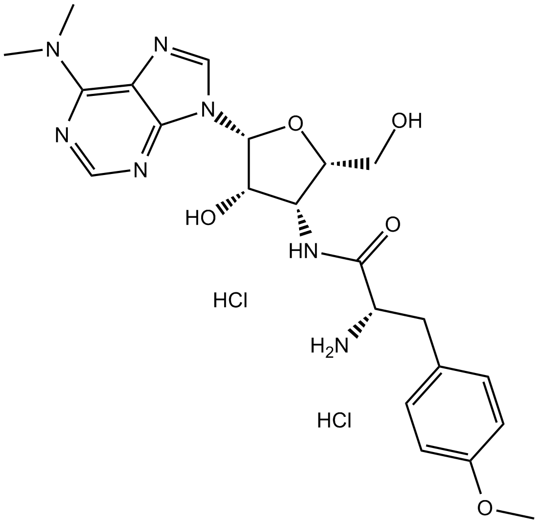
Puromycin dihydrochloride Chemical Structure

Size Price Stock Qty
10mM (in 1mL DMSO)
$45.00
In stock
20mg
$50.00
In stock
50mg
$100.00
In stock
100mg
$160.00
In stock
500mg
$640.00
In stock
1g
$1,024.00
In stock

Customer Reviews

Based on customer reviews.

Tel: (626) 353-8530 Email: sales@glpbio.com

Sample solution is provided at 25 µL, 10mM.

Product Citations

Product Documents

Quality Control & SDS

View current batch:

Protocol

Cell experiment [1]:

Cell lines

Fetal porcine somatic cells

Preparation Method

Cells were seeded in 24-well plates at a density of 2.5 x 10⁴ cells per well and cultured in medium containing 0.5–6 μg/ml puromycin dihydrochloride. Stock solution (10 mg/ml) of puromycin dihydrochloride was prepared by dissolving puromycin dihydrochloride in distilled water at the appropriate concentration. Media containing variable amounts of puromycin dihydrochloride were freshly prepared by adding the appropriate volume of puromycin dihydrochloride stock solution.

Reaction Conditions

0.5–6 μg/ml for 7 d

Applications

Puromycin dihydrochloride is an antibiotic that inhibits growth of animal cells and blocks protein synthesis by binding to 80S ribosomes at low doses. To determine the optimal concentration of puromycin dihydrochloride for selecting EGFPac-transfected cells, a puromycin dihydrochloride resistance test was performed with fetal porcine somatic cells. The puromycin-resistant gene (termed pac) encoding puromycin N-acetyl transferase was isolated from Streptomyces aboniger. If pac is introduced and expressed in animal cells, the cells can survive in the presence of puromycin dihydrochloride.

Animal experiment [2]:

Animal models

Female FVB/N mice, 8–10 weeks old.

Preparation Method

Puromycin dihydrochloride was dissolved in 100 μl of PBS. Mice were housed under a 12-h light/dark cycle with ad libitum access to food and water unless otherwise stated. Mice were anesthetized with an intraperitoneal injection of ketamine (100 mg/kg) and xylazine (10 mg/kg) before all surgical procedures.

Dosage form

Puromycin dihydrochloride was intraperitoneal injected to mice with a concentration of 0.040 μmol/g.

Applications

The antibiotic puromycin dihydrochloride (a structural analog of tyrosyl-tRNA), and anti-puromycin antibodies could be used to detect the amount of puromycin incorporation into nascent peptide chains as well as to measure changes in protein synthesis in cell cultures.

References:

[1]. Watanabe S, Iwamoto M, et al. A novel method for the production of transgenic cloned pigs: electroporation-mediated gene transfer to non-cultured cells and subsequent selection with puromycin. Biol Reprod. 2005 Feb;72(2):309-15.

[2]. Goodman CA, Mabrey DM, et al. Novel insights into the regulation of skeletal muscle protein synthesis as revealed by a new nonradioactive in vivo technique. FASEB J. 2011 Mar;25(3):1028-39.

Background

Puromycin dihydrochloride is produced by Streptomyces alboniger, a grampositive actinomycete, through a series of enzymatic reactions.[1] Puromycin dihydrochloride included a nucleoside covalently bound to an amino acid, mimicking the 30 end of aminoacylated tRNAs that participate in delivery of amino acids to elongating ribosomes.[2] It inhibits the growth of animal cells and blocks protein synthesis by binding to 80S ribosomes at low doses.[3]

In vitro study determined the optimal concentration of Puromycin dihydrochloride for selecting EGFPac-transfected cells by performing a Puromycin dihydrochloride resistance test. The puromycin-resistant gene (termed pac) encoding puromycin N-acetyl transferase was isolated from Streptomyces aboniger. If pac is introduced and expressed in animal cells, the cells can survive in the presence of Puromycin dihydrochloride. Results ahowed that it could successfully produce a somatically cloned transgenic piglet using recombinant cells obtained after gene transfer of a transgene (carrying both EGFP and pac expression units) and subsequent in vitro selection with a low concentration (2 mg/ml) of puromycin.[3]

In vivo study was conducted to determine the surface sensing of translation (SUnSET) technique could be used to measure the protein synthesis in whole tissues. Since there is currently an intense interest in identifying the molecular mechanisms that regulate skeletal muscle protein synthesis. It allows for the visualization and quantification of protein synthesis and eliminates the need for generating radioactive tissues/animals. This study also determined that the surface sensing of translation could detect relatively acute changes in protein synthesis in the absence of changes in rRNA as well as detect not only increases but also decreases in protein synthesis in vivo. [4]

References:
[1]. Tercero JA, Espinosa JC, Lacalle RA, Jiménez A. The biosynthetic pathway of the aminonucleoside antibiotic puromycin, as deduced from the molecular analysis of the pur cluster of Streptomyces alboniger. J Biol Chem 1996;271(3):1579–90.
[2]. Aviner R. et al. The science of puromycin: From studies of ribosome function to applications in biotechnology. Comput Struct Biotechnol J. 2020 Apr 24;18:1074-1083.
[3]. Watanabe S, Iwamoto M, et al. A novel method for the production of transgenic cloned pigs: electroporation-mediated gene transfer to non-cultured cells and subsequent selection with puromycin. Biol Reprod. 2005 Feb;72(2):309-15.
[4]. Goodman CA, Mabrey DM, et al. Novel insights into the regulation of skeletal muscle protein synthesis as revealed by a new nonradioactive in vivo technique. FASEB J. 2011 Mar;25(3):1028-39.

Chemical Properties

Cas No. 58-58-2 SDF
Synonyms CL13900
Chemical Name (S)-2-amino-N-((2S,3R,4S,5R)-5-(6-(dimethylamino)-9H-purin-9-yl)-4-hydroxy-2-(hydroxymethyl)tetrahydrofuran-3-yl)-3-(4-methoxyphenyl)propanamide dihydrochloride
Canonical SMILES O[C@H]1[C@H]([C@@H](CO)O[C@H]1N2C3=NC=NC(N(C)C)=C3N=C2)NC([C@H](CC(C=C4)=CC=C4OC)N)=O.Cl.Cl
Formula C22H29N7O5.2HCl M.Wt 544.43
Solubility ≥ 27.2mg/mL in DMSO, ≥ 99.4mg/mL in Water Storage Store at -20°C
General tips For obtaining a higher solubility , please warm the tube at 37 ℃ and shake it in the ultrasonic bath for a while.Stock solution can be stored below -20℃ for several months.
Shipping Condition Evaluation sample solution : ship with blue ice
All other available size: ship with RT , or blue ice upon request

In vivo Formulation Calculator (Clear solution)

Step 1: Enter information below (Recommended: An additional animal making an allowance for loss during the experiment)

mg/kg g μL

Step 2: Enter the in vivo formulation (This is only the calculator, not formulation. Please contact us first if there is no in vivo formulation at the solubility Section.)

% DMSO % % Tween 80 % saline
%DMSO %

Calculation results:

Working concentration: mg/ml;

Method for preparing DMSO master liquid: mg drug pre-dissolved in μL DMSO ( Master liquid concentration mg/mL, Please contact us first if the concentration exceeds the DMSO solubility of the batch of drug. )

Method for preparing in vivo formulation: Take μL DMSO master liquid, next addμL PEG300, mix and clarify, next addμL Tween 80, mix and clarify, next add μL saline, mix and clarify.

Method for preparing in vivo formulation: Take μL DMSO master liquid, next add μL Corn oil, mix and clarify.

Note: 1. Please make sure the liquid is clear before adding the next solvent.
2. Be sure to add the solvent(s) in order. You must ensure that the solution obtained, in the previous addition, is a clear solution before proceeding to add the next solvent. Physical methods such as vortex, ultrasound or hot water bath can be used to aid dissolving.
3. All of the above co-solvents are available for purchase on the GlpBio website.

  • Molarity Calculator

  • Dilution Calculator

Mass
=
Concentration
x
Volume
x
MW*


**When preparing stock solutions always use the batch-specific molecular weight of the product found on the vial label and MSDS / CoA (available online).

Calculate

Research Update

SUnSET, a nonradioactive method to monitor protein synthesis

We developed a nonradioactive fluorescence-activated cell sorting-based assay, called surface sensing of translation (SUnSET), which allows the monitoring and quantification of global protein synthesis in individual mammalian cells and in heterogeneous cell populations. We demonstrate here, using mouse dendritic and T cells as a model, that SUnSET offers a technical alternative to classical radioactive labeling methods for the study of mRNA translation and cellular activation.

Active Ribosome Profiling with RiboLace

Ribosome profiling, or Ribo-seq, is based on large-scale sequencing of RNA fragments protected from nuclease digestion by ribosomes. Thanks to its unique ability to provide positional information about ribosomes flowing along transcripts, this method can be used to shed light on mechanistic aspects of translation. However, current Ribo-seq approaches lack the ability to distinguish between fragments protected by either ribosomes in active translation or inactive ribosomes. To overcome this possible limitation, we developed RiboLace, a method based on an original puromycin-containing molecule capable of isolating active ribosomes by means of an antibody-free and tag-free pull-down approach. RiboLace is fast, works reliably with low amounts of input material, and can be easily and rapidly applied both in vitro and in vivo, thereby generating a global snapshot of active ribosome footprints at single nucleotide resolution.

Optical Control of Translation with a Puromycin Photoswitch

Translation is an elementary cellular process that involves a large number of factors interacting in a concerted fashion with the ribosome. Numerous natural products have emerged that interfere with the ribosomal function, such as puromycin, which mimics an aminoacyl tRNA and causes premature chain termination. Here, we introduce a photoswitchable version of puromycin that, in effect, puts translation under optical control. Our compound, termed puroswitch, features a diazocine that allows for reversible and nearly quantitative isomerization and pharmacological modulation. Its synthesis involves a new photoswitchable amino acid building block. Puroswitch shows little activity in the dark and becomes substantially more active and cytotoxic, in a graded fashion, upon irradiation with various wavelengths of visible light. In vitro translation assays confirm that puroswitch inhibits translation with a mechanism similar to that of puromycin itself. Once incorporated into nascent proteins, puroswitch reacts with standard puromycin antibodies, which allows for tracking de novo protein synthesis using western blots and immunohistochemistry. As a cell-permeable small molecule, puroswitch can be used for nascent proteome profiling in a variety of cell types, including primary mouse neurons. We envision puroswitch as a useful biochemical tool for the optical control of translation and for monitoring newly synthesized proteins in defined locations and at precise time points.

Puromycin reactivity does not accurately localize translation at the subcellular level

Puromycin is a tyrosyl-tRNA mimic that blocks translation by labeling and releasing elongating polypeptide chains from translating ribosomes. Puromycin has been used in molecular biology research for decades as a translation inhibitor. The development of puromycin antibodies and derivatized puromycin analogs has enabled the quantification of active translation in bulk and single-cell assays. More recently, in vivo puromycylation assays have become popular tools for localizing translating ribosomes in cells. These assays often use elongation inhibitors to purportedly inhibit the release of puromycin-labeled nascent peptides from ribosomes. Using in vitro and in vivo experiments in various eukaryotic systems, we demonstrate that, even in the presence of elongation inhibitors, puromycylated peptides are released and diffuse away from ribosomes. Puromycylation assays reveal subcellular sites, such as nuclei, where puromycylated peptides accumulate post-release and which do not necessarily coincide with sites of active translation. Our findings urge caution when interpreting puromycylation assays in vivo.

Instability of puromycin

Reviews

Review for Puromycin dihydrochloride

Average Rating: 5 ★★★★★ (Based on Reviews and 35 reference(s) in Google Scholar.)

5 Star
100%
4 Star
0%
3 Star
0%
2 Star
0%
1 Star
0%
Review for Puromycin dihydrochloride

GLPBIO products are for RESEARCH USE ONLY. Please make sure your review or question is research based.

Required fields are marked with *

You may receive emails regarding this submission. Any emails will include the ability to opt-out of future communications.