

Spantide I |
Catalog No.GC11280
 One-Click Copy Product Info
|

Spantide I is an analogue of substance P (SP) and a selective NK1 receptor antagonist with Ki values of 230nM and 8150nM for NK1 and NK2 receptor, respectively.
Products are for research use only. Not for human use. We do not sell to patients.
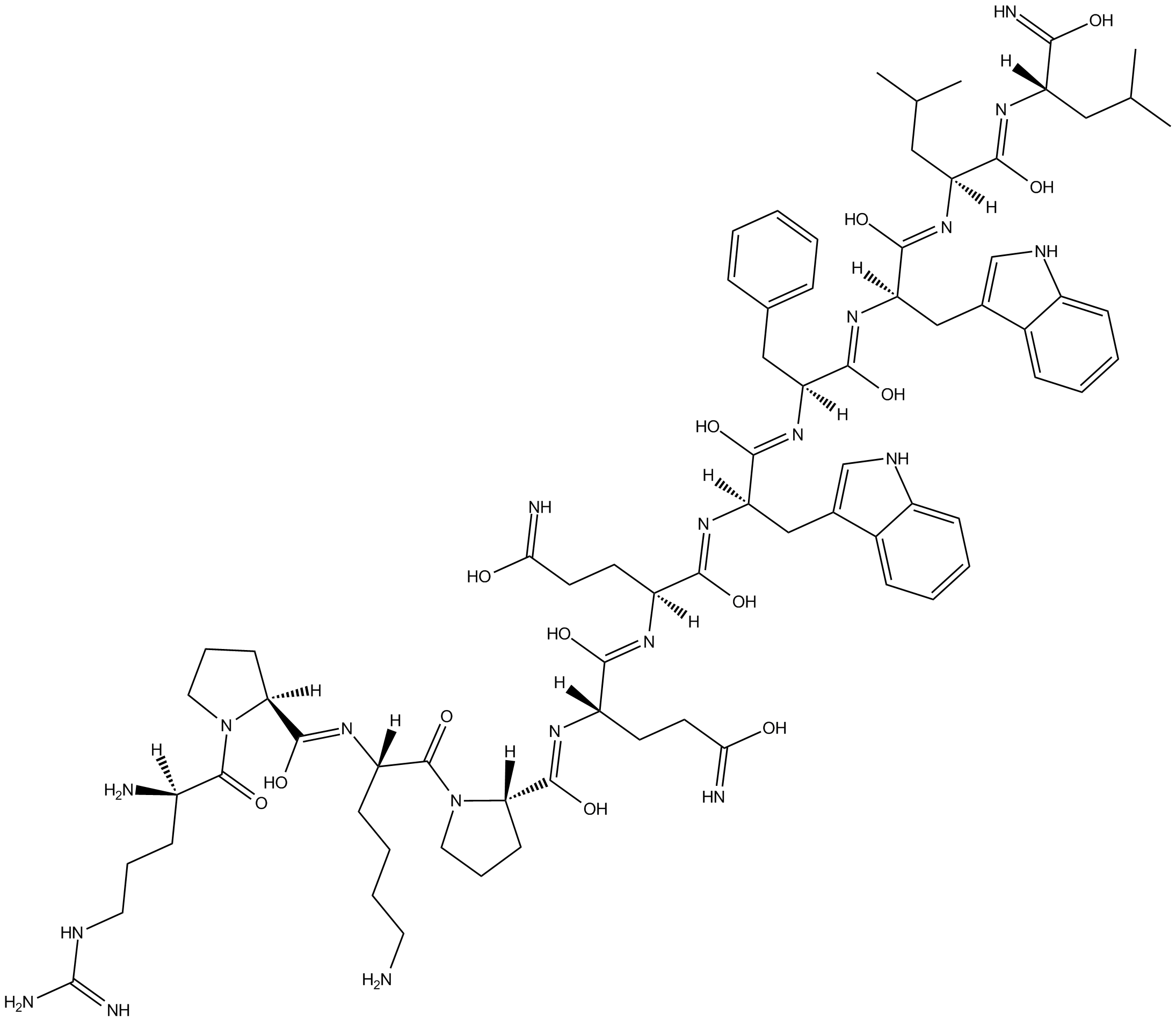
Cas No.: 91224-37-2
Sample solution is provided at 25 µL, 10mM.
_1-3.$$$39978408@51.jpg');)
_1-9.$$$38538795@65.jpg');)
_1-9.$$$39112701@51.jpg');)
_1-7.$$$37316672@70.jpg');)
_366-372.$$$36198801@70.jpg');)
.$$$38565142@65.jpg');)
_1867-1883.$$$33248023@67.jpg');)
_eads6055.$$$39977546@45.jpg');)
_eadm9805.$$$40080571@45.jpg');)
_eadj3020.$$$39666821@45.jpg');)
_1-20.$$$39757301@28.jpg');)
_1-24.$$$38898113@28.jpg');)
_273-287.$$$36806353@46.jpg');)
_1-16.$$$37156877@44.jpg');)
_1-19.$$$37460805@44.jpg');)
_1291-1307.$$$34518654@46.jpg');)
Spantide I is an analogue of substance P (SP) and a selective NK1 receptor antagonist with Ki values of 230nM and 8150nM for NK1 and NK2 receptor, respectively [1]. The NK1 is broadly expressed in the eye, in both ocular and non-ocular cells, such as leukocytes and neurons [2]. Spantide I provides an approach to reduce type 1 and enhance the type 2 cytokine IL-10 in the infected cornea, leading to a significant reduction in corneal perforation [3].
In vitro, Spantide I (10μM) treatment of regulatory T cells (Tregs) for 48 hours reversed the reduction in Foxp3, CTLA-4, and PD-1 expression levels caused by SP treatment. At the same time, Spantide I blocked the SP signal transduction and inhibited the decrease in TGF-β levels observed in SP-induced Tregs cells [4]. Treating TG neurons isolated from dry eye disease (DED) mice with a culture medium containing Spantide I (2μM) for 24 hours significantly reduced the proliferation of vascular endothelial cells (VEC) co-cultured with the TG neurons, and also eliminated the promotion of tube formation by the VECs [5].
In vivo, treating mice with Spantide I (36μg/mouse/day; i.p.) for 7 days significantly reduced the number of corneal perforations, bacterial count, and neutrophil count in mice infected with Pseudomonas aeruginosa, and also downregulated the mRNA levels of type I cytokines (such as IFN-γ) as well as MIP-2, IL-6, TNF-α, and IL-1β [3]. Treating mice exposed to UVR-B with Spantide I (72μg/mouse/day; i.p.) for 7 days significantly reduced the expression of NKR-1 in the corneas. Spantide I has a protective effect after corneal injury induction [6].
References:
[1] Zubrzycka M, Janecka A, Traczyk WZ. Comparison of antagonistic properties of substance P analogs, spantide I, II and III, on evoked tongue jerks in rats. Endocr Regul. 2000 Mar;34(1):13-8.
[2] Demirsoy I H, Ferrari G. The NK-1 receptor signaling: distribution and functional relevance in the eye[J]. Receptors, 2022, 1(1): 98-111.
[3] Hazlett LD, McClellan SA, Barrett RP, Liu J, Zhang Y, Lighvani S. Spantide I decreases type I cytokines, enhances IL-10, and reduces corneal perforation in susceptible mice after Pseudomonas aeruginosa infection. Invest Ophthalmol Vis Sci. 2007 Feb;48(2):797-807.
[4] Taketani Y, Marmalidou A, Dohlman TH, et al. Restoration of Regulatory T-Cell Function in Dry Eye Disease by Antagonizing Substance P/Neurokinin-1 Receptor. Am J Pathol. 2020;190(9):1859-1866.
[5] Liu L, Dana R, Yin J. Sensory neurons directly promote angiogenesis in response to inflammation via substance P signaling. FASEB J. 2020;34(5):6229-6243.
[6] Gross J, Wegener AR, Kronschläger M, Schönfeld CL, Holz FG, Meyer LM. UVR-B-induced NKR-1 Expression in Ocular Tissues is blocked by Substance P Receptor Antagonist Fosaprepitant in the Exposed as well as Unexposed Partner Eye. Ocul Immunol Inflamm. 2021;29(5):963-975.
| Cell experiment [1]: | |
Cell lines | Tregs |
Preparation Method | The submandibular and cervical DLNs were harvested from normal and DED mice, and single-cell suspensions were prepared as previously described. CD4CD25 Tregs were isolated by magnetic separation using a Treg isolation kit. The purity of sorted cells (>95% Foxp3 Tregs) was confirmed by using flow cytometry analysis. Tregs were resuspended in RPMI 1640–based complete medium at a concentration of 1 × 10⁶ cells/mL. Subsequently, 100μL of cell suspension was plated in each well of a 96-well plate. SP (1μM), Spantide I (10μM), or both were added to the Treg cultures. After 48 hours of incubation, cells were collected for flow cytometry analysis. The inhibitory function of the cultured Tregs was evaluated using the Treg inhibition assay. |
Reaction Conditions | 10μM; 48h |
Applications | Under the presence of SP, the in vitro culture of normal Tregs led to a significant decrease in the frequency of Tregs and their inhibitory function. The NK-1R antagonist Spantide I reversed the reduction in the expression levels of Foxp3, CTLA-4 and PD-1 in Tregs treated with SP. At the same time, Spantide I blocked the signal transduction of SP and inhibited the reduction in TGF-β production by SP-induced Tregs. |
| Animal experiment [2]: | |
Animal models | C57BL/6 mice |
Preparation Method | Copper-green Pseudomonas bacteria were used to infect B6 mice. On the -1st day and the 0th day (the day of infection), 36μg/mouse of synthetic Spantide I was injected intraperitoneally, and this injection was repeated daily within 5 days after infection. Control mice were similarly injected with vehicle (PBS <0.05EU/mL endotoxin). Infected corneas were collected at 1, 3, and 5 days pi for real-time PCR detection of mRNA levels of cytokines. Normal, uninfected corneas also were harvested to determine basal levels of the target cytokines. In separate experiments, infected corneas (n = 5/group/time/assay) were harvested at 3 and 5 days pi and used for ELISA analysis, viable bacterial enumeration, and MPO quantitation. |
Dosage form | 36μg/mouse/d for 7 days; i.p. |
Applications | At 3 and 5 days pi, Spantide I-treated mice had significantly less severe ocular disease than did the PBS-treated mice. Contained significantly fewer PMNs than the corneas of PBS-treated mice at 3 and 5 days pi. Significantly reduced levels of corneal TNF-α mRNA at 3 and 5 days pi. Significantly reduced the level of IL-18 mRNA at 1 day pi. |
References: | |
| Cas No. | 91224-37-2 | SDF | |
| Chemical Name | (S,Z)-N'1-((2R,3Z,5S,6Z,8R,9Z,11S,12Z,14S)-8-((1H-indol-3-yl)methyl)-5-benzyl-3,6,9,12-tetrahydroxy-14-(hydroxy(imino)methyl)-1-(1H-indol-3-yl)-11-isobutyl-16-methyl-4,7,10,13-tetraazaheptadeca-3,6,9,12-tetraen-2-yl)-2-((Z)-((S)-2-((Z)-(((S)-1-((S)-6-amin | ||
| Canonical SMILES | CC(C[C@@](/N=C(O)/[C@](/N=C(O)/[C@@](/N=C(O)/[C@](/N=C(O)/[C@@](/N=C(O)/[C@](/N=C(O)/[C@](/N=C(O)/[C@]1([H])CCCN1C([C@](/N=C(O)/[C@]2([H])CCCN2C([C@@](N)([H])CCCNC(N)=N)=O)([H])CCCCN)=O)([H])CCC(O)=N)([H])CCC(O)=N)([H])CC3=CNC4=CC=CC=C34)([H])CC5=CC=CC=C5 | ||
| Formula | C75H108N20O13 | M.Wt | 1497.79 |
| Solubility | Soluble to 1 mg/ml in Water | Storage | Desiccate at -20°C |
| General tips | Please select the appropriate solvent to prepare the stock solution according to the
solubility of the product in different solvents; once the solution is prepared, please store it in
separate packages to avoid product failure caused by repeated freezing and thawing.Storage method
and period of the stock solution: When stored at -80°C, please use it within 6 months; when stored
at -20°C, please use it within 1 month. To increase solubility, heat the tube to 37°C and then oscillate in an ultrasonic bath for some time. |
||
| Shipping Condition | Evaluation sample solution: shipped with blue ice. All other sizes available: with RT, or with Blue Ice upon request. | ||
| Prepare stock solution | |||

|
1 mg | 5 mg | 10 mg |
| 1 mM | 667.7 μL | 3.3383 mL | 6.6765 mL |
| 5 mM | 133.5 μL | 667.7 μL | 1.3353 mL |
| 10 mM | 66.8 μL | 333.8 μL | 667.7 μL |
Step 1: Enter information below (Recommended: An additional animal making an allowance for loss during the experiment)
 g
μL
g
μL
Step 2: Enter the in vivo formulation (This is only the calculator, not formulation. Please contact us first if there is no in vivo formulation at the solubility Section.)
Calculation results:
Working concentration: mg/ml;
Method for preparing DMSO master liquid: mg drug pre-dissolved in μL DMSO ( Master liquid concentration mg/mL, Please contact us first if the concentration exceeds the DMSO solubility of the batch of drug. )
Method for preparing in vivo formulation: Take μL DMSO master liquid, next addμL PEG300, mix and clarify, next addμL Tween 80, mix and clarify, next add μL saline, mix and clarify.
Method for preparing in vivo formulation: Take μL DMSO master liquid, next add μL Corn oil, mix and clarify.
Note: 1. Please make sure the liquid is clear before adding the next solvent.
2. Be sure to add the solvent(s) in order. You must ensure that the solution obtained, in the previous addition, is a clear solution before proceeding to add the next solvent. Physical methods such as vortex, ultrasound or hot water bath can be used to aid dissolving.
3. All of the above co-solvents are available for purchase on the GlpBio website.
Quality Control & SDS
- View current batch:
- Purity: >98.00% Appearance: A solid
- COA (Certificate of Analysis)
- SDS (Safety Data Sheet)
- Datasheet
Average Rating: 5 (Based on Reviews and 1 reference(s) in Google Scholar.)