

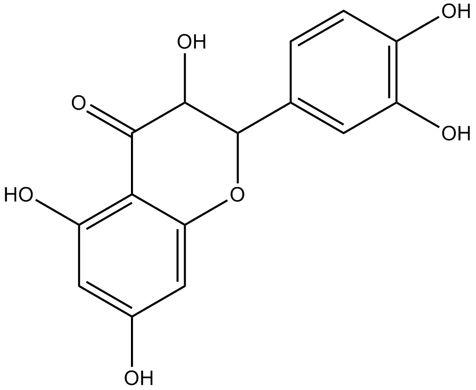
Taxifolin (Synonyms: (+)-Dihydroquercetin) |
Catalog No.GN10304
 One-Click Copy Product Info
|

Taxifolin is a tyrosinase inhibitor and has significant collagenase inhibitory activity, with an IC50 value of 193.3μM.
Products are for research use only. Not for human use. We do not sell to patients.
Cas No.: 480-18-2
Sample solution is provided at 25 µL, 10mM.
_1-3.$$$39978408@51.jpg');)
_1-9.$$$38538795@65.jpg');)
_1-9.$$$39112701@51.jpg');)
_1-7.$$$37316672@70.jpg');)
_366-372.$$$36198801@70.jpg');)
.$$$38565142@65.jpg');)
_1867-1883.$$$33248023@67.jpg');)
_eads6055.$$$39977546@45.jpg');)
_eadm9805.$$$40080571@45.jpg');)
_eadj3020.$$$39666821@45.jpg');)
_1-20.$$$39757301@28.jpg');)
_1-24.$$$38898113@28.jpg');)
_273-287.$$$36806353@46.jpg');)
_1-16.$$$37156877@44.jpg');)
_1-19.$$$37460805@44.jpg');)
_1291-1307.$$$34518654@46.jpg');)
Taxifolin is a tyrosinase inhibitor and has significant collagenase inhibitory activity, with an IC50 value of 193.3μM[1]. As a ubiquitous bioactive constituent of foods and herbs, Taxifolin is also an important natural compound with antifibrotic activity and a free radical scavenger with antioxidant capacity[2][3]. Taxifolin showed promising pharmacological activities in various diseases, including inflammation, tumors, microbial infections, oxidative stress, cardiovascular and neurodegenerative diseases[4][5].
In vitro, administration of Taxifolin(10-100μM; 48h) to human colorectal cancer HCT116 and HT29 cells induced cytotoxicity in a dose- and time-dependent manner, resulting in cell growth arrest, alterations in molecules controlling the G2 phase of the cell cycle, apoptosis in a concentration-dependent manner, and decreased expression of β-catenin, AKT, and Survivin genes and proteins[6]. Taxifolin inhibited the proliferation and migration of B16F10 melanoma cells at 200 and 400μM, and of A375 cells at 100 and 200μM after treatment for 48h[7].
In vivo, treatment of Taxifolin(60mg/kg; i.p.) suppressed tumor growth and metastasis in mouse models without causing significant toxicity[7]. In DSS-induced mice, Taxifolin(200mg/kg; p.o.; daily for 7 consecutive days) treatment decreased weight loss and diarrhea score, increased colon length, significantly upregulated the expression of GPR41 and GPR43 in the colon, inhibited the expression of TNF-α, IL-1β, and IL-6 in colon tissue, reversed the fecal metabolic pattern altered by DSS, and significantly increased fecal levels of butyric acid and isobutyric acid[8].
References:
[1] Angelis, A., Hubert, J., Aligiannis, N., Michalea, R., Abedini, A., Nuzillard, J. M., Gangloff, S. C., Skaltsounis, A. L., & Renault, J. H. (2016). Bio-Guided Isolation of Methanol-Soluble Metabolites of Common Spruce (Picea abies) Bark by-Products and Investigation of Their Dermo-Cosmetic Properties. Molecules (Basel, Switzerland), 21(11), 1586.
[2] Yang, P., Xu, F., Li, H. F., Wang, Y., Li, F. C., Shang, M. Y., Liu, G. X., Wang, X., & Cai, S. Q. (2016). Detection of 191 Taxifolin Metabolites and Their Distribution in Rats Using HPLC-ESI-IT-TOF-MS(n). Molecules (Basel, Switzerland), 21(9), 1209.
[3] Ren, L., Guo, H. N., Yang, J., Guo, X. Y., Wei, Y. S., & Yang, Z. (2021). Dissecting Efficacy and Metabolic Characteristic Mechanism of Taxifolin on Renal Fibrosis by Multivariate Approach and Ultra-Performance Liquid Chromatography Coupled With Mass Spectrometry-Based Metabolomics Strategy. Frontiers in pharmacology, 11, 608511.
[4] Sunil, C., & Xu, B. (2019). An insight into the health-promoting effects of taxifolin (dihydroquercetin). Phytochemistry, 166, 112066.
[5] Yang, R., Yang, X., & Zhang, F. (2023). New Perspectives of Taxifolin in Neurodegenerative Diseases. Current neuropharmacology, 21(10), 2097–2109.
Razak, S., Afsar, T., Ullah, A., Almajwal, A., Alkholief, M., Alshamsan, [6] A., & Jahan, S. (2018). Taxifolin, a natural flavonoid interacts with cell cycle regulators causes cell cycle arrest and causes tumor regression by activating Wnt/ β -catenin signaling pathway. BMC cancer, 18(1), 1043.
[7] Xu, L., Zhang, L., Zhang, S., Yang, J., Zhu, A., Sun, J., Kalvakolanu, D. V., Cong, X., Zhang, J., Tang, J., & Guo, B. (2024). Taxifolin inhibits melanoma proliferation/migration impeding USP18/Rac1/JNK/β-catenin oncogenic signaling. Phytomedicine : international journal of phytotherapy and phytopharmacology, 123, 155199.
[8] Li, W., Zhang, L., Xu, Q., Yang, W., Zhao, J., Ren, Y., Yu, Z., & Ma, L. (2022). Taxifolin Alleviates DSS-Induced Ulcerative Colitis by Acting on Gut Microbiome to Produce Butyric Acid. Nutrients, 14(5), 1069.
| Cell experiment [1]: | |
|
Cell lines |
HCT116 and HT29 cell lines |
|
Preparation Method |
Two human colorectal cancer cell lines HCT116 and HT29 were grown in a 5% CO2 atmosphere at 37°C in medium containing RPMI medium 1640, 10% fetal bovine serum and 1% penicillin/streptomycin. Taxifolin and β-catenin inhibitor (FH535) suspended in DMSO was applied for cell treatment. Cells with 70% confluency were induced with Taxifolin and β-catenin inhibitor at 10-100μM for 48h in cell culture medium and the dilution of DMSO applied for each treatment was 0.1% (V/V). Cell viability assay, proliferation assay and cell cycle analysis were performed according to protocols. Cells were collected for western blot and PCR analysis. |
|
Reaction Conditions |
10–100μM; 48h |
|
Applications |
Taxifolin induced cytotoxicity in a dose- and time-dependent manner, resulting in cell growth arrest, alterations in molecules controlling the G2 phase of the cell cycle, apoptosis in a concentration-dependent manner, and decreased expression of β-catenin, AKT, and Survivin genes and proteins. |
| Animal experiment [2]: | |
|
Animal models |
C57BL/6 mice |
|
Preparation Method |
C57BL/6 mice were randomly divided into 3 groups (n = 7/group): control group, DSS group, and DSS + Taxifolin group. Experimental colitis was induced by replacing the drinking water with 5% DSS for 7 days (from day 1 to day 7). While mice in the control group were orally administered ddH2O. Meanwhile, mice in the DSS + Taxifolin group were orally administered 150µL Taxifolin (200mg/kg in ddH2O) daily for 7 consecutive days (days 8–14). Mice in the control and DSS groups were given 150µL ddH2O (10mL/kg) per day. Disease severity, including the degree of body weight (BW) reduction and diarrhea severity, was recorded daily to assess colitis. Fresh feces were collected for 16S rRNA sequencing. After the mice were sacrificed, colon contents were collected for SCFA Quantification. colon tissue RNA were extraction for PCR. |
|
Dosage form |
200mg/kg/day for 7 consecutive days; p.o. |
|
Applications |
Taxifolin decreased weight loss and diarrhea score, increased colon length, significantly upregulated the expression of GPR41 and GPR43 in the colon, inhibited the expression of TNF-α, IL-1β, and IL-6 in colon tissue, reversed the fecal metabolic pattern altered by DSS, and significantly increased fecal levels of butyric acid and isobutyric acid. |
|
References: |
|
| Cas No. | 480-18-2 | SDF | |
| Synonyms | (+)-Dihydroquercetin | ||
| Chemical Name | (2R,3R)-2-(3,4-dihydroxyphenyl)-3,5,7-trihydroxy-2,3-dihydrochromen-4-one | ||
| Canonical SMILES | C1=CC(=C(C=C1C2C(C(=O)C3=C(C=C(C=C3O2)O)O)O)O)O | ||
| Formula | C15H12O7 | M.Wt | 304.25 |
| Solubility | ≥ 13.8mg/mL in DMSO | Storage | Store at -20°C,protect from light |
| General tips | Please select the appropriate solvent to prepare the stock solution according to the
solubility of the product in different solvents; once the solution is prepared, please store it in
separate packages to avoid product failure caused by repeated freezing and thawing.Storage method
and period of the stock solution: When stored at -80°C, please use it within 6 months; when stored
at -20°C, please use it within 1 month. To increase solubility, heat the tube to 37°C and then oscillate in an ultrasonic bath for some time. |
||
| Shipping Condition | Evaluation sample solution: shipped with blue ice. All other sizes available: with RT, or with Blue Ice upon request. | ||
| Prepare stock solution | |||

|
1 mg | 5 mg | 10 mg |
| 1 mM | 3.2868 mL | 16.4339 mL | 32.8677 mL |
| 5 mM | 657.4 μL | 3.2868 mL | 6.5735 mL |
| 10 mM | 328.7 μL | 1.6434 mL | 3.2868 mL |
Step 1: Enter information below (Recommended: An additional animal making an allowance for loss during the experiment)
 g
μL
g
μL
Step 2: Enter the in vivo formulation (This is only the calculator, not formulation. Please contact us first if there is no in vivo formulation at the solubility Section.)
Calculation results:
Working concentration: mg/ml;
Method for preparing DMSO master liquid: mg drug pre-dissolved in μL DMSO ( Master liquid concentration mg/mL, Please contact us first if the concentration exceeds the DMSO solubility of the batch of drug. )
Method for preparing in vivo formulation: Take μL DMSO master liquid, next addμL PEG300, mix and clarify, next addμL Tween 80, mix and clarify, next add μL saline, mix and clarify.
Method for preparing in vivo formulation: Take μL DMSO master liquid, next add μL Corn oil, mix and clarify.
Note: 1. Please make sure the liquid is clear before adding the next solvent.
2. Be sure to add the solvent(s) in order. You must ensure that the solution obtained, in the previous addition, is a clear solution before proceeding to add the next solvent. Physical methods such as vortex, ultrasound or hot water bath can be used to aid dissolving.
3. All of the above co-solvents are available for purchase on the GlpBio website.
Quality Control & SDS
- View current batch:
- Purity: >98.00% Appearance: A solid
- COA (Certificate of Analysis)
- SDS (Safety Data Sheet)
- Datasheet
Average Rating: 5 (Based on Reviews and 30 reference(s) in Google Scholar.)