

Tideglusib (Synonyms: NP031112) |
Catalog No.GC14465
 One-Click Copy Product Info
|

Tideglusib is a potent, selective, and irreversible non-ATP competitive glycogen synthase kinase-3 (GSK-3) inhibitor. The IC50 values for GSK-3βWT and GSK-3βC199A are 5nM and 60nM, respectively.
Products are for research use only. Not for human use. We do not sell to patients.
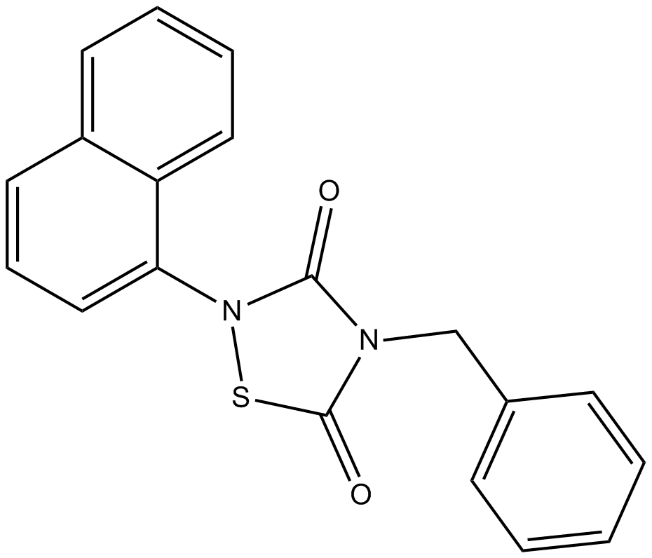
Cas No.: 865854-05-3
Sample solution is provided at 25 µL, 10mM.
Tideglusib is a potent, selective, and irreversible non-ATP competitive glycogen synthase kinase-3 (GSK-3) inhibitor. The IC50 values for GSK-3βWT and GSK-3βC199A are 5nM and 60nM, respectively [1]. GSK-3 is a serine/threonine kinase that is associated with glucose regulation, apoptosis, protein synthesis, cell signaling, cell transport, gene transcription, and cell proliferation [2]. Tideglusib can inhibit inflammation and neurodegenerative diseases and can be used to treat Alzheimer's disease [3-4].
In vitro, Tideglusib (2.5μM; 24h) completely eliminated the expression of TNF-α and COX-2 in glutamate-induced astrocyte and microglial cell cultures, and significantly reduced the number of laminin-V positive cells, exerting anti-inflammatory and neuroprotective effects on cortical neurons [5]. Different concentrations of Tideglusib (5-120μM; 48h) dose-dependently reduced the cell viability of human neuroblastoma (IMR32) cells [6].
In vivo, Tideglusib (2ng/2.5μl PBS; stereotactic injection) treatment significantly improved brain injury in kainic acid (KA) induced focal excitotoxicity model rats and reduced TNF-α positive staining in astrocytes and microglia [5]. Tideglusib (200mg/kg/day; 3 months) oral treatment reduced tau protein phosphorylation levels in APP/tau double transgenic mice, decreased amyloid protein deposition and plaque-related astrocyte proliferation, and protected the entorhinal cortex and prevented hippocampal CA1 zone neuron death [7].
References:
[1] Domínguez JM, Fuertes A, Orozco L, et al. Evidence for irreversible inhibition of glycogen synthase kinase-3β by tideglusib. J Biol Chem. 2012;287(2):893-904.
[2] Pandey M K, DeGrado T R. Glycogen synthase kinase-3 (GSK-3)-targeted therapy and imaging[J]. Theranostics, 2016, 6(4): 571.
[3] Serenóa L, Coma M, Rodríguez M, et al. A novel GSK-3β inhibitor reduces Alzheimer's pathology and rescues neuronal loss in vivo. Neurobiol Dis. 2009 Sep; 35(3): 359-67.
[4] Lovestone S, Boada M, Dubois B, et al. A phase II trial of tideglusib in Alzheimer's disease[J]. Journal of Alzheimer’s Disease, 2015, 45(1): 75-88.
[5] Luna-Medina R, Cortes-Canteli M, Sanchez-Galiano S, et al. NP031112, a thiadiazolidinone compound, prevents inflammation and neurodegeneration under excitotoxic conditions: potential therapeutic role in brain disorders[J]. Journal of Neuroscience, 2007, 27(21): 5766-5776.
[6] Mathuram, Theodore Lemuel et al. “Tideglusib induces apoptosis in human neuroblastoma IMR32 cells, provoking sub-G0/G1 accumulation and ROS generation.” Environmental toxicology and pharmacology vol. 46 (2016): 194-205.
[7] Serenó, L et al. “A novel GSK-3beta inhibitor reduces Alzheimer's pathology and rescues neuronal loss in vivo.” Neurobiology of disease vol. 35,3 (2009): 359-67.
| Kinase experiment [1]: | |
Preparation Method | Kinase assay was carried out at 25°C in a final volume of 10μl in 384-well low volume round bottom black plates with 50mm Hepes, pH 7.5, 10mm MgCl2, 1mm EGTA, and 0.01% Brij-35 as assay buffer. Enzyme concentration ranged from 2 to 5nm. ATP and peptide concentrations were 12.5 and 2μm, respectively. The assays were run for 1h in the presence or absence of compounds in a final DMSO concentration of 1%, and samples were processed. When double titrations of ATP and Tideglusib were performed, varying both ATP and Tideglusib concentrations while keeping the concentration of peptide substrate fixed. Data analysis was performed by fitting the experimental data to the appropriate equations for competitive, uncompetitive, and non-competitive inhibition. |
Reaction Conditions | 0.1nM-1μM; 1h |
Applications | The IC50 value of Tideglusib in inhibiting GSK-3β is 5nm in the pre-incubation condition and 105nm in the non-pre-incubation condition. |
| Cell experiment [2]: | |
Cell lines | Rat primary astrocytes, microglia, and neurons |
Preparation Method | Tideglusib (2.5μm) was added to the culture medium of astrocytes and microglia 1 h before exposure to glutamate (500μm), cells were incubated for 24 h before tissue culture medium was collected, and the cells were evaluated for tumor necrosis factor-α (TNF-α) and cyclooxygenase type 2 (COX-2) expression. |
Reaction Conditions | 2.5μM; 24h |
Applications | Tideglusib completely abolished the expression of TNF-α and COX-2 in the astrocyte and microglial cell cultures induced by glutamate. |
| Animal experiment [2]: | |
Animal models | Wistar rats (focal excitotoxic model) |
Preparation Method | Each group of rats (n ≥ 5) was anesthetized by intraperitoneal injection of ketamine (60mg/kg) and Domtor (5μg/kg), and then placed in a stereotactic apparatus. kainic acid (KA) (1μg dissolved in 2.5μL PBS) or in combination with Tideglusib (2ng dissolved in 2.5μL PBS) was injected separately into the hippocampus. The control animals were injected with solvent. Each injection was administered using a micro-pump and lasted for more than 2.5 minutes. Afterward, these rats were housed individually for recovery care. Behavioral analysis was conducted. After stereotactic injection, the animals were anesthetized at different times and perfused with 4% paraformaldehyde solution via the heart. The brain was removed and placed in the same solution overnight at 4°C, subjected to cryoprotection in a 30% sucrose polyformaldehyde solution, frozen, and obtained 30μm coronal sections under a cryostat. The floating sections were processed with the diaminobenzidine method or double immunofluorescence analysis for Nissl staining or immunohistochemical treatment. |
Dosage form | 2ng/2.5μl PBS; stereotactic injection |
Applications | Tideglusib treatment significantly improved brain damage in rats, reduced the loss of hippocampal cells, and significantly decreased the TNF-α positive staining in astrocytes and microglia. |
References: | |
| Cas No. | 865854-05-3 | SDF | |
| Synonyms | NP031112 | ||
| Chemical Name | 4-benzyl-2-naphthalen-1-yl-1,2,4-thiadiazolidine-3,5-dione | ||
| Canonical SMILES | C1=CC=C(C=C1)CN2C(=O)N(SC2=O)C3=CC=CC4=CC=CC=C43 | ||
| Formula | C19H14N2O2S | M.Wt | 334.39 |
| Solubility | ≥ 16.7 mg/mL in DMSO with gentle warming | Storage | Store at -20°C |
| General tips | Please select the appropriate solvent to prepare the stock solution according to the
solubility of the product in different solvents; once the solution is prepared, please store it in
separate packages to avoid product failure caused by repeated freezing and thawing.Storage method
and period of the stock solution: When stored at -80°C, please use it within 6 months; when stored
at -20°C, please use it within 1 month. To increase solubility, heat the tube to 37°C and then oscillate in an ultrasonic bath for some time. |
||
| Shipping Condition | Evaluation sample solution: shipped with blue ice. All other sizes available: with RT, or with Blue Ice upon request. | ||
| Prepare stock solution | |||

|
1 mg | 5 mg | 10 mg |
| 1 mM | 2.9905 mL | 14.9526 mL | 29.9052 mL |
| 5 mM | 598.1 μL | 2.9905 mL | 5.981 mL |
| 10 mM | 299.1 μL | 1.4953 mL | 2.9905 mL |
Step 1: Enter information below (Recommended: An additional animal making an allowance for loss during the experiment)
 g
μL
g
μL
Step 2: Enter the in vivo formulation (This is only the calculator, not formulation. Please contact us first if there is no in vivo formulation at the solubility Section.)
Calculation results:
Working concentration: mg/ml;
Method for preparing DMSO master liquid: mg drug pre-dissolved in μL DMSO ( Master liquid concentration mg/mL, Please contact us first if the concentration exceeds the DMSO solubility of the batch of drug. )
Method for preparing in vivo formulation: Take μL DMSO master liquid, next addμL PEG300, mix and clarify, next addμL Tween 80, mix and clarify, next add μL saline, mix and clarify.
Method for preparing in vivo formulation: Take μL DMSO master liquid, next add μL Corn oil, mix and clarify.
Note: 1. Please make sure the liquid is clear before adding the next solvent.
2. Be sure to add the solvent(s) in order. You must ensure that the solution obtained, in the previous addition, is a clear solution before proceeding to add the next solvent. Physical methods such as vortex, ultrasound or hot water bath can be used to aid dissolving.
3. All of the above co-solvents are available for purchase on the GlpBio website.
Quality Control & SDS
- View current batch:
- Purity: >99.50% Appearance: A solid
- COA (Certificate of Analysis)
- SDS (Safety Data Sheet)
- Datasheet
Average Rating: 5 (Based on Reviews and 8 reference(s) in Google Scholar.)