

APETx2 |
Catalog No.GC17168
 One-Click Copy Product Info
|

APETx2 is a peptide toxin derived from Anthopleura elegantissimaand serves as a selective, reversible inhibitor of the acid-sensing ion channel 3 (ASIC3).
Products are for research use only. Not for human use. We do not sell to patients.
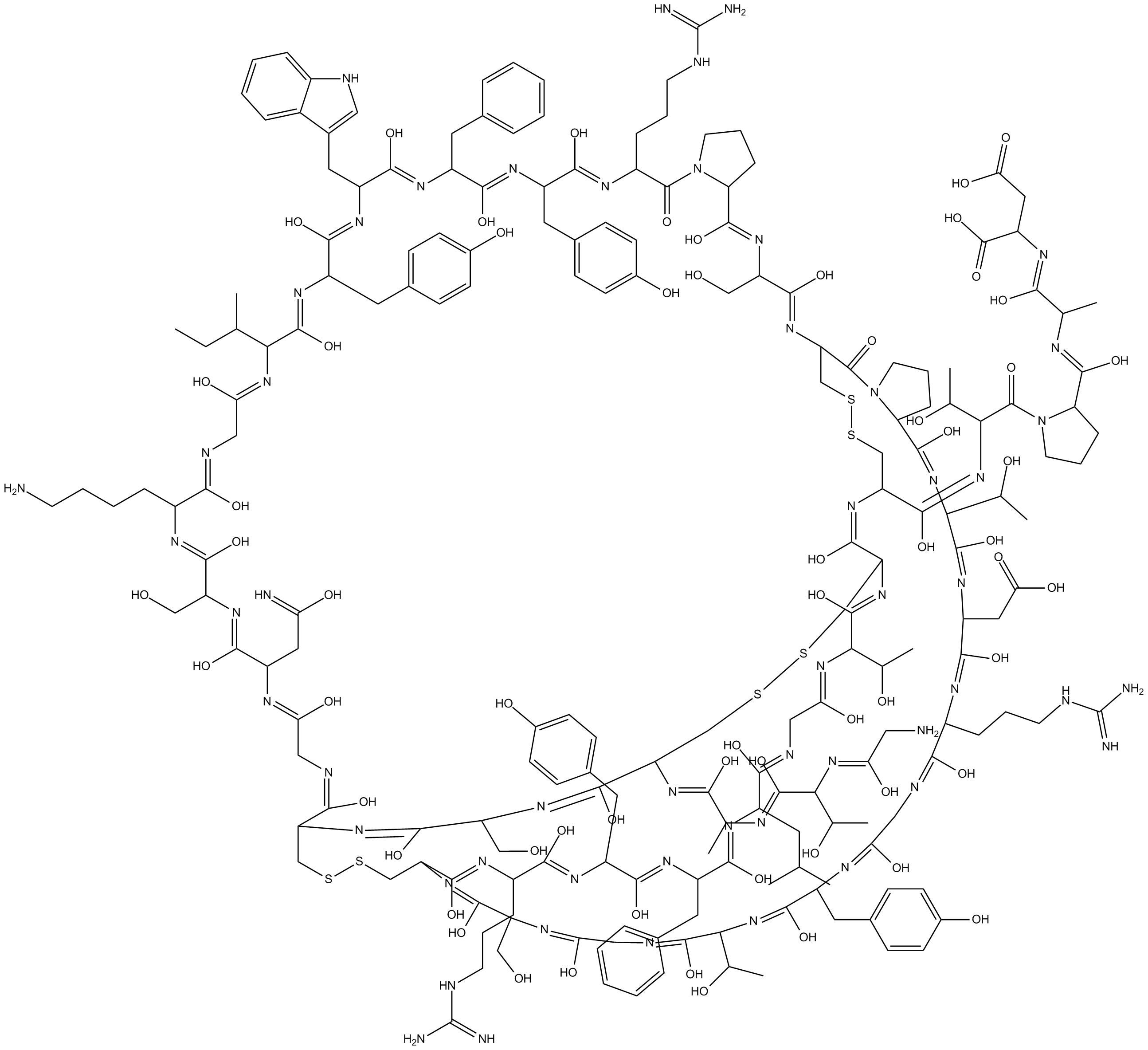
Cas No.: 713544-47-9
Sample solution is provided at 25 µL, 10mM.
_1-3.$$$39978408@51.jpg');)
_1-9.$$$38538795@65.jpg');)
_1-9.$$$39112701@51.jpg');)
_1-7.$$$37316672@70.jpg');)
_366-372.$$$36198801@70.jpg');)
.$$$38565142@65.jpg');)
_1867-1883.$$$33248023@67.jpg');)
_eads6055.$$$39977546@45.jpg');)
_eadm9805.$$$40080571@45.jpg');)
_eadj3020.$$$39666821@45.jpg');)
_1-20.$$$39757301@28.jpg');)
_1-24.$$$38898113@28.jpg');)
_273-287.$$$36806353@46.jpg');)
_1-16.$$$37156877@44.jpg');)
_1-19.$$$37460805@44.jpg');)
_1291-1307.$$$34518654@46.jpg');)
APETx2 is a peptide toxin derived from Anthopleura elegantissimaand serves as a selective, reversible inhibitor of the acid-sensing ion channel 3 (ASIC3)[1-2]. By inhibiting the activity of the ASIC3 channel, APETx2 blocks acid-induced pain signaling. APETx2 can be used in research related to inflammatory pain, post-infectious irritable bowel syndrome, and gastric mucosal injury[3-4].
In vitro, rat articular chondrocytes were pretreated with APETx2 (3µM) and PcTx1 (100ng/ml) for 1 hour, followed by incubation under acidic conditions (pH=6.0) for 3 hours. APETx2 significantly alleviated acidosis-induced cytotoxicity and apoptosis, exerting a chondroprotective effect by reducing intracellular Ca²⁺ levels and inhibiting the phosphorylation of the p38/ERK1/2 MAPK signaling pathway[5]. Under magnesium-free medium conditions (used to induce epileptiform discharge), primary hippocampal neurons were incubated with APETx2 (63nM) for 1 hour. APETx2 significantly increased the frequency of burst discharges induced by the magnesium-free medium[6].
In vivo, APETx2 (20pmol per mouse) was injected into the Zusanli acupoint (ST36) of a fibromyalgia mouse model. By antagonizing ASIC3, APETx2 significantly reduced mechanical hyperalgesia on day 14 post-induction and decreased the overexpression of pain-related channel proteins ASIC3, Nav1.7, and Nav1.8 in the dorsal root ganglia, spinal cord, and thalamus[7]. In a rat model of osteoarthritis, daily continuous intra-articular injections of APETx2 (2.5µg/kg) for 7 days, APETx2 significantly inhibited weight-bearing asymmetry and secondary mechanical hyperalgesia in the knee joint. APETx2 also effectively prevented articular cartilage damage by reducing chondrocyte loss and matrix destruction[8].
References:
[1] Yuan L, Xiao H, Li H, et al. APETx2 regulates intestinal motility and visceral sensitivity in post-infectious irritable bowel syndrome mice through 5-HT signalling pathway. J Pharm Pharmacol. 2023 Apr 17;75(5):712-717.
[2] Li J, Wei Y, Wang Y, et al. Metabolomics study of APETx2 post-conditioning on myocardial ischemia-reperfusion injury. Front Pharmacol. 2024 Dec 6;15:1470142.
[3] Diochot S, Baron A, Rash LD, et al. A new sea anemone peptide, APETx2, inhibits ASIC3, a major acid-sensitive channel in sensory neurons. EMBO J. 2004 Apr 7;23(7):1516-25.
[4] Chagot B, Escoubas P, Diochot S, et al. Solution structure of APETx2, a specific peptide inhibitor of ASIC3 proton-gated channels. Protein Sci. 2005 Aug;14(8):2003-10.
[5] Zhou RP, Ni WL, Dai BB, et al. ASIC2a overexpression enhances the protective effect of PcTx1 and APETx2 against acidosis-induced articular chondrocyte apoptosis and cytotoxicity. Gene. 2018 Feb 5;642:230-240.
[6] Cao Q, Xiao ZM, Wang X, et al. Inhibition of Acid Sensing Ion Channel 3 Aggravates Seizures by Regulating NMDAR Function. Neurochem Res. 2018 Jun;43(6):1227-1241.
[7] Yen LT, Hsieh CL, Hsu HC, et al. Preventing the induction of acid saline-induced fibromyalgia pain in mice by electroacupuncture or APETx2 injection. Acupunct Med. 2020 Jun;38(3):188-193.
[8] Izumi M, Ikeuchi M, Ji Q, et al. Local ASIC3 modulates pain and disease progression in a rat model of osteoarthritis. J Biomed Sci. 2012 Aug 21;19(1):77.
| Cell experiment [1]: | |
Cell lines | Articular chondrocytes |
Preparation Method | Articular chondrocytes were cultured in DMEM supplemented with 10% fetal bovine serum (FBS) at 37°C, 5% CO₂. Cells were pretreated with ASIC3-specific blocker APETx2 (3μM) for 1 hour, and exposure to pH 6.0 solution for 3 hours. |
Reaction Conditions | 3μM; 1-hour pretreatment. |
Applications | Pretreatment with APETx2, in combination with the ASIC1a-specific blocker PcTx1, significantly attenuated acidosis (pH=6.0)-induced chondrocyte cytotoxicity and apoptosis. This protective effect was characterized by increased cell viability, reduced LDH release, decreased intracellular Ca²⁺ concentration ([Ca²⁺]i) elevation, reduced apoptotic rate, and inhibition of the p38 and ERK1/2 MAPK signaling pathways. Additionally, the combination treatment (PcTx1 and APETx2) reversed the acid-induced decrease in type II collagen levels. |
| Animal experiment [2]: | |
Animal models | Male Sprague-Dawley rats (with MIA-induced osteoarthritis model) |
Preparation Method | A single dose of monosodium iodoacetate (MIA, 3mg dissolved in 50μl saline) was injected into the left knee joint to induce osteoarthritis. Rats were treated with intra-articular injections of the selective ASIC3 blocker APETx2 (2.5μg/kg) daily, either during the early phase (Days 1-7 after MIA) or the late phase (Days 7-13 after MIA). Pain-related behaviors (weight distribution asymmetry and mechanical hyperalgesia) were assessed at specified time points, and histological evaluation of knee joints was performed at Day 14. |
Dosage form | 2.5μg/kg; intra-articular injection; Daily continuous injection. |
Applications | Continuous administration of APETx2 inhibited the upregulation of ASIC3 in knee joint afferent neurons. APETx2 significantly reduced mechanical hyperalgesia (secondary hyperalgesia) in both early- and late-phase treatment groups. APETx2 also significantly inhibited weight distribution asymmetry in the early-phase group at Day 3. Furthermore, early administration of APETx2 (Days 1-7) prevented cartilage damage and reduced the severity of osteoarthritis, as evidenced by improved histological scores (modified Mankin score). |
References: | |
| Cas No. | 713544-47-9 | SDF | |
| Canonical SMILES | CCC(C(/N=C(O)/C/N=C(O)/C(/N=C(O)/C(/N=C(O)/C(/N=C(O)/C/N=C(O)\C1CSSCC(N=C(O)C(N=C(O)CN=C(O)C(N=C(O)C(N=C(O)CN=C(O)C(N=C(O)C(N=C(O)C(N=C(O)C2CCCN23)C(O)C)CC(O)=O)CCCNC(N)=N)CC4=CC=C(O)C=C4)C(O)C)CO)/C(O)=N/C(/C(O)=N/C(/C(O)=N/C(/C(O)=N/C(/C(O)=N/C/C(O)=N/C | ||
| Formula | C196H280N54O61S6 | M.Wt | 4561.06 |
| Solubility | Soluble to 5 mg/ml in Water | Storage | Store at -20°C |
| General tips | Please select the appropriate solvent to prepare the stock solution according to the
solubility of the product in different solvents; once the solution is prepared, please store it in
separate packages to avoid product failure caused by repeated freezing and thawing.Storage method
and period of the stock solution: When stored at -80°C, please use it within 6 months; when stored
at -20°C, please use it within 1 month. To increase solubility, heat the tube to 37°C and then oscillate in an ultrasonic bath for some time. |
||
| Shipping Condition | Evaluation sample solution: shipped with blue ice. All other sizes available: with RT, or with Blue Ice upon request. | ||
| Prepare stock solution | |||

|
1 mg | 5 mg | 10 mg |
| 1 mM | 219.2 μL | 1.0962 mL | 2.1925 mL |
| 5 mM | 43.8 μL | 219.2 μL | 438.5 μL |
| 10 mM | 21.9 μL | 109.6 μL | 219.2 μL |
Step 1: Enter information below (Recommended: An additional animal making an allowance for loss during the experiment)
 g
μL
g
μL
Step 2: Enter the in vivo formulation (This is only the calculator, not formulation. Please contact us first if there is no in vivo formulation at the solubility Section.)
Calculation results:
Working concentration: mg/ml;
Method for preparing DMSO master liquid: mg drug pre-dissolved in μL DMSO ( Master liquid concentration mg/mL, Please contact us first if the concentration exceeds the DMSO solubility of the batch of drug. )
Method for preparing in vivo formulation: Take μL DMSO master liquid, next addμL PEG300, mix and clarify, next addμL Tween 80, mix and clarify, next add μL saline, mix and clarify.
Method for preparing in vivo formulation: Take μL DMSO master liquid, next add μL Corn oil, mix and clarify.
Note: 1. Please make sure the liquid is clear before adding the next solvent.
2. Be sure to add the solvent(s) in order. You must ensure that the solution obtained, in the previous addition, is a clear solution before proceeding to add the next solvent. Physical methods such as vortex, ultrasound or hot water bath can be used to aid dissolving.
3. All of the above co-solvents are available for purchase on the GlpBio website.
Quality Control & SDS
- View current batch:
- Purity: >98.00% Appearance: A solid
- COA (Certificate of Analysis)
- SDS (Safety Data Sheet)
- Datasheet
Average Rating: 5 (Based on Reviews and 19 reference(s) in Google Scholar.)