

Bavachin (Synonyms: Corylifolin) |
Catalog No.GC17588
 One-Click Copy Product Info
|

estrogen receptors ERα and ERβ activator
Products are for research use only. Not for human use. We do not sell to patients.
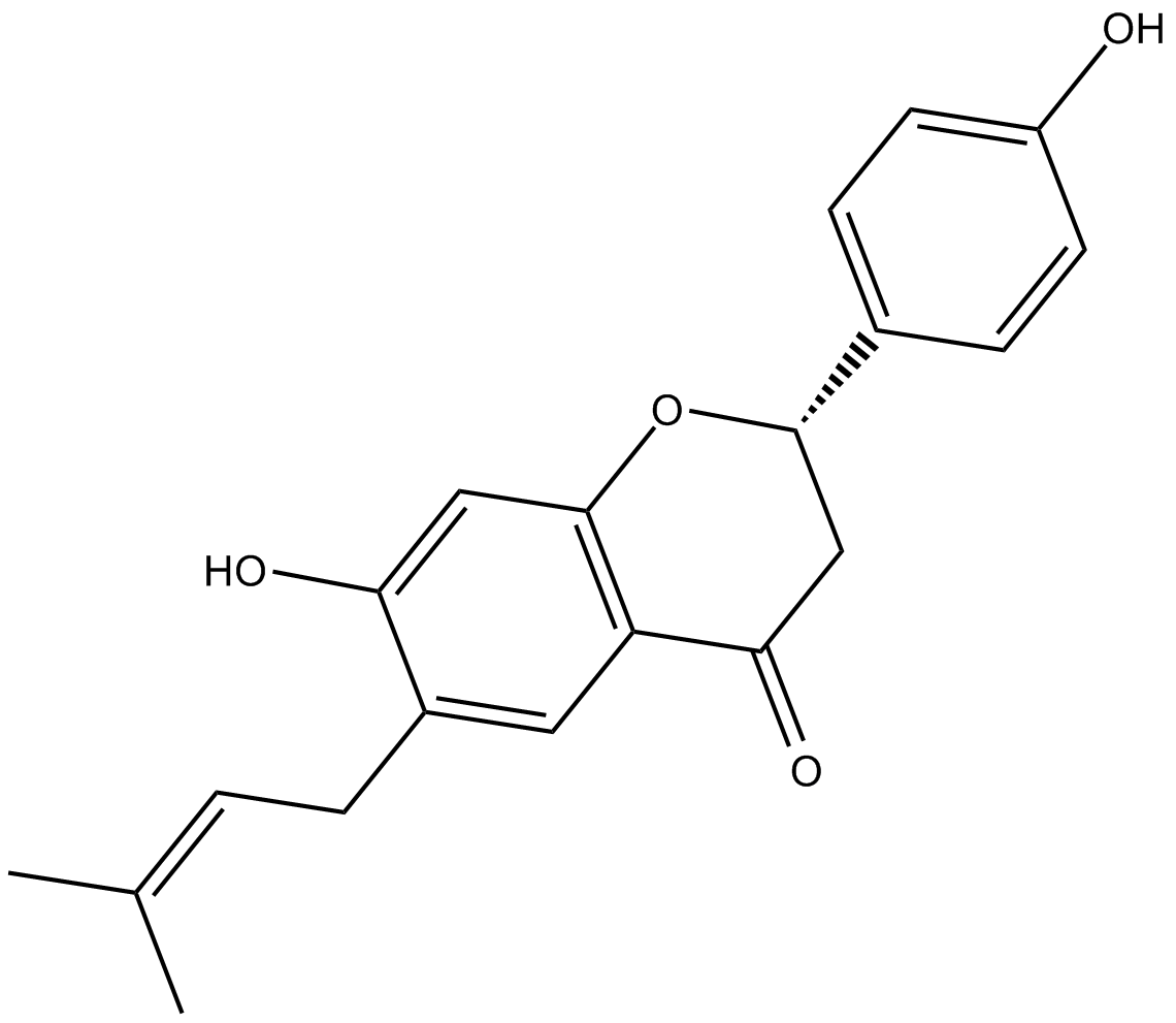
Cas No.: 19879-32-4
Sample solution is provided at 25 µL, 10mM.
_1-3.$$$39978408@51.jpg');)
_1-9.$$$38538795@65.jpg');)
_1-9.$$$39112701@51.jpg');)
_1-7.$$$37316672@70.jpg');)
_366-372.$$$36198801@70.jpg');)
.$$$38565142@65.jpg');)
_1867-1883.$$$33248023@67.jpg');)
_eads6055.$$$39977546@45.jpg');)
_eadm9805.$$$40080571@45.jpg');)
_eadj3020.$$$39666821@45.jpg');)
_1-20.$$$39757301@28.jpg');)
_1-24.$$$38898113@28.jpg');)
_273-287.$$$36806353@46.jpg');)
_1-16.$$$37156877@44.jpg');)
_1-19.$$$37460805@44.jpg');)
_1291-1307.$$$34518654@46.jpg');)
Bavachin is an acyl-coenzyme A: cholesterol acyltransferase inhibitor [1].
Acyl-coenzyme A: cholesterol acyl transferase (ACAT) is an enzyme responsible for the intracellular esterification of free cholesterol with fatty acids and plays dominant roles in intestinal absorption of cholesterol, hepatic production of lipoproteins and accumulation of cholesteryl ester within macrophages and smooth muscle cells. [1].
Bavachin showed a significant inhibition of ACAT enzyme. The IC50 value of bavachin was 86.0 μM in the ACAT assay system using rat liver microsome [1]. Bavachin is a flavonoid first isolated from Psoralea corylifolia that has been used as a traditional medicine in Asia. In CV-1 cells transfected with plasmids ERα or ERβ, bavachin showed ER ligand binding activity with an EC50 of 320 nM and 680 nM, respectively. Bavachin increased the mRNA levels of estrogen-responsive genes such as pS2 and PR, and decreased the protein level of ERα by proteasomal pathway [2]. Bavachin activated gene expression of proliferator-activated receptorγ (PPARγ), adipogenic transcriptional factors, and CCAAT/enhancer binding protein-α (C/EBPα). Bavachin increased adiponectin expression and secretion in adipocytes. Bavachin increased insulin-induced glucose uptake by differentiated adipocytes and myoblasts. In differentiated adipocytes, bavachin enhanced glucose uptake [3].
References:
[1] Choi J H, Rho M C, Lee S W, et al. Bavachin and isobavachalcone, acyl-coenzyme A: cholesterol acyltransferase inhibitors from Psoralea corylifolia[J]. Archives of pharmacal research, 2008, 31(11): 1419-1423.
[2] Park J, Kim D H, Ahn H N, et al. Activation of estrogen receptor by bavachin from Psoralea corylifolia[J]. Biomolecules & therapeutics, 2012, 20(2): 183-188.
[3] Lee H, Li H, Noh M, et al. Bavachin from Psoralea corylifolia improves insulin-dependent glucose uptake through insulin signaling and AMPK activation in 3T3-L1 adipocytes[J]. International journal of molecular sciences, 2016, 17(4): 527.
|
Kinase experiment: |
The chemiluminescent assay is used to confirm PCSEE MAO-A and MAO-B inhibitory effects and to test BNN and BVN hMAO-A and hMAO-B inhibition using MAO-Glo kit. Each enzyme's Arbitrary Light Unit (ALU) is measured in the presence of PCSEE, BNN, BVN, and standard DEP as an MAO-BI positive control. Briefly, hMAO-A and hMAO-B isozymes are diluted to 2× with reaction buffer (pH 7.4) and preincubated with 4× PCSEE, BNN, BVN, or DEP working solutions at RT for 30 min in white opaque 96-well plates. For determining activity inhibition, final 8.5 μg/mL concentrations of PCSEE, BNN, BVN, and DEP are used. For IC50 determination, 8× PCSEE and BNN working solutions are serially diluted using reaction buffers (pH 7.4) to make a 4× concentration. Ten points' range of PCSEE (1.0 to 250.0 μg/mL) and BNN (up to 400 μM (135.4 μg/mL)) final concentrations is used. Controls used are with and without ethanol. Ethanol solvent in controls is kept to a maximum final (volume) of ≤2%. Each isozyme is substituted with the reaction buffer for the blank. Based on our preliminary optimizations and Valley's method, the reaction is initiated by adding 4× luciferin derivative substrate (LDS) for a final (concentration) of 40 and 4 μM for hMAO-A and hMAO-B reactions, respectively. The final volume per well of each reaction is 50 μL. The reaction is optimized for the amount of A and B enzyme used to be incubated for less than 3.5 h at RT. To stop the reaction and produce the luminescence signal RLDR is added to all wells, 50 μL to each well, and incubated for a further 30 min. |
|
Cell experiment: |
MTT solution (20 µL) is added to each well of the 96-well plates, the cells are cultured for 4 h, the solution is discarded, and the purple crystal is dissolved in the wells with 150 µL DMSO solution, agitated in a 37°C incubator shaker for 10 min, and the optical density (OD) is measured at 490 nm by the microplate reader. |
|
References: [1]. Wang JH, et al. Effects of bavachin and its regulation of melanin synthesis in A375 cells. Biomed Rep. 2016 Jul;5(1):87-92. Epub 2016 May 20. |
|
| Cas No. | 19879-32-4 | SDF | |
| Synonyms | Corylifolin | ||
| Chemical Name | (2S)-2,3-dihydro-7-hydroxy-2-(4-hydroxyphenyl)-6-(3-methyl-2-butenyl)-4H-1-benzopyran-4-one | ||
| Canonical SMILES | OC1=CC(O[C@H](C2=CC=C(O)C=C2)CC3=O)=C3C=C1C/C=C(C)/C | ||
| Formula | C20H20O4 | M.Wt | 324.4 |
| Solubility | ≤20mg/ml in ethanol;30mg/ml in DMSO;50mg/ml in dimethyl formamide | Storage | Store at -20°C,protect from light |
| General tips | Please select the appropriate solvent to prepare the stock solution according to the
solubility of the product in different solvents; once the solution is prepared, please store it in
separate packages to avoid product failure caused by repeated freezing and thawing.Storage method
and period of the stock solution: When stored at -80°C, please use it within 6 months; when stored
at -20°C, please use it within 1 month. To increase solubility, heat the tube to 37°C and then oscillate in an ultrasonic bath for some time. |
||
| Shipping Condition | Evaluation sample solution: shipped with blue ice. All other sizes available: with RT, or with Blue Ice upon request. | ||
| Prepare stock solution | |||

|
1 mg | 5 mg | 10 mg |
| 1 mM | 3.0826 mL | 15.4131 mL | 30.8261 mL |
| 5 mM | 616.5 μL | 3.0826 mL | 6.1652 mL |
| 10 mM | 308.3 μL | 1.5413 mL | 3.0826 mL |
Step 1: Enter information below (Recommended: An additional animal making an allowance for loss during the experiment)
 g
μL
g
μL
Step 2: Enter the in vivo formulation (This is only the calculator, not formulation. Please contact us first if there is no in vivo formulation at the solubility Section.)
Calculation results:
Working concentration: mg/ml;
Method for preparing DMSO master liquid: mg drug pre-dissolved in μL DMSO ( Master liquid concentration mg/mL, Please contact us first if the concentration exceeds the DMSO solubility of the batch of drug. )
Method for preparing in vivo formulation: Take μL DMSO master liquid, next addμL PEG300, mix and clarify, next addμL Tween 80, mix and clarify, next add μL saline, mix and clarify.
Method for preparing in vivo formulation: Take μL DMSO master liquid, next add μL Corn oil, mix and clarify.
Note: 1. Please make sure the liquid is clear before adding the next solvent.
2. Be sure to add the solvent(s) in order. You must ensure that the solution obtained, in the previous addition, is a clear solution before proceeding to add the next solvent. Physical methods such as vortex, ultrasound or hot water bath can be used to aid dissolving.
3. All of the above co-solvents are available for purchase on the GlpBio website.
Quality Control & SDS
- View current batch:
- Purity: >98.00% Appearance: A solid
- COA (Certificate of Analysis)
- SDS (Safety Data Sheet)
- Datasheet
Average Rating: 5 (Based on Reviews and 5 reference(s) in Google Scholar.)