Home>>Signaling Pathways>> GPCR/G protein>> Endothelin Receptor>>Endothelin 1 swine, human

Endothelin 1 swine, human

Catalog No.: GC30485

Endothelin 1 swine, human is a synthetic peptide with the sequence of human and swine Endothelin 1, which is a potent endogenous vasoconstrictor.

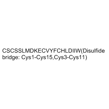
Endothelin 1 swine, human Chemical Structure

Size Price Stock Qty
500μg
$125.00
In stock
1mg
$220.00
In stock
5mg
$883.00
In stock
10mg
$1,471.00
In stock

Customer Reviews

Based on customer reviews.

Tel: (626) 353-8530 Email: sales@glpbio.com

Sample solution is provided at 25 µL, 10mM.

Product Documents

Quality Control & SDS

View current batch:

Protocol

Cell experiment [1]:

Cell lines

Immortalized mouse podocytes

Preparation Method

Immortalized mouse podocytes were incubated with RPMI 1640 medium alone or with 100 nM Endothelin 1 (swine, human) for 5, 15, or 30 minutes.

Reaction Conditions

100 nM for 5, 15, 30 minutes, 16, 24 hours.

Applications

Endothelin 1 (swine, human) reduced synaptopodin and increased a-SMA expression at 6 hours and even more so at 24 hours

Animal experiment [2]:

Animal models

Male severe combined immunodeficiency (SCID) mice

Preparation Method

The mice were inoculated with PPC-1 (4×10⁵ cells/25 µl/paw) subcutaneously in the left paw using a microsyringe. After acclimation, 10 µl of vehicle alone or vehicle with Endothelin 1 (swine, human) (10 pmol/paw) or sarafotoxin S6c was subcutaneously injected into the left hind paw.

Dosage form

10 pmol/paw, s.c.

Applications

Endothelin 1 (swine, human) (10 pmol/paw) induced pain responses in the sham-operated animals and potentiated responses in the PPC-1 inoculated animals

References:

[1]: Buelli S, RosanÒ L, Gagliardini E, et al. β-Arrestin-1 drives endothelin-1-mediated podocyte activation and sustains renal injury[J]. Journal of the American Society of Nephrology, 2014, 25(3): 523-533.
[2]: Yuyama H, Koakutsu A, Fujiyasu N, et al. Effects of selective endothelin ETA receptor antagonists on endothelin-1-induced potentiation of cancer pain[J]. European journal of pharmacology, 2004, 492(2-3): 177-182.

Background

Endothelin 1 (swine, human) is a 21aa peptide vasoconstrictor and agonist of endothelin (ET) receptors ETA and ETB (IC50s = 0.15 and 0.12 nM, respectively) [1]. Endothelin 1 (swine, human) is the endothelin generated in the endothelium, where it acts in a paracrine or autocrine manner on ETA and ETB receptors on adjacent endothelial or smooth muscle cells [2].

Endothelin 1 (swine, human) activats endothelin-A receptor (ETAR) and drives epithelial-to-mesenchymal transition in ovarian tumor cells through b-arrestin signaling. In cultured mouse podocytes, Endothelin 1 (swine, human) caused loss of the podocyte differentiation marker synaptopodin and acquisition of the mesenchymal marker a-smooth muscle actin. Endothelin 1 (swine, human) promoted podocyte migration via ETAR activation and increased b-arrestin-1 expression [3].

The Endothelin 1 (swine, human) (1nmol/kg) produced strong pressor responses in the anesthetized rats in vivo [4]. Mice received an intradermal injection of 1-30 pmol Endothelin 1 (swine, human) and were caused dose-dependent scratching bouts [5]. A subpressor dose of ET-1 administered to rats was found to increase glomerular permeability and inflammation as well as the excretion of the glomerular slit-diaphragm protein nephrin, effects that could be blocked by an ETA receptor antagonist [6]. The magnitude of the ET-1 rise during antiangiogenic treatment may be useful biomarker of the efficacy of treatment [7].

References:
[1]. Kikuchi T, Kubo K, Ohtaki T, et al. Endothelin-1 analogues substituted at both position 18 and 19: highly potent endothelin antagonists with no selectivity for either receptor subtype ETA or ETB[J]. Journal of medicinal chemistry, 1993, 36(25): 4087-4093.
[2]. Schiffrin E L. Role of endothelin-1 in hypertension and vascular disease[J]. American journal of hypertension, 2001, 14(S3): 83S-89S.
[3]. Buelli S, RosanÒ L, Gagliardini E, et al. β-Arrestin-1 drives endothelin-1-mediated podocyte activation and sustains renal injury[J]. Journal of the American Society of Nephrology, 2014, 25(3): 523-533.
[4]. Inoue A, Yanagisawa M, Kimura S, et al. The human endothelin family: three structurally and pharmacologically distinct isopeptides predicted by three separate genes[J]. Proceedings of the national academy of sciences, 1989, 86(8): 2863-2867.
[5]. Trentin P G, Fernandes M B, D'OrlÉans-Juste P, et al. Endothelin-1 causes pruritus in mice[J]. Experimental biology and medicine, 2006, 231(6): 1146-1151.
[6]. Saleh M A, Pollock J S, Pollock D M. Distinct actions of endothelin A-selective versus combined endothelin A/B receptor antagonists in early diabetic kidney disease[J]. Journal of Pharmacology and Experimental Therapeutics, 2011, 338(1): 263-270.
[7]. Lankhorst S, Jan Danser A H, van den Meiracker A H. Endothelin-1 and antiangiogenesis[J]. American Journal of Physiology-Regulatory, Integrative and Comparative Physiology, 2016, 310(3): R230-R234.

Chemical Properties

Cas No. 117399-94-7 SDF
Canonical SMILES Cys-Ser-Cys-Ser-Ser-Leu-Met-Asp-Lys-Glu-Cys-Val-Tyr-Phe-Cys-His-Leu-Asp-Ile-Ile-Trp (Disulfide bridge: Cys1-Cys15, Cys3-Cys11)
Formula C109H159N25O32S5 M.Wt 2491.9
Solubility H2O : 1.8 mg/mL (0.72 mM; ultrasonic and adjust pH to 3 with HCl) Storage Store at -20°C
General tips For obtaining a higher solubility , please warm the tube at 37 ℃ and shake it in the ultrasonic bath for a while.Stock solution can be stored below -20℃ for several months.
Shipping Condition Evaluation sample solution : ship with blue ice
All other available size: ship with RT , or blue ice upon request

In vivo Formulation Calculator (Clear solution)

Step 1: Enter information below (Recommended: An additional animal making an allowance for loss during the experiment)

mg/kg g μL

Step 2: Enter the in vivo formulation (This is only the calculator, not formulation. Please contact us first if there is no in vivo formulation at the solubility Section.)

% DMSO % % Tween 80 % saline
%DMSO %

Calculation results:

Working concentration: mg/ml;

Method for preparing DMSO master liquid: mg drug pre-dissolved in μL DMSO ( Master liquid concentration mg/mL, Please contact us first if the concentration exceeds the DMSO solubility of the batch of drug. )

Method for preparing in vivo formulation: Take μL DMSO master liquid, next addμL PEG300, mix and clarify, next addμL Tween 80, mix and clarify, next add μL saline, mix and clarify.

Method for preparing in vivo formulation: Take μL DMSO master liquid, next add μL Corn oil, mix and clarify.

Note: 1. Please make sure the liquid is clear before adding the next solvent.
2. Be sure to add the solvent(s) in order. You must ensure that the solution obtained, in the previous addition, is a clear solution before proceeding to add the next solvent. Physical methods such as vortex, ultrasound or hot water bath can be used to aid dissolving.
3. All of the above co-solvents are available for purchase on the GlpBio website.

  • Molarity Calculator

  • Dilution Calculator

Mass
=
Concentration
x
Volume
x
MW*


**When preparing stock solutions always use the batch-specific molecular weight of the product found on the vial label and MSDS / CoA (available online).

Calculate

Research Update

Hyperglycemia Augments Endothelin-1-Induced Constriction of Human Retinal Venules

Purpose: Endothelin-1 (ET-1) is a potent vasoactive factor implicated in development of diabetic retinopathy, which is commonly associated with retinal edema and hyperglycemia. Although the vasomotor activity of venules contributes to the regulation of tissue fluid homeostasis, responses of human retinal venules to ET-1 under euglycemia and hyperglycemia remain unknown and the ET-1 receptor subtype corresponding to vasomotor function has not been determined. Herein, we addressed these issues by examining the reactivity of isolated human retinal venules to ET-1, and results from porcine retinal venules were compared. Methods: Retinal tissues were obtained from patients undergoing enucleation. Human and porcine retinal venules were isolated and pressurized to assess diameter changes in response to ET-1 after exposure to 5 mM control glucose or 25 mM high glucose for 2 hours. Results: Both human and porcine retinal venules exposed to control glucose developed similar basal tone and constricted comparably to ET-1 in a concentration-dependent manner. ET-1-induced constrictions of human and porcine retinal venules were abolished by ETA receptor antagonist BQ123. During high glucose exposure, basal tone of human and porcine retinal venules was unaltered but ET-1-induced vasoconstrictions were enhanced. Conclusions: ET-1 elicits comparable constriction of human and porcine retinal venules by activation of ETA receptors. In vitro hyperglycemia augments human and porcine retinal venular responses to ET-1. Translational relevance: Similarities in vasoconstriction to ET-1 between human and porcine retinal venules support the latter as an effective model of the human retinal microcirculation to help identify vascular targets for the treatment of retinal complications in patients with diabetes.

Endothelin-1 regulates oligodendrocyte development

In the postnatal brain, oligodendrocyte progenitor cells (OPCs) arise from the subventricular zone (SVZ) and migrate into the developing white matter, where they differentiate into oligodendrocytes and myelinate axons. The mechanisms regulating OPC migration and differentiation are not fully defined. The present study demonstrates that endothelin-1 (ET-1) is an astrocyte-derived signal that regulates OPC migration and differentiation. OPCs in vivo and in culture express functional ET(A) and ET(B) receptors, which mediate ET-1-induced ERK (extracellular signal-regulated kinase) and CREB (cAMP response element-binding protein) phosphorylation. ET-1 exerts both chemotactic and chemokinetic effects on OPCs to enhance cell migration; it also prevents lineage progression from the O4(+) to the O1(+) stage without affecting cell proliferation. Astrocyte-conditioned medium stimulates OPC migration in culture through ET receptor activation, whereas multiphoton time-lapse imaging shows that selective ET receptor antagonists or anti-ET-1 antibodies inhibit OPC migration from the SVZ. Inhibition of ET receptor activity also derepresses OPC differentiation in the corpus callosum in slice cultures. Our findings indicate that ET-1 is a soluble astrocyte-derived signal that regulates OPC migration and differentiation during development.

Human polymorphonuclear leukocytes have dual effects on endothelin-1: the induction of endothelin-1 mRNA expression in vascular endothelial cells and modification of the endothelin-1 molecule

The effect of human polymorphonuclear leukocytes (PMNs) on the expression of the endothelin-1 (ET-1) gene and the production of ET-1 peptide was investigated. Human PMNs were separated from venous blood with Mono-Poly Resolving Medium and activated by incubation with formyl-methionyl-lencylphenylalanine (FMLP) (1 microM). Then PMN suspension was added to cultured porcine endothelial cell monolayers and coincubated for various periods. Following the coincubation, ET-1 mRNA in endothelial cells was examined by Northern blotting and immunoreactive ET-1 (irET-1) peptide levels in the conditioned media were measured by an enzyme-linked immunosorbent assay (ELISA). Similar experiments were also carried out with cell-free PMN supernatant. Untreated and activated PMNs led to a 1.4-fold and 6.3-fold increase in ET-1 mRNA levels in endothelial cells, respectively, at 6h, while irET-1 peptide levels did not significantly increase as compared with control. In contrast, when PMNs were coincubated in the presence of an Intercell chamber without direct contact to endothelial cells, PMNs did not induce ET-1 mRNA expression in endothelial cells, and significantly decreased irET-1 peptide levels in the conditioned media. Cell-free PMN supernatant did not have all these effects on ET-1. These findings suggest that direct PMN-endothelial cell contact was essential for PMN-induced expression of the ET-1 gene and that PMNs may decrease irET-1 through some modification of the ET-1 molecule.

Localisation of endothelin-1 mRNA expression and immunoreactivity in the retina and optic nerve from human and porcine eye. Evidence for endothelin-1 expression in astrocytes

We have investigated the localisation of endothelin-1 (ET-1) mRNA and ET-1-like immunoreactivity in retina and anterior portion of optic nerve from human and porcine eyes. In situ hybridisation method revealed expression of ET-1 mRNA mainly in the innermost layers of the retinas, in the retinal pigment epithelium cells as well as in the astrocytes of the optic nerve. Immunohistochemical studies showed that ET-1-like immunoreactivity appeared in the same regions where ET-1 mRNA was expressed as well as in the inner nuclear layer and in the inner segments of photoreceptors. In the nerve fibre and ganglion cell layers, astrocytes expressed both glial fibrillary acidic protein and ET-1 proteins suggesting that these cells may secrete ET-1. Expression of ETA and ETB receptors in human retina were demonstrated by reverse transcription-polymerase chain reaction. Our results demonstrated expression of ET-1 in glial, neural and vascular components of retina and optic nerve from human and porcine eyes.

Interleukin 1 increases the production of endothelin-1 by cultured endothelial cells

We examined the effect of human recombinant interleukin 1 (IL-1) on the production of endothelin-1 by cultured porcine endothelial cells. The induction of endothelin-1 mRNA began within 1 hr of exposure to IL-1, showed twin peaks at 4 and 24 hr, and declined thereafter. Enzyme-linked immunosorbent assay revealed that the amount of endothelin-1 peptide in conditioned media was also increased by IL-1 in a dose- and time-dependent manner. Our results suggested that IL-1, a macrophage-derived cytokine, may affect the contraction and proliferation of vascular smooth muscle cells by stimulating the production of endothelin by endothelial cells.

Reviews

Review for Endothelin 1 swine, human

Average Rating: 5 ★★★★★ (Based on Reviews and 29 reference(s) in Google Scholar.)

5 Star
100%
4 Star
0%
3 Star
0%
2 Star
0%
1 Star
0%
Review for Endothelin 1 swine, human

GLPBIO products are for RESEARCH USE ONLY. Please make sure your review or question is research based.

Required fields are marked with *

You may receive emails regarding this submission. Any emails will include the ability to opt-out of future communications.