CY7 |
Catalog No.GC35772 |
CY7 is a cyanine-containing fluorescent dye that is commonly used to label proteins, antibodies, and small molecule compounds. Its maximum excitation/emission wavelengths are 745/800nm, respectively.
Products are for research use only. Not for human use. We do not sell to patients.
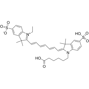
Cas No.: 943298-08-6
Sample solution is provided at 25 µL, 10mM.
CY7 is a cyanine-containing fluorescent dye that is commonly used to label proteins, antibodies, and small molecule compounds. Its maximum excitation/emission wavelengths are 745/800nm, respectively[1]. The basic structure of CY7 is a cyclic compound with a conjugated double bond system. It has excellent fluorescence properties in the far-red region and good photostability[2]. CY7 is a cyanine compound with the characteristics of long wavelength, adjustable absorption and emission, high extinction coefficient, good water solubility, and relatively simple synthesis[3]. CY7 usually exists in the form of its active ester or azide, which enables it to be labeled with biological molecules such as proteins and nucleic acids through covalent bonds[4]. The optimal molar ratio of CY dye to protein is about 10:1[5]. CY7 has a wide range of applications. It is not only suitable for biomarkers and cell imaging, but also for nucleic acid detection, protein analysis, and drug screening[6]. The molecular weight of this product is 682.85. It is for research purposes only and is not suitable for food, drugs, medical devices, or cosmetics.
References:
[1] Zhang R, Yang J, Radford D C, et al. FRET Imaging of Enzyme‐Responsive HPMA Copolymer Conjugate[J]. Macromolecular bioscience, 2017, 17(1): 1600125.
[2] Yuan L, Lin W, Zheng K, et al. Far-red to near infrared analyte-responsive fluorescent probes based on organic fluorophore platforms for fluorescence imaging[J]. Chemical Society Reviews, 2013, 42(2): 622-661.
[3] Pandey R K, James N, Chen Y, et al. Cyanine dye-based compounds for tumor imaging with and without photodynamic therapy[J]. Heterocyclic Polymethine Dyes: Synthesis, Properties and Applications, 2008: 41-74.
[4] Gerowska M, Hall L, Richardson J, et al. Efficient reverse click labeling of azide oligonucleotides with multiple alkynyl Cy-Dyes applied to the synthesis of HyBeacon probes for genetic analysis[J]. Tetrahedron, 2012, 68(3): 857-864.
[5] Yu H, Chao J, Patek D, et al. Cyanine dye dUTP analogs for enzymatic labeling of DNA probes[J]. Nucleic Acids Research, 1994, 22(15): 3226-3232.
[6] Ntziachristos V, Schellenberger E A, Ripoll J, et al. Visualization of antitumor treatment by means of fluorescence molecular tomography with an annexin V–CY7. 5 conjugate[J]. Proceedings of the National Academy of Sciences, 2004, 101(33): 12294-12299.
This protocol only provides a guide, please modify it to meet your specific needs.
1. Solution preparation
(1) Working solution: Dissolve CY7 solid in DMSO to a final concentration of 10mM.
Note: The working solution must be prepared and used immediately. Before use, 500μg/mL condensation solution (Cat. No.: GA11126) must be used for activation before subsequent labeling experiments can be performed.
2. Experimental steps for labeling proteins with CY7
(1) Protein preparation: Dissolve the protein in a pH 8.5 buffer without primary amines (such as Tris or glycine) and ammonium ions, and prepare the concentration to 2-10mg/mL to obtain the best labeling efficiency. If the pH is lower than 8.0, adjust with 1M sodium bicarbonate.
(2) Calculation of the amount of CY7 working solution: The amount of CY7 (MW=682.85) required for the labeling reaction depends on the amount of protein to be labeled. The optimal molar ratio of CY dye to protein is about 10:1.
(3) Labeling reaction: Take the calculated volume of CY7 working solution and slowly add it to the protein sample solution, gently shake to mix, and then briefly centrifuge to collect the sample at the bottom of the reaction tube. Avoid violent mixing to prevent the protein sample from denaturing and inactivating.
(4) Incubation reaction: Place the reaction tube in a dark place and incubate at room temperature or 4°C with gentle shaking for 60min. Every 10-15min, gently invert the reaction tube several times to fully mix the two reactants and improve the labeling efficiency. The labeling time is 2-18h, and the labeling ratio and time are adjusted according to the labeling effect.
Note:
(1)This product is an unactivated fluorescent dye. To use this product to label biological molecules such as peptides and proteins, it must first be activated with carboxylic acid. If an activated form is required, sulfo-Cyanine5 NHS Ester (Cat. No.: GC59174) is recommended.
(2)This protocol provides guidance for using CY7 for protein labeling experiments. It can be adjusted according to other literature and specific experimental requirements. The operation should be carried out in a sterile environment to prevent contaminants from interfering with the reaction. Avoid direct contact with the reaction reagents.
Cas No. | 943298-08-6 | SDF | |
Canonical SMILES | CC(/C(N1CCCCCC(O)=O)=C\C=C\C=C\C=C\C2=[N+](CC)C(C=CC(S(=O)([O-])=O)=C3)=C3C2(C)C)(C)C4=C1C=CC(S(=O)(O)=O)=C4 | ||
Formula | C35H42N2O8S2 | M.Wt | 682.85 |
Solubility | DMSO: ≥ 33 mg/mL (48.33 mM) | Storage | Store at -20°C, protect from light |
General tips | Please select the appropriate solvent to prepare the stock solution according to the
solubility of the product in different solvents; once the solution is prepared, please store it in
separate packages to avoid product failure caused by repeated freezing and thawing.Storage method
and period of the stock solution: When stored at -80°C, please use it within 6 months; when stored
at -20°C, please use it within 1 month. To increase solubility, heat the tube to 37°C and then oscillate in an ultrasonic bath for some time. |
||
Shipping Condition | Evaluation sample solution: shipped with blue ice. All other sizes available: with RT, or with Blue Ice upon request. |
Prepare stock solution | |||
![]() |
1 mg | 5 mg | 10 mg |
1 mM | 1.4645 mL | 7.3223 mL | 14.6445 mL |
5 mM | 0.2929 mL | 1.4645 mL | 2.9289 mL |
10 mM | 0.1464 mL | 0.7322 mL | 1.4645 mL |
Step 1: Enter information below (Recommended: An additional animal making an allowance for loss during the experiment)
Step 2: Enter the in vivo formulation (This is only the calculator, not formulation. Please contact us first if there is no in vivo formulation at the solubility Section.)
Calculation results:
Working concentration: mg/ml;
Method for preparing DMSO master liquid: mg drug pre-dissolved in μL DMSO ( Master liquid concentration mg/mL, Please contact us first if the concentration exceeds the DMSO solubility of the batch of drug. )
Method for preparing in vivo formulation: Take μL DMSO master liquid, next addμL PEG300, mix and clarify, next addμL Tween 80, mix and clarify, next add μL ddH2O, mix and clarify.
Method for preparing in vivo formulation: Take μL DMSO master liquid, next add μL Corn oil, mix and clarify.
Note: 1. Please make sure the liquid is clear before adding the next solvent.
2. Be sure to add the solvent(s) in order. You must ensure that the solution obtained, in the previous addition, is a clear solution before proceeding to add the next solvent. Physical methods such as vortex, ultrasound or hot water bath can be used to aid dissolving.
3. All of the above co-solvents are available for purchase on the GlpBio website.
Quality Control & SDS
- View current batch:
- Purity: >95.00%
- COA (Certificate Of Analysis)
- SDS (Safety Data Sheet)
- Datasheet
Average Rating: 5
(Based on Reviews and 40 reference(s) in Google Scholar.)GLPBIO products are for RESEARCH USE ONLY. Please make sure your review or question is research based.
Required fields are marked with *