

SX-682 |
Catalog No.GC38204
 One-Click Copy Product Info
|

SX-682 is an orally bioavailable small-molecule inhibitor of CXCR1 and CXCR2 with IC50 values of 31 and 21nM, respectively.
Products are for research use only. Not for human use. We do not sell to patients.
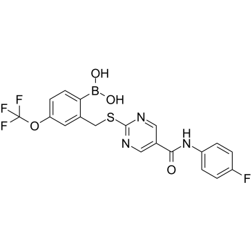
Cas No.: 1648843-04-2
Sample solution is provided at 25 µL, 10mM.
_1-3.$$$39978408@51.jpg');)
_1-9.$$$38538795@65.jpg');)
_1-9.$$$39112701@51.jpg');)
_1-7.$$$37316672@70.jpg');)
_366-372.$$$36198801@70.jpg');)
.$$$38565142@65.jpg');)
_1867-1883.$$$33248023@67.jpg');)
_eads6055.$$$39977546@45.jpg');)
_eadm9805.$$$40080571@45.jpg');)
_eadj3020.$$$39666821@45.jpg');)
_1-20.$$$39757301@28.jpg');)
_1-24.$$$38898113@28.jpg');)
_273-287.$$$36806353@46.jpg');)
_1-16.$$$37156877@44.jpg');)
_1-19.$$$37460805@44.jpg');)
_1291-1307.$$$34518654@46.jpg');)
SX-682 is an orally bioavailable small-molecule inhibitor of CXCR1 and CXCR2 with IC50 values of 31 and 21nM, respectively [1]. SX-682 effective at promoting a switch from a mesenchymal to an epithelial tumor phenotype, while simultaneously enhancing the tumor cells’ susceptibility to immune-mediated cytotoxicity [2]. SX-682 has been widely used as an anti-cancer agent to inhibit the progression of colorectal cancer[3].
In vitro, SX-682 (5μM) treatment for 4 days significantly inhibited the growth of B16F0 and B16F10 cells, and reduced the production of Cxcl1 and vascular endothelial growth factor (VEGF)[4]. The combined treatment of 2.5µM SX-682 and 25µg/mL bintrafusp alfa for 3 days led to a significant aggregation of 4T1 cells, upregulated the expression of E-cadherin, and decreased the expressions of mesenchymal cell vimentin, as well as mesenchymal transcription factors Snail and Zeb-1[5]. 10µM SX-682 pretreatment for 72 hours significantly enhanced the sensitivity of UM-SCC-11B cells to docetaxel, and greatly reduced cell viability[6].
In vivo, SX-682 treatment via oral administration at a dose of 200mg/kg dose twice daily for 2 weeks significantly inhibited the growth of lung tumors in mice, without reducing the content of tumor-associated neutrophils[7]. The combined treatment with SX-682 (0.756g/kg/day; p.o.) and anti-PD-1 antibody for 3 weeks significantly inhibited the growth of tumors in Rich1.1 melanoma cell-xenograft mouse model, reduced the myeloid-derived suppressor cells (MDSC) within the tumors, and increased the B1b cells expressing CXCL11 within the tumors[8].
References:
[1] Maeda D Y, Peck A M, Schuler A D, et al. Boronic acid-containing CXCR1/2 antagonists: Optimization of metabolic stability, in vivo evaluation, and a proposed receptor binding model[J]. Bioorganic & medicinal chemistry letters, 2015, 25(11): 2280-2284.
[2] Horn L, Hamilton D, Maeda D, et al. Abstract B04: Multimodal cancer immunotherapy combining IL-8 inhibition, adenovirus vaccine, IL-15 super agonist, and anti-PD-L1/TGFβRII agent reduces mesenchymalization and enhances anti-tumor efficacy[J]. Cancer Immunology Research, 2020, 8(4_Supplement): B04-B04.
[3] Sherry C, Dadgar N, Liu Z, et al. The Interleukin-8-CXCR1/2 Axis as a Therapeutic Target in Peritoneal Carcinomatosis[J]. Current Oncology, 2025, 32(9): 496.
[4] Yang J, Bergdorf K, Yan C, et al. CXCR2 expression during melanoma tumorigenesis controls transcriptional programs that facilitate tumor growth[J]. Molecular Cancer, 2023, 22(1): 92.
[5] Horn L A, Riskin J, Hempel H A, et al. Simultaneous inhibition of CXCR1/2, TGF-β, and PD-L1 remodels the tumor and its microenvironment to drive antitumor immunity[J]. Journal for immunotherapy of cancer, 2020, 8(1): e000326.
[6] Horn L A, Lind H, Fousek K, et al. Inhibition of the chemokine receptors CXCR1 and CXCR2 synergizes with docetaxel for effective tumor control and remodeling of the immune microenvironment of HPV-negative head and neck cancer models[J]. Journal of Experimental & Clinical Cancer Research, 2024, 43(1): 318
[7] Kwak J W, Nguyen H Q, Camai A, et al. CXCR1/2 antagonism inhibits neutrophil function and not recruitment in cancer[J]. Oncoimmunology, 2024, 13(1): 2384674.
[8] Yang J, Yan C, Vilgelm A E, et al. Targeted deletion of CXCR2 in myeloid cells alters the tumor immune environment to improve antitumor immunity[J]. Cancer immunology research, 2021, 9(2): 200-213.
| Cell experiment [1]: | |
Cell lines | 4T1 cells |
Preparation Method | The 4T1 cells were respectively cultured in IMDM medium containing 10% FBS for 72h with 2.5µM SX-682, 25µg/ml bintrafusp alfa, or 2.5µM SX-682 combined with 25µg/ml bintrafusp alfa. The expression of E-cadherin and mesenchymal transcription factors in the cells were analyzed. |
Reaction Conditions | 2.5µM; 72h |
Applications | SX-682 treatment upregulated the expression of E-cadherin and reduced mesenchymal transcription factors Snail and Zeb-1 in 4T1 cells. |
| Animal experiment [2]: | |
Animal models | C57BL/6 mice |
Preparation Method | The homologous Rich1.1 melanoma cells BrafV637E PtenR74X, Q396X were subcutaneously implanted into immunocompetent C57BL/6 mice. The C57BL/6 mice were fed with feed containing SX-682 (0.756g/kg), and were simultaneously intraperitoneally injected with anti-PD-1 antibody (100μg per mouse), or control IgG, or fed with vehicle control feed without SX-682, and were intraperitoneally injected with anti-PD-1 antibody or the same type of control IgG every other day. After 3 weeks of treatment, the lung tissues of the mice were removed for analysis. |
Dosage form | 0.756g/kg/day for 3 weeks; p.o. |
Applications | The combined treatment with SX-682 and anti-PD-1 antibody significantly inhibited the growth of lung tumors in mice, reduced the myeloid-derived suppressor cells (MDSC) within the tumors, and increased the B1b cells expressing CXCL11 within the tumors. |
References: | |
| Cas No. | 1648843-04-2 | SDF | |
| Canonical SMILES | O=C(NC1=CC=C(F)C=C1)C(C=N2)=CN=C2SCC3=CC(OC(F)(F)F)=CC=C3B(O)O | ||
| Formula | C19H14BF4N3O4S | M.Wt | 467.2 |
| Solubility | DMSO: 250 mg/mL (535.10 mM) | Storage | Store at -20°C |
| General tips | Please select the appropriate solvent to prepare the stock solution according to the
solubility of the product in different solvents; once the solution is prepared, please store it in
separate packages to avoid product failure caused by repeated freezing and thawing.Storage method
and period of the stock solution: When stored at -80°C, please use it within 6 months; when stored
at -20°C, please use it within 1 month. To increase solubility, heat the tube to 37°C and then oscillate in an ultrasonic bath for some time. |
||
| Shipping Condition | Evaluation sample solution: shipped with blue ice. All other sizes available: with RT, or with Blue Ice upon request. | ||
| Prepare stock solution | |||

|
1 mg | 5 mg | 10 mg |
| 1 mM | 2.1404 mL | 10.7021 mL | 21.4041 mL |
| 5 mM | 428.1 μL | 2.1404 mL | 4.2808 mL |
| 10 mM | 214 μL | 1.0702 mL | 2.1404 mL |
Step 1: Enter information below (Recommended: An additional animal making an allowance for loss during the experiment)
 g
μL
g
μL
Step 2: Enter the in vivo formulation (This is only the calculator, not formulation. Please contact us first if there is no in vivo formulation at the solubility Section.)
Calculation results:
Working concentration: mg/ml;
Method for preparing DMSO master liquid: mg drug pre-dissolved in μL DMSO ( Master liquid concentration mg/mL, Please contact us first if the concentration exceeds the DMSO solubility of the batch of drug. )
Method for preparing in vivo formulation: Take μL DMSO master liquid, next addμL PEG300, mix and clarify, next addμL Tween 80, mix and clarify, next add μL saline, mix and clarify.
Method for preparing in vivo formulation: Take μL DMSO master liquid, next add μL Corn oil, mix and clarify.
Note: 1. Please make sure the liquid is clear before adding the next solvent.
2. Be sure to add the solvent(s) in order. You must ensure that the solution obtained, in the previous addition, is a clear solution before proceeding to add the next solvent. Physical methods such as vortex, ultrasound or hot water bath can be used to aid dissolving.
3. All of the above co-solvents are available for purchase on the GlpBio website.
Quality Control & SDS
- View current batch:
- Purity: >98.00% Appearance: A solid
- COA (Certificate of Analysis)
- SDS (Safety Data Sheet)
- Datasheet
Average Rating: 5 (Based on Reviews and 30 reference(s) in Google Scholar.)