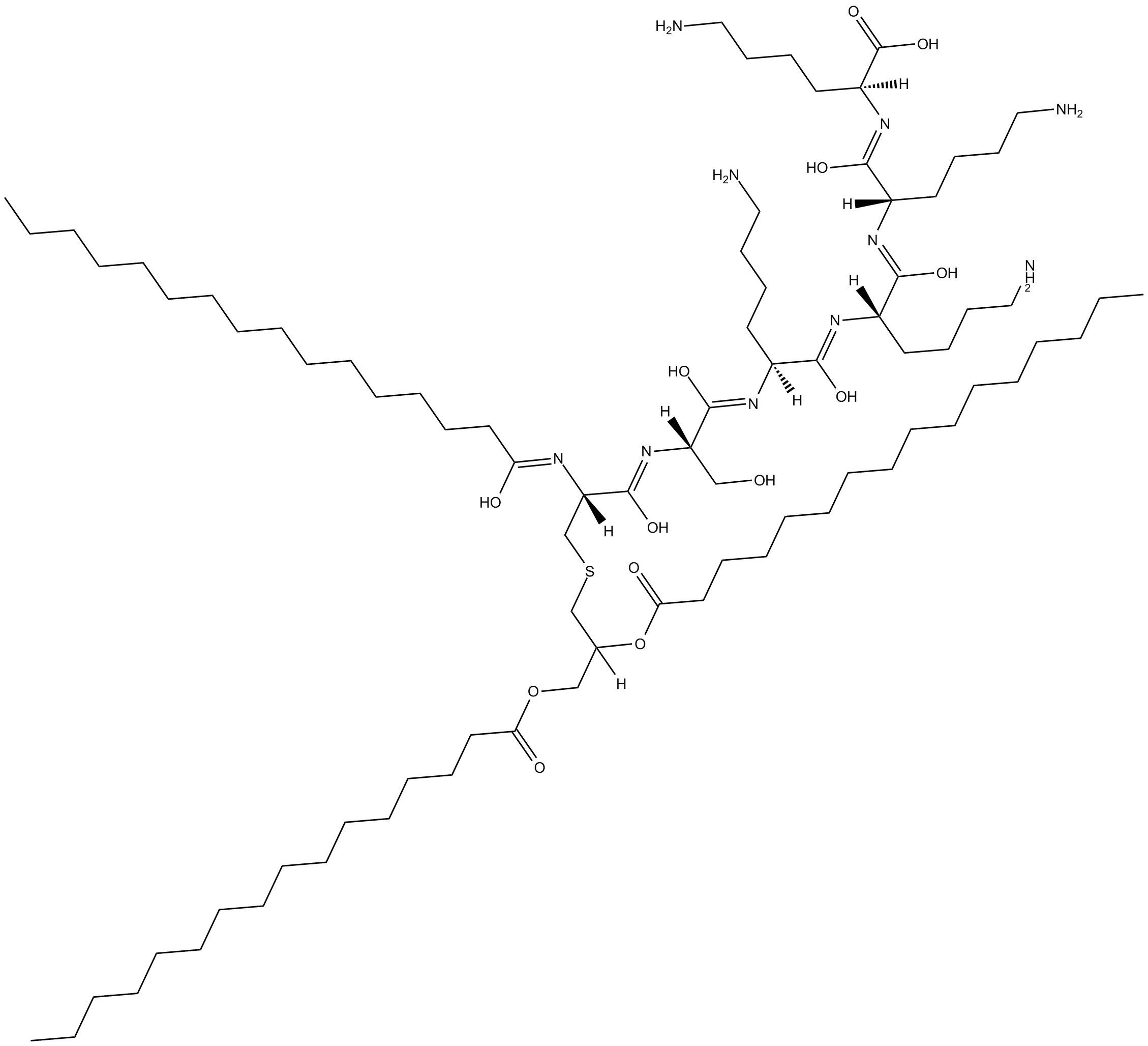
Pam3CSK4 |
Catalog No.: GC10273 |
Pam3CSK4 (Pam3CysSerLys4) is a synthetic triacylated lipopeptide (LP) and a TLR2/TLR1 ligand?agonist with an EC50 of 0.47 ng/mL.
Sample solution is provided at 25 µL, 10mM.
Quality Control & SDS
- View current batch:
-
Purity: >98.00%
- COA (Certificate Of Analysis)
- SDS (Safety Data Sheet)
- Datasheet
Cell experiment [1]: | |
Cell lines |
HepaRG cell |
Preparation Method |
Indicated differentiated HepaRG cell lines were infected with HBV with 100 vge/cell. At day-7 post-infection , cells were treated twice with either Pam3CSK4 (100 ng/mL), IFNα (500 IU/mL), RG7834 (0.1 μM), or the nucleoside analogue lamivudine (3 TC; 1 μM) for a total exposure time of 6 days. Cells and supernatants were harvested for analyses at day-13 post-infection. |
Reaction Conditions |
100 ng/mL; 6 days |
Applications |
Pam3CSK4 was capable to inhibit HBV (genotype D) replication in dHepaRG cells. The levels of all HBV parameters analyzed, (including intracellular total HBV RNAs, viremia, and secretion of viral antigens) were significantly reduced even at very low concentration of Pam3CSK4. Pam3CSK4 was also capable to inhibit the replication of another HBV genotype (genotype C). thus suggesting a broad antiviral activity against HBV. Pam3CSK4 as a TLR2 agonist, is a potent anti-HBV agents. Pam3CSK4 inhibits RNA accumulation by both impairing HBV transcription and RNA stability. Pam3CSK4 decreases also cccDNA level via a FEN-1-dependent mechanism. |
Animal experiment [2]: | |
Animal models |
C57 BL/6 mice |
Preparation Method |
To analyse the influence of TLR2 heterodimer activation on IL-13-induced itch-like behaviour, C57 BL/6 mice were pre-administered with Pam3CSK4 (20 μg/100 μl sterilized water) by gavage every three days for 3 times . In another experiment, FSL-1 (0.3 mg/kg, 60 μl sterilized water) was intraperitoneally injected 14 h before IL-13 injection. Control mice were given sterilized water only. All of these mice were then intradermally injected with IL-13 (1 μg/10 μl) or vehicle into the left cheek. All the mice were video-recorded for 1 h for the analysis of scratching bouts. |
Dosage form |
Pam3CSK4 (20 μg/100 μl sterilized water) by gavage every three days for 3 times |
Applications |
As a cutaneous regulator, IL-13 activates sensory neurons and participates in the initiation of AD and itch. Pam3CSK4 enhances IL-13-induced calcium transient in sensory neurons and elevates IL-13-induced Itch-like behaviours in mice。 |
References: [1]. Desmares M, Delphin M,et,al. Insights on the antiviral mechanisms of action of the TLR1/2 agonist Pam3CSK4 in hepatitis B virus (HBV)-infected hepatocytes. Antiviral Res. 2022 Aug 10;206:105386. doi: 10.1016/j.antiviral.2022.105386. Epub ahead of print. PMID: 35963549. [2]. Xiao S, Lu Z, et,al. Innate immune regulates cutaneous sensory IL-13 receptor alpha 2 to promote atopic dermatitis. Brain Behav Immun. 2021 Nov;98:28-39. doi: 10.1016/j.bbi.2021.08.211. Epub 2021 Aug 13. PMID: 34391816. |
Pam3CSK4 (Pam3CysSerLys4) is a synthetic triacylated lipopeptide (LP) and a TLR2/TLR1 ligand agonist with an EC50 of 0.47 ng/mL[10]. Pam3CSK4 mimics the acylated amino terminus of bacterial LPs. Bacterial LPs are a family of pro-inflammatory cell wall components found in both Gram-positive and Gram-negative bacteria. These bacterial LPs are recognized by TLR2, a receptor that plays a pivotal role in detecting a diverse range of pathogen-associated molecular patterns (PAMPs).Recognition of Pam3CSK4, a triacylated LP, is mediated by TLR2 which cooperates with TLR1 through their cytoplasmic domain to induce the signaling cascade leading to the activation of NF-κB [1].
In HepaRG cells, Pam3CSK4 shows a broad antiviral activity against HBV. Pam3CSK4 is a well-characterized and specific TLR1/2 ligand[2]. The anti-HBV effect of Pam4CSK4 is dependent on TLR1/2 and its adaptor MyD88, targeting TLR1 or TLR6 with specific siRNA do affect the feed-forward expression of TLR2 in the context of Pam3CSK4 activation ; but invalidation of TLR1 led to a reduced inhibitory effect of Pam3CSK4. The low functional level of TLR2 was sufficient to produce the therapeutic effect of Pam3CSK4[4,5].Thus confirming that Pam3CSK4 likely signals through TLR1/2 heterodimers in hepatocytes[3].
Activation of TLR2/1 heterodimers by Pam3CSK4 mediates pain and itching, whereas activation of TLR2/6 heterodimers by lipoteichoic acid (LTA) or yeast glycan leads to itching[9]. As a cutaneous regulator, IL-13 activates sensory neurons and participates in the initiation of AD and itch[7]. IL-13 alone did not lead to a significant increase in scratching number,TLR2 promotes IL-13 signaling in sensory neurons. Innate TLR2 signaling not only promotes itch and pain but convert transient TH2 cell-mediated dermatitis into persistent inflammation which is linked to chronic human AD[8]. In mice pretreated with Pam3CSK4, IL-13 injection caused approximately twice as many scratches as vehicle injection[6]. Pam3CSK4 enhances IL-13-induced calcium transient in sensory neurons and elevates IL-13-induced Itch-like behaviours in mice.
[1]: Ozinsky A, Underhill DM, et,al.The repertoire for pattern recognition of pathogens by the innate immune system is defined by cooperation between toll-like receptors. Proc Natl Acad Sci U S A. 2000 Dec 5;97(25):13766-71. doi: 10.1073/pnas.250476497. PMID: 11095740; PMCID: PMC17650.
[2]: Lucifora J, Xia Y, et,al. Specific and nonhepatotoxic degradation of nuclear hepatitis B virus cccDNA. Science. 2014 Mar 14;343(6176):1221-8. doi: 10.1126/science.1243462. Epub 2014 Feb 20. PMID: 24557838; PMCID: PMC6309542.
[3]: K. Visvanathan, N.A. Skinner, et al. Regulation of Toll-like receptor-2 expression in chronic hepatitis B by the precore protein. Hepatology. doi:10.1002. 2006.
[4]: Xiao S, Lu Z, et,al. Innate immune regulates cutaneous sensory IL-13 receptor alpha 2 to promote atopic dermatitis. Brain Behav Immun. 2021 Nov;98:28-39. doi: 10.1016/j.bbi.2021.08.211. Epub 2021 Aug 13. PMID: 34391816.
[5]: Erickson S, Heul AV, et,al. New and emerging treatments for inflammatory itch. Ann Allergy Asthma Immunol. 2021 Jan;126(1):13-20. doi: 10.1016/j.anai.2020.05.028. Epub 2020 Jun 1. PMID: 32497711.
[6]: Oetjen LK, Mack MR, et,al. Sensory Neurons Co-opt Classical Immune Signaling Pathways to Mediate Chronic Itch. Cell. 2017 Sep 21;171(1):217-228.e13. doi: 10.1016/j.cell.2017.08.006. Epub 2017 Sep 7. PMID: 28890086; PMCID: PMC5658016.
[7]: Kaesler S, Volz T, et,al. Toll-like receptor 2 ligands promote chronic atopic dermatitis through IL-4-mediated suppression of IL-10. J Allergy Clin Immunol. 2014 Jul;134(1):92-9. doi: 10.1016/j.jaci.2014.02.017. Epub 2014 Apr 1. PMID: 24698321.
[8]: Liu T, Gao YJ, et,al. Emerging role of Toll-like receptors in the control of pain and itch. Neurosci Bull. 2012 Apr;28(2):131-44. doi: 10.1007/s12264-012-1219-5. PMID: 22466124; PMCID: PMC3347759.
[9]:Wang TT, Xu XY, et,al. Activation of Different Heterodimers of TLR2 Distinctly Mediates Pain and Itch. Neuroscience. 2020 Mar 1;429:245-255. doi: 10.1016/j.neuroscience.2020.01.010. Epub 2020 Jan 16. PMID: 31954829.
[10]:Irvine KL, Hopkins LJ, Gangloff M, Bryant CE. The molecular basis for recognition of bacterial ligands at equine TLR2, TLR1 and TLR6. Vet Res. 2013 Jul 4;44(1):50. doi: 10.1186/1297-9716-44-50. PMID: 23826682; PMCID: PMC3716717.
Cas No. | 112208-00-1 | SDF | |
Synonyms | Pam3Cys-Ser-(Lys)4 | ||
Chemical Name | (2S,3Z,5S,6Z,8S,9Z,11S,12Z,14S,15Z,17R)-2,5,8,11-tetrakis(4-aminobutyl)-4,7,10,13,16-pentahydroxy-17-((Z)-(1-hydroxyhexadecylidene)amino)-14-(hydroxymethyl)-24-oxo-21-(palmitoyloxy)-23-oxa-19-thia-3,6,9,12,15-pentaazanonatriaconta-3,6,9,12,15-pentaen-1-oi | ||
Canonical SMILES | CCCCCCCCCCCCCCC/C(O)=N/[C@@](/C(O)=N/[C@@](/C(O)=N/[C@@](/C(O)=N/[C@@](/C(O)=N/[C@@](/C(O)=N/[C@@](C(O)=O)([H])CCCCN)([H])CCCCN)([H])CCCCN)([H])CCCCN)([H])CO)([H])CSCC(OC(CCCCCCCCCCCCCCC)=O)([H])COC(CCCCCCCCCCCCCCC)=O | ||
Formula | C81H156N10O13S | M.Wt | 1510.24 |
Solubility | 1mg/mL in formic acid | Storage | Store at -20°C |
General tips | For obtaining a higher solubility , please warm the tube at 37 ℃ and shake it in the ultrasonic bath for a while.Stock solution can be stored below -20℃ for several months. | ||
Shipping Condition | Evaluation sample solution : ship with blue ice All other available size: ship with RT , or blue ice upon request |
Step 1: Enter information below (Recommended: An additional animal making an allowance for loss during the experiment)
Step 2: Enter the in vivo formulation (This is only the calculator, not formulation. Please contact us first if there is no in vivo formulation at the solubility Section.)
Calculation results:
Working concentration: mg/ml;
Method for preparing DMSO master liquid: mg drug pre-dissolved in μL DMSO ( Master liquid concentration mg/mL, Please contact us first if the concentration exceeds the DMSO solubility of the batch of drug. )
Method for preparing in vivo formulation: Take μL DMSO master liquid, next addμL PEG300, mix and clarify, next addμL Tween 80, mix and clarify, next add μL ddH2O, mix and clarify.
Method for preparing in vivo formulation: Take μL DMSO master liquid, next add μL Corn oil, mix and clarify.
Note: 1. Please make sure the liquid is clear before adding the next solvent.
2. Be sure to add the solvent(s) in order. You must ensure that the solution obtained, in the previous addition, is a clear solution before proceeding to add the next solvent. Physical methods such as vortex, ultrasound or hot water bath can be used to aid dissolving.
3. All of the above co-solvents are available for purchase on the GlpBio website.
Pam3CSK4 Induces MMP-9 Expression in Human Monocytic THP-1 Cells
Background: Matrix metalloproteinase (MMP)-9 is known to degrade the extracellular matrix and increased MMP-9 levels are related with the pathogenesis of many inflammatory conditions including obesity. Pam3CSK4 is a synthetic triacylated lipopeptide (LP) which is a potent activator of immune cells and induces cytokine production. However, it is unclear whether Pam3CSK4 is able to induce MMP-9 expression in monocytic cells. We, therefore, determined MMP-9 production by Pam3CSK4-treated THP-1 cells and also investigated the signal transduction pathway(s) involved. Methods: MMP-9 expression was determined by real-time qPCR and ELISA. MMP-9 activity was assessed by zymography. THP-1 cells, THP1-XBlueTM cells, THP1-XBlueTM-defMyD cells, anti-TLR2 mAb and selective pharmacological inhibitors were used to study signaling pathways involved. Phosphorylated and total proteins were detected by western blotting. Results: Pam3CSK4 induced MMP-9 expression (P<0.05) at both mRNA and protein levels in human monocytic THP-1 cells. Increased NF-κB/AP-1 activity was detected in Pam3CSK4-treated THP-1 cells and MMP-9 production in these cells was significantly suppressed by pre-treatment with anti-TLR2 neutralizing antibody or by inhibition of clathrin-dependent endocytosis. Also, MyD88-/- THP-1 cells did not express MMP-9 following treatment with Pam3CSK4. Inhibition of JNK, MEK/ERK, p38 MAPK and NF-κB significantly suppressed MMP-9 gene expression (P<0.05). Conclusion: Pam3CSK4 induces MMP-9 production in THP-1 cells through the TLR-2/MyD88-dependent mechanism involving MEK/ERK, JNK, p38 MAPK and NF-κB/AP-1 activation.
Prostaglandin E2 promotes Pam3CSK4-induced inflammation in endometrial epithelial cells of cattle
Bacterial contamination often impairs uterine function in cattle leading to uterine diseases such as endometritis. Inflammatory responses to bacterial infections in the uterus of cattle are generated through pattern recognition receptors, including Toll-like receptor 2 (TLR2), which is responsible for Pam3CSK4 recognition. This cellular response induces inflammatory responses through stimulation of mitogen-activated protein kinases (MAPKs) and nuclear factor (NF)-κB signaling activation, stimulating the expression of inflammatory mediators. Prostaglandin (PG) E2 has important actions in bacterial endometritis, although details through which these mechanisms regulate Pam3CSK4-induced inflammatory responses in cattle endometrial epithelial cells (bEECs) remain unclear. In the present study there was examination of the actions of exogenous PGE2 in Pam3CSK4-induced inflammatory responses. The bEECs pre-treated with exogenous PGE2 prior to Pam3CSK4 treatment had an augmented Pam3CSK4-stimulated phosphorylation of protein kinase A (PKA), extracellular signal-regulated kinase (ERK), and IκB-α; stimulation of TLR2, cyclooxygenase-2, and interleukin-6 functions; and suppression of the activation of PGE2 receptor 4. Thus, Pam3CSK4-induced inflammatory responses through TLR2 signaling in bEECs were enhanced by exogenous PGE2 pre-treatment.
Pam3CSK4 adjuvant given intranasally boosts anti-Leishmania immunogenicity but not protective immune responses conferred by LaAg vaccine against visceral leishmaniasis
The use of adjuvants in vaccine formulations is a well-established practice to improve immunogenicity and protective immunity against diseases. Previously, we have demonstrated the feasibility of intranasal vaccination with the antigen of killed Leishmania amazonensis promastigotes (LaAg) against experimental leishmaniasis. In this work, we sought to optimize the immunogenic effect and protective immunity against murine visceral leishmaniasis conferred by intranasal delivery of LaAg in combination with a synthetic TLR1/TLR2 agonist (Pam3CSK4). Intranasal vaccination with LaAg/PAM did not show toxicity or adverse effects, induced the increase of delayed-type hypersensitivity response and the production of inflammatory cytokines after parasite antigen recall. However, mice vaccinated with LaAg/PAM and challenged with Leishmania infantum presented significant reduction of parasite burden in both liver and spleen, similar to those vaccinated with LaAg. Although LaAg/PAM intranasal vaccination had induced higher frequencies of specific CD4+ and CD8+ T cells and increased levels of IgG2a antibody isotype in serum, both LaAg and LaAg/PAM groups presented similar levels of IL-4 and IFN-y and decreased production of IL-10 when compared to controls. Our results provide the first evidence of the feasibility of intranasal immunization with antigens of killed Leishmania in association with a TLR agonist, which may be explored for developing an effective and alternative strategy for vaccination against visceral leishmaniasis.
A20 is critical for the induction of Pam3CSK4-tolerance in monocytic THP-1 cells
A20 functions to terminate Toll-like receptor (TLR)-induced immune response, and play important roles in the induction of lipopolysacchride (LPS)-tolerance. However, the molecular mechanism for Pam3CSK4-tolerance is uncertain. Here we report that TLR1/2 ligand Pam3CSK4 induced tolerance in monocytic THP-1 cells. The pre-treatment of THP-1 cells with Pam3CSK4 down-regulated the induction of pro-inflammatory cytokines induced by Pam3CSK4 re-stimulation. Pam3CSK4 pre-treatment also down-regulated the signaling transduction of JNK, p38 and NF-κB induced by Pam3CSK4 re-stimulation. The activation of TLR1/2 induced a rapid and robust up-regulation of A20, suggesting that A20 may contribute to the induction of Pam3CSK4-tolerance. This hypothesis was proved by the observation that the over-expression of A20 by gene transfer down-regulated Pam3CSK4-induced inflammatory responses, and the down-regulation of A20 by RNA interference inhibited the induction of tolerance. Moreover, LPS induced a significant up-regulation of A20, which contributed to the induction of cross-tolerance between LPS and Pam3CSK4. A20 was also induced by the treatment of THP-1 cells with TNF-α and IL-1β. The pre-treatment with TNF-α and IL-1β partly down-regulated Pam3CSK4-induced activation of MAPKs. Furthermore, pharmacologic inhibition of GSK3 signaling down-regulated Pam3CSK4-induced A20 expression, up-regulated Pam3CSK4-induced inflammatory responses, and partly reversed Pam3CSK4 pre-treatment-induced tolerance, suggesting that GSK3 is involved in TLR1/2-induced tolerance by up-regulation of A20 expression. Taken together, these results indicated that A20 is a critical regulator for TLR1/2-induced pro-inflammatory responses.
Insights on the antiviral mechanisms of action of the TLR1/2 agonist Pam3CSK4 in hepatitis B virus (HBV)-infected hepatocytes
Objectives: Pegylated-interferon-alpha (Peg-IFNα), an injectable innate immune protein, is still used to treat chronically HBV-infected patients, despite its poor tolerability. Peg-IFNα has the advantage over nucleos(t)ide analogues (NAs) to be administrated in finite regimen and to lead to a higher HBsAg loss rate. Yet it would be interesting to improve the efficacy (i.e. while decreasing doses), or replace, this old medicine by novel small molecules/stimulators able to engage innate immune receptors in both HBV replicating hepatocytes and relevant innate immune cells. We have previously identified the Toll-Like-Receptor (TLR)-2 agonist Pam3CSK4 as such a potential novel immune stimulator. The aim of this study was to gain insights on the antiviral mechanisms of action of this agonist in in vitro cultivated human hepatocytes.
Design: We used in vitro models of HBV-infected cells, based on both primary human hepatocytes (PHH) and the non-transformed HepaRG cell line to investigate the MoA of Pam3SCK4 and identify relevant combinations with other approved or investigational drugs.
Results: We exhaustively described the inhibitory anti-HBV phenotypes induced by Pam3CSK4, which include a strong decrease in HBV RNA production (inhibition of synthesis and acceleration of decay) and cccDNA levels. We confirmed the long-lasting anti-HBV activity of this agonist, better described the kinetics of antiviral events, and demonstrated the specificity of action through the TLR1/2- NF-κB canonical-pathway. Moreover, we found that FEN-1 could be involved in the regulation and inhibitory phenotype on cccDNA levels. Finally, we identified the combination of Pam3CSK4 with IFNα or an investigational kinase inhibitor (called 1C8) as valuable strategies to reduce cccDNA levels and obtain a long-lasting anti-HBV effect in vitro.
Conclusions: TLR2 agonists represent possible assets to improve the rate of HBV cure in patients. Further evaluations, including regulatory toxicity studies, are warranted to move toward clinical trials.
Average Rating: 5
(Based on Reviews and 38 reference(s) in Google Scholar.)GLPBIO products are for RESEARCH USE ONLY. Please make sure your review or question is research based.
Required fields are marked with *