

PF-2771 |
Catalog No.GC34297
 One-Click Copy Product Info
|

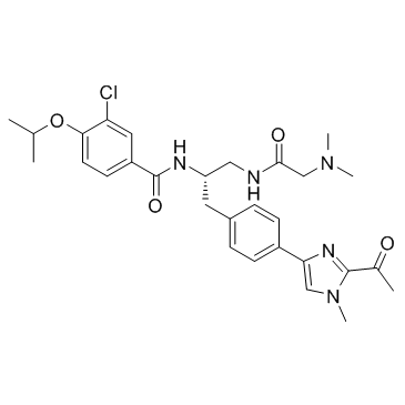
PF-2771 is a potent and selective centromere protein E (CENP-E) inhibitor, inhibiting CENP-E motor activity with an IC50 of 16.1 nM; PF-2771 is used as an anticancer agent.
Products are for research use only. Not for human use. We do not sell to patients.
Cas No.: 2070009-55-9
Sample solution is provided at 25 µL, 10mM.
_1-3.$$$39978408@51.jpg');)
_1-9.$$$38538795@65.jpg');)
_1-9.$$$39112701@51.jpg');)
_1-7.$$$37316672@70.jpg');)
_366-372.$$$36198801@70.jpg');)
.$$$38565142@65.jpg');)
_1867-1883.$$$33248023@67.jpg');)
_eads6055.$$$39977546@45.jpg');)
_eadm9805.$$$40080571@45.jpg');)
_eadj3020.$$$39666821@45.jpg');)
_1-20.$$$39757301@28.jpg');)
_1-24.$$$38898113@28.jpg');)
_273-287.$$$36806353@46.jpg');)
_1-16.$$$37156877@44.jpg');)
_1-19.$$$37460805@44.jpg');)
_1291-1307.$$$34518654@46.jpg');)
PF-2771 is a potent and selective centromere protein E (CENP-E) inhibitor, inhibiting CENP-E motor activity with an IC50 of 16.1 nM; PF-2771 is used as an anticancer agent.
PF-2771 is a potent and selective CENP-E inhibitor, inhibiting CENP-E motor activity with an IC50 of 16.1 nM. PF-2771 shows no inhibitory effect on the ATPase activities of highly related kinesins (0% inhibition of Eg5/KSP, chromokinesin, and MCAK at both 1 or 10 μM PF-2771). PF-2771 exhibits inactive activity against 74 protein kinases (all 5 μM). PF-2771 (100 nM) reusults in a chromosomal congression defect in MDA-MB-468 cells[1].
PF-2771 (100 mg/kg, every day i.p.) potently inhibits CENP-E motor function, and causes tumor regression in SCID mice bearing AA1077 mammary tumors[1].
[1]. Kung PP, et al. Chemogenetic evaluation of the mitotic kinesin CENP-E reveals a critical role in triple-negative breast cancer. Mol Cancer Ther. 2014 Aug;13(8):2104-15.
Kinase experiment: | Microtubule-activated CENP-E kinesin ATPase activity is measured spectrophotometrically by coupling the hydrolysis of ATP to ADP to NADH oxidation (decrease in 340 nm absorbance) through the activities of pyruvate kinase (PK) and lactate dehydrogenase (LDH). Reactions contained 2 mM phosphoenolpyruvate, 0.28 mM NADH, 5 mM MgCl2, 1 mM DTT, 15 μM taxol, 0.7 μM MT (preformed porcine microtubules), 50 μM ATP, 10 U/mL PK, and 10 U/mL LDH in 15 mM PIPES buffer (pH 7.0). Reactions are initiated with a 20 nM CENP-E addition (Human: 1-342; WT) at 30°C. The IC50 values are determined by a nonlinear, least squares fit of the data to the four-parameter dose-response curve equation. PF-2771 kinesin biochemical selectivity toward other kinesins is tested in triplicate at both 1 and 10 μM PF-2771 in a variant of the CENP-E enzymatic assays that have the following enzyme concentrations: 200 nM chromokinesin motor domain, 200 nM Eg5 motor domain, 226 nM MCAK motor domain. PF-2771 biochemical mechanism is determined by measuring CENP-E enzymatic activity as a function of ATP and PF-2771 concentrations (0, 2.5, 5, 10, 20, 30, 45 nM PF-2771; 1,000, 500, 250, 125, 62.5, 31.2, 15.6, 7.81, 3.90, 1.95, 0.977 μM ATP)[1]. |
Cell experiment: | PF-2771 is added to cells seeded in 96-well plates. The number of cells seeded (1,000-3,000) depended on growth characteristics of each cell type and normalized proliferation rates. Ten different concentrations of compound (PF-2771) used are based on a half-log increment between 1 nM and either 1 or 25 μM. Cells are incubated at 37°C for 7 days before assessing viability with the CellTiter-Glo reagent. Untreated control cells are 80% to 90% confluent after 7 days of culture. Data are fitted into a sigmoidal curve-fitting program to calculate IC50 values[1]. |
Animal experiment: | HCC1806 tumor cells (3 × 10⁶) are implanted in the mammary fat pad of CB17/lcr.Cg-PrkdcscidLystbg female mice. PF-2771 is administered intraperitoneally (i.p.) to groups of 12 mice at 3, 10, and 30 mg/kg every day for 14 days and at 100 mg/kg every day for 4 days followed by 3 days off and then another 4-day cycle. Tumor volumes are recorded twice weekly by calipers with the final measurement taken 3 days after the last dose. Tumor growth inhibition (TGI) is calculated using the formula 100 × (1 − ΔT/ΔC), where ΔT (treated) and ΔC (control) are the mean tumor volume changes between 1 day after the last dose and the first-day treatment. Time-to-progression endpoint and associated tumor growth delay determinations are calculated using median days to reach to two doublings of initial tumor size. Statistical comparisons are made using one-way ANOVA with Dunnett posttests. Other tumor models had similar methodology except where noted. PDX-AA1077 is a patient-derived xenograft model developed from tumor tissue from a triple-negative patient engrafted to the mammary glands of NSG female mice to create passage 1 tumor-bearing mice. Surgically resected tumor tissue is cut into 2- to 4-mm3 fragments and subcutaneously implanted into the flank of SCID-bg mice. Mice with palpable tumors are randomized before dosing. An HCC1599 tumor cell line xenograft model is established from tumor fragments of established tumors and reimplanted into flank of female SCID mice. When average tumor size reached 250 mm3, animals are randomized into five groups of 10 mice. Animals are treated with vehicle (daily dosing), 10 mg/kg docetaxel (once per week dosing), 25 mg/kg docetaxel (once per week dosing), 20 mg/kg paclitaxel (twice per week dosing), or 100 mg/kg of PF-2771 (every day, i.p.)[1]. |
References: [1]. Kung PP, et al. Chemogenetic evaluation of the mitotic kinesin CENP-E reveals a critical role in triple-negative breast cancer. Mol Cancer Ther. 2014 Aug;13(8):2104-15. | |
| Cas No. | 2070009-55-9 | SDF | |
| Canonical SMILES | ClC1=CC(C(N[C@@H](CC2=CC=C(C3=CN(C)C(C(C)=O)=N3)C=C2)CNC(CN(C)C)=O)=O)=CC=C1OC(C)C | ||
| Formula | C29H36ClN5O4 | M.Wt | 554.08 |
| Solubility | DMSO : ≥ 32 mg/mL (57.75 mM) | Storage | Store at -20°C |
| General tips | Please select the appropriate solvent to prepare the stock solution according to the
solubility of the product in different solvents; once the solution is prepared, please store it in
separate packages to avoid product failure caused by repeated freezing and thawing.Storage method
and period of the stock solution: When stored at -80°C, please use it within 6 months; when stored
at -20°C, please use it within 1 month. To increase solubility, heat the tube to 37°C and then oscillate in an ultrasonic bath for some time. |
||
| Shipping Condition | Evaluation sample solution: shipped with blue ice. All other sizes available: with RT, or with Blue Ice upon request. | ||
| Prepare stock solution | |||

|
1 mg | 5 mg | 10 mg |
| 1 mM | 1.8048 mL | 9.024 mL | 18.0479 mL |
| 5 mM | 361 μL | 1.8048 mL | 3.6096 mL |
| 10 mM | 180.5 μL | 902.4 μL | 1.8048 mL |
Step 1: Enter information below (Recommended: An additional animal making an allowance for loss during the experiment)
 g
μL
g
μL
Step 2: Enter the in vivo formulation (This is only the calculator, not formulation. Please contact us first if there is no in vivo formulation at the solubility Section.)
Calculation results:
Working concentration: mg/ml;
Method for preparing DMSO master liquid: mg drug pre-dissolved in μL DMSO ( Master liquid concentration mg/mL, Please contact us first if the concentration exceeds the DMSO solubility of the batch of drug. )
Method for preparing in vivo formulation: Take μL DMSO master liquid, next addμL PEG300, mix and clarify, next addμL Tween 80, mix and clarify, next add μL saline, mix and clarify.
Method for preparing in vivo formulation: Take μL DMSO master liquid, next add μL Corn oil, mix and clarify.
Note: 1. Please make sure the liquid is clear before adding the next solvent.
2. Be sure to add the solvent(s) in order. You must ensure that the solution obtained, in the previous addition, is a clear solution before proceeding to add the next solvent. Physical methods such as vortex, ultrasound or hot water bath can be used to aid dissolving.
3. All of the above co-solvents are available for purchase on the GlpBio website.
Quality Control & SDS
- View current batch:
- Purity: >99.50% Appearance: A solid
- COA (Certificate of Analysis)
- SDS (Safety Data Sheet)
- Datasheet
Average Rating: 5 (Based on Reviews and 10 reference(s) in Google Scholar.)