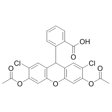
H2DCFDA (DCFH-DA) (Synonyms: DCFH, DCFHDA) |
| Catalog No.GC30006 |
H2DCFDA (DCFH-DA) (DCFH-DA) is a cell-permeable probe used to detect intracellular reactive oxygen species (ROS) (Ex/Em=488/525 nm).
Products are for research use only. Not for human use. We do not sell to patients.

Cas No.: 4091-99-0
Sample solution is provided at 25 µL, 10mM.
H2DCFDA(DCFH-DA) is a redox-sensitive fluorescent probe, which could be used to measure intracellular reactive oxygen species levels.[1] The most popular method used to measure the level of cellular ROS formation is 2′,7′-dichlorodihydrofluorescein diacetate (H2DCFDA(DCFH-DA)) assay.
The fluorogenic dye H2DCFDA(DCFH-DA) was used to detect ROS production. Usually, after diffusion into the cell, H2DCFDA(DCFH-DA) is deacetylated by cellular esterases into a non-fluorescent compound that is subsequently oxidized by ROS into 2′,7′-dichlorofluorescein (DCF). The in vitro experiment to determine the ability of TBBPA alone to stimulate the conversion of H2DCFDA(DCFH-DA) to its fluorescent product DCF was conducted in a cell-free model. Dilution of 5 μM H2DCFDA(DCFH-DA) and increasing concentrations of TBBPA (0.1–100 μM) were added to 96-well plates containing PBS buffer without Ca2+ and Mg2+ or serum-free DMEM/F12 or DMEM/F12 supplemented with 5 % FBS in the final volume of 100 μL. The fluorescence was measured 30 and 60 min after the addition of TBBPA. The deacetylated and oxidized version of H2DCFDA(DCFH-DA): DCF ‘s fluorescence was detected at 485 and 535 nm of maximum excitation and emission spectra, respectively. This in vitro study examined the impact of TBBPA on H2DCFDA(DCFH-DA) fluorescence without cells in PBS buffer, DMEM/F12, and DMEM/F12 with 5 % of FBS media. The obtained results showed that TBBPA in all tested concentrations interacted with H2DCFDA(DCFH-DA) in PBS buffer and caused a significant increase in fluorescence. H2DCFDA(DCFH-DA) assay cannot be used in cell culture experiments with TBBPA. Results suggested that the data regarding TBBPA-stimulated ROS production in cell culture models using the H2DCFDA(DCFH-DA) assay should be revised using a different method. [3]
References:
[1]. Park JH, Moon S-H, Kang DH, et al. Diquafosol sodium inhibits apoptosis and inflammation of corneal epithelial cells via activation of Erk1/2 and RSK: in vitro and in vivo dry eye model. Invest Ophthalmol Vis Sci. 2018;59:5108–5115. doi.org/ 10.1167/iovs.17-22925.
[2]. Szychowski KA, Rybczyńska-Tkaczyk K, et al. Tetrabromobisphenol A (TBBPA)-stimulated reactive oxygen species (ROS) production in cell-free model using the 2',7'-dichlorodihydrofluorescein diacetate (H2DCFDA) assay-limitations of method. Environ Sci Pollut Res Int. 2016 Jun;23(12):12246-52.
[3]. Gomes A, Fernandes E, at al. Fluorescence probes used for detection of reactive oxygen species. J Biochem Biophys Methods JLFC (2005) 65:45–80
Average Rating: 5 (Based on Reviews and 36 reference(s) in Google Scholar.)
GLPBIO products are for RESEARCH USE ONLY. Please make sure your review or question is research based.
Required fields are marked with *


