

Bindarit (Synonyms: AF 2838) |
Catalog No.GC13335
 One-Click Copy Product Info
|

Bindarit is an indazolic derivative that exhibits anti-inflammatory activity in different models.
Products are for research use only. Not for human use. We do not sell to patients.
Cas No.: 130641-38-2
Sample solution is provided at 25 µL, 10mM.
_1-3.$$$39978408@51.jpg');)
_1-9.$$$38538795@65.jpg');)
_1-9.$$$39112701@51.jpg');)
_1-7.$$$37316672@70.jpg');)
_366-372.$$$36198801@70.jpg');)
.$$$38565142@65.jpg');)
_1867-1883.$$$33248023@67.jpg');)
_eads6055.$$$39977546@45.jpg');)
_eadm9805.$$$40080571@45.jpg');)
_eadj3020.$$$39666821@45.jpg');)
_1-20.$$$39757301@28.jpg');)
_1-24.$$$38898113@28.jpg');)
_273-287.$$$36806353@46.jpg');)
_1-16.$$$37156877@44.jpg');)
_1-19.$$$37460805@44.jpg');)
_1291-1307.$$$34518654@46.jpg');)
Bindarit is an indazolic derivative that exhibits anti-inflammatory activity in different models[1]. Bindarit specifically inhibits p65- and p65/p50-mediated activation of the MCP-1 promoter, and directly interacts with FABP4 by increasing expression and nuclear localization, thereby affecting peroxisome proliferator-activated receptor γ (PPARγ) and lipopolysaccharide (LPS) -dependent kinase signaling pathways[2]. Bindarit has been widely used to prevent bone loss caused by Chikungunya virus infection[3].
In vitro, Bindarit treatment (100µM) for 48 hours significantly inhibited the proliferation and migration of coronary artery smooth muscle cells (CASMCs) induced by TNF-α (30ng/ml)[4]. Pretreatment with 300µM Bindarit for 1 hour significantly reduced Angiotensin II, endothelin-1, and TGFβ-induced contraction of human renal mesangial cells (HRMCs) and phosphorylation of vinculin protein[5].
In vivo, Bindarit treatment via oral administration at a dose of 100mg/kg twice daily for 10 days significantly alleviated trinitro-benzene sulfonic acid (TNBS)-induced colitis in mice, reduced leukocyte infiltration, and downregulated MCP-1 synthesis[6]. Daily intraperitoneal injection of Bindarit at a dose of 100mg/kg for 12 weeks significantly inhibited tumor growth and metastasis in the PC-3M-Luc2 xenograft mouse model, and improved survival rate[7]. Subcutaneous injection of Bindarit at a dose of 100mg/kg daily for 28 days alleviated brain injury, maintained blood-brain barrier integrity, and mitigated neuroinflammation in mice with status epilepticus[8].
References:
[1] Grassia G, Maddaluno M, Guglielmotti A, et al. The anti-inflammatory agent bindarit inhibits neointima formation in both rats and hyperlipidaemic mice[J]. Cardiovascular research, 2009, 84(3): 485-493.
[2] Oddi S, Scipioni L, Totaro A, et al. The anti-inflammatory agent bindarit acts as a modulator of fatty acid-binding protein 4 in human monocytic cells[J]. Scientific reports, 2019, 9(1): 15155.
[3] Chen W, Foo S S, Taylor A, et al. Bindarit, an inhibitor of monocyte chemotactic protein synthesis, protects against bone loss induced by chikungunya virus infection[J]. Journal of virology, 2015, 89(1): 581-593.
[4] Maddaluno M, Grassia G, Di Lauro M V, et al. Bindarit inhibits human coronary artery smooth muscle cell proliferation, migration and phenotypic switching[J]. Plos one, 2012, 7(10): e47464.
[5] Paccosi S, Giachi M, Di Gennaro P, et al. The chemokine (CC motif) ligand protein synthesis inhibitor bindarit prevents cytoskeletal rearrangement and contraction of human mesangial cells[J]. Cytokine, 2016, 85: 92-100.
[6] Bhatia M, Landolfi C, Basta F, et al. Treatment with bindarit, an inhibitor of MCP-1 synthesis, protects mice against trinitrobenzene sulfonic acid-induced colitis[J]. Inflammation research, 2008, 57(10): 464-471.
[7] Zollo M, Di Dato V, Spano D, et al. Targeting monocyte chemotactic protein-1 synthesis with bindarit induces tumor regression in prostate and breast cancer animal models[J]. Clinical & experimental metastasis, 2012, 29(6): 585-601.
[8] Chen J, Hu Y, Xie J, et al. Bindarit attenuates brain injury and neuroinflammation in status epilepticus by disrupting C3/C3aR signaling and modulating microglia-astrocyte interactions[J]. Cell Communication and Signaling, 2026.
| Cell experiment [1]: | |
|
Cell lines |
Human coronary artery smooth muscle cells (CASMCs) |
|
Preparation Method |
CASMCs were cultured in smooth muscle basal medium supplemented with 0.5mg/ml hEGF, 5mg/ml insulin, 1mg/ml hFGF, 50mg/ml gentamicin/amphotericin-B, 5% fetal bovine serum (FBS) at 37°C in 5% CO2/atmosphere. Cells cultured were seeded onto 24-well microplates (1.5×10⁴ cells/well) and cultured for 24h. After the induction of quiescence, cells were stimulated with TNF-α (30ng/ml) or FBS (5%) for 48 hours in the presence or absence of Bindarit (10, 25, 50, 100, 200, and 300µM), and cell proliferation was assessed. |
|
Reaction Conditions |
10, 25, 50, 100, 200, and 300µM; 48h |
|
Applications |
Bindarit treatment reduced the cell proliferation of CASMCs induced by TNF-α in a dose-dependent manner. |
| Animal experiment [2]: | |
|
Animal models |
Female BALB/c mice |
|
Preparation Method |
Female BALB/c mice (6 weeks old) were housed under controlled temperature (21-23°C), humidity (30-35%), and light cycle (7:00 AM to 7:00 PM) and maintained on a standard rodent diet. 2.5×10⁵ 4T1-Luc cells per 100μl PBS were orthotopically transplanted into the mammary fat pads of 10-week-old BALB/c mice. 100mg/kg Bindarit was administered by oral gavage, once a day, for five consecutive days per week, for 2 weeks. Tumor growth was monitored by bioluminescent (BLI). |
|
Dosage form |
100mg/kg; five times a week, 2 weeks; p.o. |
|
Applications |
Bindarit treatment significantly inhibited local tumorigenesis and tumor growth in mice with 4T1-Luc xenografts. |
References: [1] Maddaluno M, Grassia G, Di Lauro M V, et al. Bindarit inhibits human coronary artery smooth muscle cell proliferation, migration and phenotypic switching[J]. Plos one, 2012, 7(10): e47464. [2] Zollo M, Di Dato V, Spano D, et al. Targeting monocyte chemotactic protein-1 synthesis with bindarit induces tumor regression in prostate and breast cancer animal models[J]. Clinical & experimental metastasis, 2012, 29(6): 585-601. | |
| Cas No. | 130641-38-2 | SDF | |
| Synonyms | AF 2838 | ||
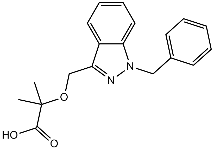
| Chemical Name | 2-[(1-benzylindazol-3-yl)methoxy]-2-methylpropanoic acid | ||
| Canonical SMILES | CC(C)(C(=O)O)OCC1=NN(C2=CC=CC=C21)CC3=CC=CC=C3 | ||
| Formula | C19H20N2O3 | M.Wt | 324.37 |
| Solubility | ≥ 16.2mg/mL in DMSO | Storage | Store at -20°C |
| Shipping Condition | Evaluation sample solution: shipped with blue ice. All other sizes available: with RT, or with Blue Ice upon request. | ||
| Prepare stock solution | |||

|
1 mg | 5 mg | 10 mg |
| 1 mM | 3.0829 mL | 15.4145 mL | 30.829 mL |
| 5 mM | 616.6 μL | 3.0829 mL | 6.1658 mL |
| 10 mM | 308.3 μL | 1.5414 mL | 3.0829 mL |
Step 1: Enter information below (Recommended: An additional animal making an allowance for loss during the experiment)
 g
μL
g
μL
Step 2: Enter the in vivo formulation (This is only the calculator, not formulation. Please contact us first if there is no in vivo formulation at the solubility Section.)
Calculation results:
Working concentration: mg/ml;
Method for preparing DMSO master liquid: mg drug pre-dissolved in μL DMSO ( Master liquid concentration mg/mL, Please contact us first if the concentration exceeds the DMSO solubility of the batch of drug. )
Method for preparing in vivo formulation: Take μL DMSO master liquid, next addμL PEG300, mix and clarify, next addμL Tween 80, mix and clarify, next add μL saline, mix and clarify.
Method for preparing in vivo formulation: Take μL DMSO master liquid, next add μL Corn oil, mix and clarify.
Note: 1. Please make sure the liquid is clear before adding the next solvent.
2. Be sure to add the solvent(s) in order. You must ensure that the solution obtained, in the previous addition, is a clear solution before proceeding to add the next solvent. Physical methods such as vortex, ultrasound or hot water bath can be used to aid dissolving.
3. All of the above co-solvents are available for purchase on the GlpBio website.
Quality Control & SDS
- View current batch:
- Purity: >98.00% Appearance: A solid
- COA (Certificate of Analysis)
- SDS (Safety Data Sheet)
- Datasheet
Average Rating: 5 (Based on Reviews and 26 reference(s) in Google Scholar.)