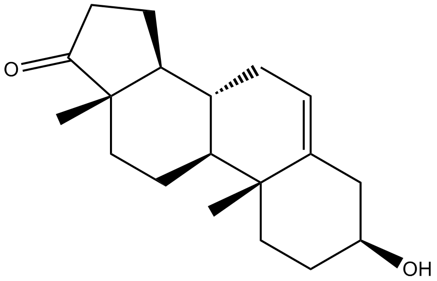
Dehydroepiandrosterone (DHEA) |
Catalog No.: GC11070 |
Dehydroepiandrosterone (DHEA) and its sulfate ester, DHEAS, together represent the most abundant steroid hormones in the human body .
Sample solution is provided at 25 µL, 10mM.
Quality Control & SDS
- View current batch:
-
Purity: >99.00%
- COA (Certificate Of Analysis)
- SDS (Safety Data Sheet)
- Datasheet
Cell experiment [1]: | |
Cell lines |
HCC1 cell line |
Preparation Method |
Transferred cells to serum-free dehydroepiandrosterone (DHEA) containing either 0.1% ethanol which was added to all control cultures or 10-12-10-6 M DHEA. The cell culture supernatant was harvested after 72 h. |
Reaction Conditions |
10-12-10-6 M for 72 hours |
Applications |
Co-treatment with DEX (10-7 M)/ANDI (10-7 M) or DEX (10-7 M)/DHEA (10-7 M) reversed the increase in RANKL mRNA expression |
Animal experiment [2]: | |
Animal models |
female BALB/c mice |
Preparation Method |
Effects of treatment with dehydroepiandrosterone (DHEA) were assessed on either ovaries with functional corpora lutea (CL) or regressing CL by two s.c. injections of 60 mg DHEA/kg body weight (DHEA group), 24 h apart on days 3 and 4 after ovulation, followed by decapitation on day 5 (functional CL) or on day 7 and 8, followed by decapitation on day 9 (regressing CL). |
Dosage form |
s.c., 60 mg/kg |
Applications |
In mice with functional CL (day 5), the hyperandrogenization with dehydroepiandrosterone (DHEA) decreased both serum P and estradiol (E2) levels when compared to controls |
References: [1]: Harding G, Mak YT, Evans B, Cheung J, MacDonald D, Hampson G: The effects of dexamethasone and dehydroepiandrosterone (DHEA) on cytokines and receptor expression in a human osteoblastic cell line: potential steroid-sparing role of DHEA. Cytokine. 2006, 36: 57-68. 10.1016/j.cyto.2006.10.012. |
Dehydroepiandrosterone (DHEA) and its sulfate ester, DHEAS, together represent the most abundant steroid hormones in the human body [1].
Dehydroepiandrosterone (DHEA) (10-8 and 10-6 M) or DHEAS pretreated rat cerebral cortical cultures was increased neuronal survival when the cultures subjected to anoxia for 2 h [2]. When rat cerebral cortical cultures were subjected to anoxia for 2 h in an anaerobic chamber and pretreated with dehydroepiandrosterone (DHEA) (10-8 and 10-6 M) or DHEAS (10-6 M), there was increased neuronal survival. Using cultured neural precursors from rat embryonic forebrains, dehydroepiandrosterone (DHEA) (50 and 100 nM) activated the serine-threonine protein kinase Akt, which is widely implicated in cell survival signaling [3].
Dehydroepiandrosterone (DHEA) treating had better locomotor recovery on CD-1 female mice, after contusive spinal cord injury (SCI), and also left-right coordination, and fine motor control than control animals [4]. Mice treated with dehydroepiandrosterone (DHEA) also had significantly more white matter spared at the epicenter of the injury and reduced area of reactive gliosis surrounding the lesion. Dehydroepiandrosterone (DHEA) treatment was intensive and consisted of three different modes of administration: a DHEA Matrigel patch (10-10 M) applied to the spinal cord before closure of the wound, followed by 12 days of i.p. injections of saline containing Dehydroepiandrosterone (DHEA) (10-6 M or 0.02 mg/kg/day) after SCI, and Dehydroepiandrosterone (DHEA) (10-6 M) in the drinking water for 42 days [4].
References:
[1]. Rice SP, Zhang L, Grennan-Jones F, Agarwal N, Lewis MD, Rees DA, Ludgate M: Dehydroepiandrosterone (DHEA) treatment in vitro inhibits adipogenesis in human omental but not subcutaneous adipose tissue. Mol Cell Endocrinol 2010, 320: 51-57. 10.1016/j.mce.2010.02.017
[2]. C.E. Marx, L.F. Jarskog, J.M. Lauder, J.H. Gilmore, J.A. Lieberman, A.L. Morrow. Neurosteroid modulation of embryonic neuronal survival in vitro following anoxia. Brain Res., 871 (2000), pp. 104-112
[3]. L. Zhang, B. Li, W. Ma, J.L. Barker, Y.H. Chang, W. Zhao, D.R. Rubinow. Dehydroepiandrosterone (DHEA) and its sulfated derivative (DHEAS) regulate apoptosis during neurogenesis by triggering the Akt signaling pathway in opposing ways. Brain Res. Mol. Brain Res., 98 (2002), pp. 58-66
[4]. C. Fiore, D.M. Inman, S. Hirose, L.J. Noble, T. Igarashi, N.A. Compagnone. Treatment with the neurosteroid dehydroepiandrosterone promotes recovery of motor behavior after moderate contusive spinal cord injury in the mouse. J. Neurosci. Res., 75 (2004), pp. 391-400
Cas No. | 53-43-0 | SDF | |
Chemical Name | (3S,8R,9S,10R,13S,14S)-3-hydroxy-10,13-dimethyl-1,2,3,4,7,8,9,11,12,14,15,16-dodecahydrocyclopenta[a]phenanthren-17-one | ||
Canonical SMILES | CC12CCC3C(C1CCC2=O)CC=C4C3(CCC(C4)O)C | ||
Formula | C19H28O2 | M.Wt | 288.42 |
Solubility | ≥ 13.7 mg/mL in DMSO, ≥ 58.6 mg/mL in ETOH | Storage | Store at RT |
General tips | For obtaining a higher solubility , please warm the tube at 37 ℃ and shake it in the ultrasonic bath for a while.Stock solution can be stored below -20℃ for several months. | ||
Shipping Condition | Evaluation sample solution : ship with blue ice All other available size: ship with RT , or blue ice upon request |
Step 1: Enter information below (Recommended: An additional animal making an allowance for loss during the experiment)
Step 2: Enter the in vivo formulation (This is only the calculator, not formulation. Please contact us first if there is no in vivo formulation at the solubility Section.)
Calculation results:
Working concentration: mg/ml;
Method for preparing DMSO master liquid: mg drug pre-dissolved in μL DMSO ( Master liquid concentration mg/mL, Please contact us first if the concentration exceeds the DMSO solubility of the batch of drug. )
Method for preparing in vivo formulation: Take μL DMSO master liquid, next addμL PEG300, mix and clarify, next addμL Tween 80, mix and clarify, next add μL saline, mix and clarify.
Method for preparing in vivo formulation: Take μL DMSO master liquid, next add μL Corn oil, mix and clarify.
Note: 1. Please make sure the liquid is clear before adding the next solvent.
2. Be sure to add the solvent(s) in order. You must ensure that the solution obtained, in the previous addition, is a clear solution before proceeding to add the next solvent. Physical methods such as vortex, ultrasound or hot water bath can be used to aid dissolving.
3. All of the above co-solvents are available for purchase on the GlpBio website.
Dehydroepiandrosterone (DHEA): hypes and hopes
Dehydroepiandrosterone (DHEA) and its sulfated form dehydroepiandrosterone sulfate (DHEAS) are the most abundant circulating steroid hormones in humans. In animal studies, their low levels have been associated with age-related involuntary changes, including reduced lifespan. Extrapolation of animal data to humans turned DHEA into a 'superhormone' and an 'anti-aging' panacea. It has been aggressively marketed and sold in large quantities as a dietary supplement. Recent double-blind, placebo-controlled human studies provided evidence to support some of these claims. In the elderly, DHEA exerts an immunomodulatory action, increasing the number of monocytes, T cells expressing T-cell receptor gamma/delta (TCRγδ) and natural killer (NK) cells. It improves physical and psychological well-being, muscle strength and bone density, and reduces body fat and age-related skin atrophy stimulating procollagen/sebum production. In adrenal insufficiency, DHEA restores DHEA/DHEAS and androstenedione levels, reduces total cholesterol, improves well-being, sexual satisfaction and insulin sensitivity, and prevents loss of bone mineral density. Normal levels of CD4+CD25(hi) and FoxP3 (forkhead box P3) are restored. In systemic lupus erythematosus, DHEA is steroid-sparing. In an unblinded study, it induced remission in the majority of patients with inflammatory bowel disease. DHEA modulates cardiovascular signalling pathways and exerts an anti-inflammatory, vasorelaxant and anti-remodelling effect. Its low levels correlate with increased cardiovascular disease and all-cause mortality. DHEA/DHEAS appear protective in asthma and allergy. It attenuates T helper 2 allergic inflammation, and reduces eosinophilia and airway hyperreactivity. Low levels of DHEAS accompany adrenal suppression. It could be used to screen for the side effects of steroids. In women, DHEA improves sexual satisfaction, fertility and age-related vaginal atrophy. Many factors are responsible for the inconsistent/negative results of some studies. Overreliance on animal models (DHEA is essentially a human molecule), different dosing protocols with non-pharmacological doses often unachievable in humans, rapid metabolism of DHEA, co-morbidities and organ-specific differences render data interpretation difficult. Nevertheless, a growing body of evidence supports the notion that DHEA is not just an overrated dietary supplement but a useful drug for some, but not all, human diseases. Large-scale randomised controlled trials are needed to fine-tune the indications and optimal dosing protocols before DHEA enters routine clinical practice.
Effect of Dehydroepiandrosterone (DHEA) on Diabetes Mellitus and Obesity
Type 2 diabetes is a metabolic disorder that is characterized by an impaired capacity to secrete insulin, insulin resistance, or both. Dehydroepiandrosterone (DHEA), a steroid hormone produced by the adrenal cortex, has been reported to have beneficial effects on diabetes mellitus and obesity in animal models. DHEA and DHEA-sulfate (DHEA-S) have been reported to increase not only insulin secretion of the pancreas but also insulin sensitivity of the liver, adipose tissue, and muscle. We investigated the effects of DHEA on glucose metabolism in animal models and reported decrease of liver gluconeogenesis. Recently, we reported the effect of DHEA on the liver and muscle by using insulin-stimulated insulin receptor substrate 1 and 2 (IRS1 and IRS2)-deficient mice. DHEA increased Akt phosphorylation in the liver of C57BL6 IRS1- and IRS2-deficient mice fed with a high-fat diet (HFD), which suggests that the increase in DHEA-induced Akt signaling is sufficient in the presence of IRS1 or IRS2. In addition, other studies have also reported the effect of DHEA on diabetes mellitus in the liver, muscle, adipose tissue, and pancreatic β-cell and its effect on obesity in animal models. A meta-analysis in elderly men and women has found that DHEA supplementation has no effects on blood glucose levels. However, DHEA supplementation to patients with type 2 diabetes has not been fully elucidated. Therefore, further studies are needed to provide greater insight into the effect of DHEA on diabetes and obesity in animal and human models.
Dehydroepiandrosterone (DHEA) supplementation and IVF outcome in poor responders
Ovarian stimulation of poor ovarian responders still remains a challenging issue. The incidence of poor responders among infertile women is reported in 9-24% IVF cycles and is associated with very low clinical pregnancy rates. Different treatments have been reported in the literature in an attempt to identify the best stimulation protocol for those patients. Administration of dehydroepiandrosterone acetate (DHEA) was suggested as a promising treatment. It is well known that androgens can influence ovarian follicular growth, augment steroidogenesis, promote follicular recruitment and increase the number of primary and pre-antral follicles. The purpose of this review is to evaluate the effect of DHEA supplementation on women with diminished ovarian reserve. Because of the uncertainty of published data, we suggest that well-designed multicentre RCTs are required to provide more insight on the effectiveness of DHEA. The absence of significant side effects should not be considered as an argument to support DHEA treatment.
[Dehydroepiandrosterone(DHEA)and bone metabolism]
Dehydroepiandrosterone(DHEA), an adrenal androgen, has attracted much attention as an anti-aging hormone as well as a marker for senescence because of its unique change along with aging. DHEA is reported to have beneficial effects such as anti-diabetes, anti-obesity, and anti-atherosclerosis. It is also shown that DHEA has anti-osteoporosis effects to increase bone mineral density in randomized controlled trials(RCTs). As osteoblasts express aromatase which will convert androgen to estrogen, DHEA may act protectively against osteoporosis through its metabolites. Because there is no report on fracture risk by DHEA administration, further studies are required to clarify DHEA effects on human bone metabolism.
DHEA: dehydroepiandrosterone
Average Rating: 5
(Based on Reviews and 28 reference(s) in Google Scholar.)GLPBIO products are for RESEARCH USE ONLY. Please make sure your review or question is research based.
Required fields are marked with *