

Propofol |
Catalog No.GC39424
 One-Click Copy Product Info
|

Propofol, a widely used intravenous anesthetic for induction and maintenance, modulates autophagy and exhibits anti-tumor or carcinogenic activity.
Products are for research use only. Not for human use. We do not sell to patients.
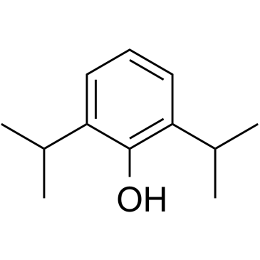
Cas No.: 2078-54-8
Sample solution is provided at 25 µL, 10mM.
Propofol, a widely used intravenous anesthetic for induction and maintenance, modulates autophagy and exhibits anti-tumor or carcinogenic activity[1-2]. Propofol exerts its hypnotic actions by activating the central inhibitory neurotransmitter gamma-aminobutyric acid (GABA) in the central nervous system[2]. In addition, Propofol possesses cyclooxygenase (COX)-inhibiting activity[3].
In vitro, exposure of H9c2 embryonal rat heart-derived cells to escalating concentrations of Propofol (12.5-100µmol/L) for 4h produced a dose-dependent, significant reduction in hypoxia/reoxygenation (H/R)-induced early-apoptotic cells while simultaneously elevating LC3-II levels in a dose-dependent manner. H/R alone reduced p62 protein abundance, and Propofol further amplified this H/R-induced decline in p62[4]. In neuronal PC12 cells, a 10min exposure to Propofol (10, 20, or 50µM) dose-dependently attenuated autophagy by down-regulating the autophagy-related proteins LC3-II, Beclin-1, and Beclin-1-phosphatidylinositol-3 kinase class III (class III PI3K)[5].
In vivo, in male Sprague-Dawley rats, continuous intravenous infusion of Propofol at 12mg/kg/h for 30min via the femoral vein markedly attenuated myocardial injury caused by ischemia–reperfusion (I/R) and significantly reduced the formation of cytosolic vacuoles and myofibrillar lysis in cardiomyocytes[6]. Nude mice received intraperitoneal injections of Propofol (35mg/kg) every 2 days for 14 days, resulting in significantly smaller pheochromocytoma (PCC) tumor volumes and weights and markedly enhanced tumor-cell apoptosis compared with the sham group[7].
References:
[1] Xu Y, Pan S, Jiang W, et al. Effects of propofol on the development of cancer in humans. Cell Prolif. 2020;53(8):e12867.
[2] Guo XN, Ma X. The Effects of Propofol on Autophagy. DNA Cell Biol. 2020;39(2):197-209.
[3] Inada T, Kubo K, Shingu K. Possible link between cyclooxygenase-inhibiting and antitumor properties of propofol. J Anesth. 2011;25(4):569-575.
[4] Li H, Zhang X, Tan J, et al. Propofol postconditioning protects H9c2 cells from hypoxia/reoxygenation injury by inducing autophagy via the SAPK/JNK pathway. Mol Med Rep. 2018;17(3):4573-4580.
[5] Cui D, Wang L, Qi A, et al. Propofol prevents autophagic cell death following oxygen and glucose deprivation in PC12 cells and cerebral ischemia-reperfusion injury in rats [retracted in: PLoS One. 2022 Sep 28;17(9):e0275548. doi: 10.1371/journal.pone.0275548.]. PLoS One. 2012;7(4):e35324.
[6] Noh HS, Shin IW, Ha JH, et al. Propofol protects the autophagic cell death induced by the ischemia/reperfusion injury in rats. Mol Cells. 2010;30(5):455-460.
[7] Wang H, Zhang S, Zhang A, et al. Propofol Prevents the Progression of Malignant Pheochromocytoma In Vitro and In Vivo. DNA Cell Biol. 2018;37(4):308-315.
| Cell experiment [1]: | |
Cell lines | H9c2 embryonal rat heart-derived (cardiac muscle) cell line |
Preparation Method | Hypoxia was modeled in the H9c2 embryonal rat heart-derived (cardiac muscle) cell line. Hypoxia was terminated by exposing the cells to fresh Dulbecco's modified Eagle's medium (DMEM) containing 10% FBS and incubating them in a normal incubator (95% air; 5% CO2) for 4h at 37°C to simulate reperfusion (reoxygenation). Propofol (12.5-100µmol/L) was added to fresh medium at the onset of reoxygenation. After reoxygenation, cells were harvested by 0.25% trypsinization at 37°C, centrifuged at 800×g for 3min at room temperature, and washed twice with PBS. |
Reaction Conditions | 12.5, 25, 50, 100µmol/L; 4h |
Applications | Treatment with 12.5-50µmol/L Propofol caused a dose-dependent and significant reduction in the proportion of early apoptotic cells induced by hypoxia/reoxygenation (H/R). Post-treatment with Propofol dose-dependently enhanced 1A/1B-light chain 3 (LC3)-II levels. H/R exposure alone already decreased p62 protein abundance, and Propofol further intensified this H/R-induced reduction in p62. |
| Animal experiment [2]: | |
Animal models | Male Sprague-Dawley rats |
Preparation Method | The animals were anesthetized with ketamine 50mg/kg and xylazine 5mg/kg into the gluteus maximus muscle, and were ventilated artificially with room air using a rodent ventilator. The chest was opened via a left thoracotomy, followed by a pericardiotomy. A 4-0 black silk suture was passed around the left ascending (LAD) coronary artery, and the ends were pulled through a small vinyl tube to form a snare and then tightened. Coronary artery occlusion was verified by epicardial cyanosis. After 25min of ischemia, the myocardium was reperfused for 24h. The rats were randomized into three groups as follows. Saline (1.2ml/kg/h) and Propofol (12mg/kg/h) were administered via a femoral vein, beginning 15 minutes before reperfusion and continuing for 30 minutes into reperfusion. Group 1 rats (sham), surgically operated on, but no tightening of the coronary sutures. Group 2 rats (ischemia–reperfusion (I/R)) received saline and were subjected to 25min of ischemia followed by reperfusion. Group 3 rats (I/R with Propofol) received Propofol and were subjected to 25min of ischemia followed by reperfusion. |
Dosage form | 12mg/kg/h, 30min; administered via femoral vein |
Applications | The weight of the infarction area of the left ventricle obtained from I/R- injured rats was 0.14±0.01g, whereas the infarction weight of I/R-injured rats treated with Propofol was significantly decreased to 0.06±0.02g (P<0.05). Propofol treatment during I/R dramatically decreased the formation of cytosolic vacuoles and myofibrillar lysis in cardiomyocytes of I/R- damaged rats. |
References: | |
| Cas No. | 2078-54-8 | SDF | |
| Canonical SMILES | OC1=C(C(C)C)C=CC=C1C(C)C | ||
| Formula | C12H18O | M.Wt | 178.27 |
| Solubility | DMSO: 100 mg/mL (560.95 mM); Water: 1 mg/mL (5.61 mM) | Storage | Store at 2-8°C |
| General tips | Please select the appropriate solvent to prepare the stock solution according to the
solubility of the product in different solvents; once the solution is prepared, please store it in
separate packages to avoid product failure caused by repeated freezing and thawing.Storage method
and period of the stock solution: When stored at -80°C, please use it within 6 months; when stored
at -20°C, please use it within 1 month. To increase solubility, heat the tube to 37°C and then oscillate in an ultrasonic bath for some time. |
||
| Shipping Condition | Evaluation sample solution: shipped with blue ice. All other sizes available: with RT, or with Blue Ice upon request. | ||
| Prepare stock solution | |||

|
1 mg | 5 mg | 10 mg |
| 1 mM | 5.6095 mL | 28.0473 mL | 56.0947 mL |
| 5 mM | 1.1219 mL | 5.6095 mL | 11.2189 mL |
| 10 mM | 560.9 μL | 2.8047 mL | 5.6095 mL |
Step 1: Enter information below (Recommended: An additional animal making an allowance for loss during the experiment)
 g
μL
g
μL
Step 2: Enter the in vivo formulation (This is only the calculator, not formulation. Please contact us first if there is no in vivo formulation at the solubility Section.)
Calculation results:
Working concentration: mg/ml;
Method for preparing DMSO master liquid: mg drug pre-dissolved in μL DMSO ( Master liquid concentration mg/mL, Please contact us first if the concentration exceeds the DMSO solubility of the batch of drug. )
Method for preparing in vivo formulation: Take μL DMSO master liquid, next addμL PEG300, mix and clarify, next addμL Tween 80, mix and clarify, next add μL saline, mix and clarify.
Method for preparing in vivo formulation: Take μL DMSO master liquid, next add μL Corn oil, mix and clarify.
Note: 1. Please make sure the liquid is clear before adding the next solvent.
2. Be sure to add the solvent(s) in order. You must ensure that the solution obtained, in the previous addition, is a clear solution before proceeding to add the next solvent. Physical methods such as vortex, ultrasound or hot water bath can be used to aid dissolving.
3. All of the above co-solvents are available for purchase on the GlpBio website.
Quality Control & SDS
- View current batch:
- Purity: >99.50% Appearance: A solid
- COA (Certificate of Analysis)
- SDS (Safety Data Sheet)
- Datasheet
Average Rating: 5 (Based on Reviews and 33 reference(s) in Google Scholar.)