

Spautin-1 |
Catalog No.GC13526
 One-Click Copy Product Info
|

Spautin-1 inhibited the deubiquitinating activity of USP10 and USP13 with IC50 values of 0.6-0.7µM.
Products are for research use only. Not for human use. We do not sell to patients.
Cas No.: 1262888-28-7
Sample solution is provided at 25 µL, 10mM.
_1-3.$$$39978408@51.jpg');)
_1-9.$$$38538795@65.jpg');)
_1-9.$$$39112701@51.jpg');)
_1-7.$$$37316672@70.jpg');)
_366-372.$$$36198801@70.jpg');)
.$$$38565142@65.jpg');)
_1867-1883.$$$33248023@67.jpg');)
_eads6055.$$$39977546@45.jpg');)
_eadm9805.$$$40080571@45.jpg');)
_eadj3020.$$$39666821@45.jpg');)
_1-20.$$$39757301@28.jpg');)
_1-24.$$$38898113@28.jpg');)
_273-287.$$$36806353@46.jpg');)
_1-16.$$$37156877@44.jpg');)
_1-19.$$$37460805@44.jpg');)
_1291-1307.$$$34518654@46.jpg');)
Spautin-1 inhibited the deubiquitinating activity of USP10 and USP13 with IC50 values of 0.6-0.7µM [1]. As an autophagy inhibitor, Spautin-1 has been widely used to induce cell death in a variety of cancer cell models[2].
In vitro, Spautin-1 treatment for 72 hours significantly inhibited the growth of melanoma cells, with the IC50 values of 1.830 and 2.062μM against A375 and SK-Mel-28, respectively[3]. Treatment of U87MG cells with 10μM Spautin-1 for 48h significantly inhibited cell viability, the RAF-ERK pathway, and glycolytic function in the cells[4]. Pretreatment of PC12 cells with 10μM Spautin-1 for 24 hours reduced the accumulation of reactive oxygen species (ROS) and the number of autophagic microsomes induced by oxygen-glucose deprivation/reoxygenation (OGD/R) treatment, and increased cell viability[5].
In vivo, Spautin-1 treatment via intraperitoneal injection at a dose of 20mg/kg/day for 14 days significantly inhibited tumor volume growth in a mouse model of the A20 cell xenograft, and resulted in tumor cell shrinkage, fragmentation, and disorganization, as well as reduced PCNA expression[6]. In a mouse model of acute lung injury (ALI), a single dose of Spautin-1 (0.5mg/kg) intratracheal administration for 72 hours effectively reversed lipopolysaccharide (LPS)-induced lung tissue structural damage, alveolar septum thickening, and peripheral neutrophil infiltration[7]. In a mouse model of prostate cancer, Spautin-1 treatment (20mg/kg/day; i.p.) for 30 days resulted in a significant reduction in tumor weight and volume without affecting mouse body weight[8].
References:
[1] Liu J, Xia H, Kim M, et al. Beclin1 controls the levels of p53 by regulating the deubiquitination activity of USP10 and USP13[J]. Cell, 2011, 147(1): 223-234.
[2] Schott C R, Ludwig L, Mutsaers A J, et al. The autophagy inhibitor spautin-1, either alone or combined with doxorubicin, decreases cell survival and colony formation in canine appendicular osteosarcoma cells[J]. PloS one, 2018, 13(10): e0206427.
[3] Guo J, Zhang J L, Liang L, et al. Potent USP10/13 antagonist spautin‐1 suppresses melanoma growth via ROS‐mediated DNA damage and exhibits synergy with cisplatin[J]. Journal of Cellular and Molecular Medicine, 2020, 24(7): 4324-4340.
[4] Kona S V, Kalivendi S V. The USP10/13 inhibitor, spautin-1, attenuates the progression of glioblastoma by independently regulating RAF-ERK mediated glycolysis and SKP2[J]. Biochimica et Biophysica Acta (BBA)-Molecular Basis of Disease, 2024, 1870(7): 167291.
[5] Liu H, Zhao Z, Wu T, et al. Inhibition of autophagy‐dependent pyroptosis attenuates cerebral ischaemia/reperfusion injury[J]. Journal of Cellular and Molecular Medicine, 2021, 25(11): 5060-5069.
[6] Wu J, Deng Y, Gao Y, et al. Spautin-1 inhibits the growth of diffuse large B-cell lymphoma by inducing mitochondrial damage-mediated PANoptosis and anti-tumor immunity[J]. Cancer Immunology, Immunotherapy, 2025, 74(9): 293.
[7] Wen H, Miao W, Liu B, et al. SPAUTIN-1 alleviates LPS-induced acute lung injury by inhibiting NF-κB pathway in neutrophils[J]. International Immunopharmacology, 2024, 130: 111741.
[8] Liao Y, Guo Z, Xia X, et al. Inhibition of EGFR signaling with Spautin-1 represents a novel therapeutics for prostate cancer[J]. Journal of Experimental & Clinical Cancer Research, 2019, 38(1): 157.
| Cell experiment [1]: | |
Cell lines | A375 cells |
Preparation Method | A375 cells (2×10³/well) were seeded in 96-well plates and cultured in medium containing 10% fetal bovine serum overnight. The cells were then treated with different concentrations of Spautin-1 (control, 2.5, 5, 10, 20μM for 24, 48, and 72 hours, respectively. MTS assay was used to test the effect of Spautin-1 on cell proliferation. |
Reaction Conditions | 2.5, 5, 10, 20μM; 24, 48, and 72h |
Applications | Spautin-1 treatment inhibited the proliferation of A375 cells in a dose- and time-dependent manner. |
| Animal experiment [2]: | |
Animal models | Female BALB/c nude mice |
Preparation Method | Female BALB/c nude mice (5 weeks of age) were subcutaneously injected with A20 cells (100μL PBS, 1 × 10⁷ cells). When the tumor volume reached 50mm3, Spautin-1 (20mg/kg) was intraperitoneally injected daily. The whole experiment lasted for 14 days. The tumor size (longest/shortest axis) was recorded with a precision caliper, and the volume was calculated by using the 0.5 × longest × shortest2 formula. Tumor specimens were collected and analyzed by histochemical staining and H&E staining. |
Dosage form | 20mg/kg/day for 14 days; i.p. |
Applications | Spautin-1 treatment significantly inhibited tumor volume growth in mice, and resulted in tumor cell shrinkage, fragmentation and disorganization, as well as reduced PCNA expression. |
References: | |
| Cas No. | 1262888-28-7 | SDF | |
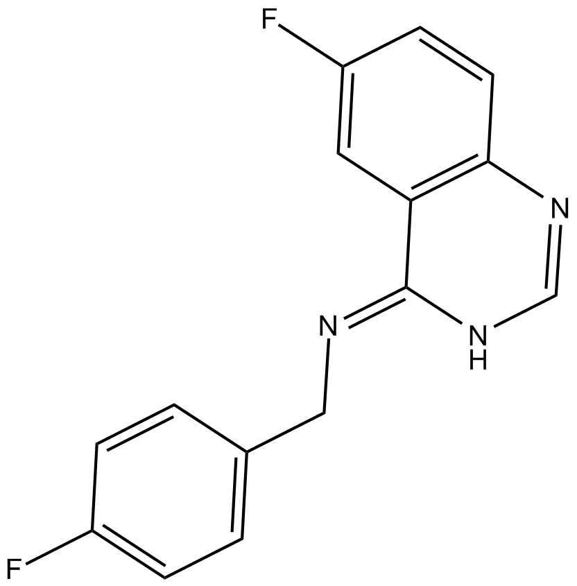
| Chemical Name | (Z)-1-(4-fluorophenyl)-N-(6-fluoroquinazolin-4(3H)-ylidene)methanamine | ||
| Canonical SMILES | FC1=CC=C(C/N=C2C3=C(N=CN/2)C=CC(F)=C3)C=C1 | ||
| Formula | C15H11F2N3 | M.Wt | 271.26 |
| Solubility | ≥ 13.5mg/mL in DMSO | Storage | Store at -20°C |
| General tips | Please select the appropriate solvent to prepare the stock solution according to the
solubility of the product in different solvents; once the solution is prepared, please store it in
separate packages to avoid product failure caused by repeated freezing and thawing.Storage method
and period of the stock solution: When stored at -80°C, please use it within 6 months; when stored
at -20°C, please use it within 1 month. To increase solubility, heat the tube to 37°C and then oscillate in an ultrasonic bath for some time. |
||
| Shipping Condition | Evaluation sample solution: shipped with blue ice. All other sizes available: with RT, or with Blue Ice upon request. | ||
| Prepare stock solution | |||

|
1 mg | 5 mg | 10 mg |
| 1 mM | 3.6865 mL | 18.4325 mL | 36.865 mL |
| 5 mM | 737.3 μL | 3.6865 mL | 7.373 mL |
| 10 mM | 368.7 μL | 1.8433 mL | 3.6865 mL |
Step 1: Enter information below (Recommended: An additional animal making an allowance for loss during the experiment)
 g
μL
g
μL
Step 2: Enter the in vivo formulation (This is only the calculator, not formulation. Please contact us first if there is no in vivo formulation at the solubility Section.)
Calculation results:
Working concentration: mg/ml;
Method for preparing DMSO master liquid: mg drug pre-dissolved in μL DMSO ( Master liquid concentration mg/mL, Please contact us first if the concentration exceeds the DMSO solubility of the batch of drug. )
Method for preparing in vivo formulation: Take μL DMSO master liquid, next addμL PEG300, mix and clarify, next addμL Tween 80, mix and clarify, next add μL saline, mix and clarify.
Method for preparing in vivo formulation: Take μL DMSO master liquid, next add μL Corn oil, mix and clarify.
Note: 1. Please make sure the liquid is clear before adding the next solvent.
2. Be sure to add the solvent(s) in order. You must ensure that the solution obtained, in the previous addition, is a clear solution before proceeding to add the next solvent. Physical methods such as vortex, ultrasound or hot water bath can be used to aid dissolving.
3. All of the above co-solvents are available for purchase on the GlpBio website.
Quality Control & SDS
- View current batch:
- Purity: >99.00% Appearance: A solid
- COA (Certificate of Analysis)
- SDS (Safety Data Sheet)
- Datasheet
Average Rating: 5 (Based on Reviews and 34 reference(s) in Google Scholar.)