

Substance P |
Catalog No.GC15649
 One-Click Copy Product Info
|

Substance P, an important neuropeptide neurotransmitter, stimulates phosphatidylinositol turnover with an EC50 value of 0.36nM.
Products are for research use only. Not for human use. We do not sell to patients.
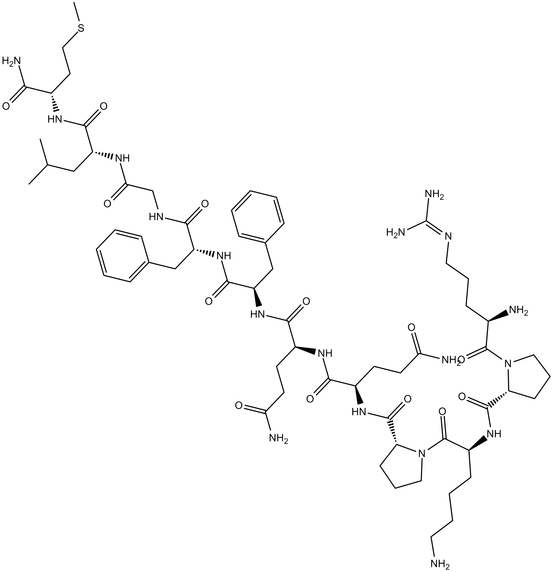
Cas No.: 33507-63-0
Sample solution is provided at 25 µL, 10mM.
Substance P, an important neuropeptide neurotransmitter, stimulates phosphatidylinositol turnover with an EC50 value of 0.36nM[1]. Substance P has been widely used in the research of pain regulation, emotion regulation and bone metabolism regulation[2-3].
In vitro, Substance P (10nM) treatment induced Akt phosphorylation at Ser-473 in a time-dependent manner and persisted over the 60-minute incubation period in colonic epithelial NCM460 colonocytes[4]. Treatment with 10nM Substance P for 8 hours inhibited tamoxifen (10μM; 8h) induced cell death of NCM460 colonocytes, and cell proliferation was stimulated by treatment with 10nM Substance P for 8h in the absence of tamoxifen[4]. Treatment of buffalo fetal fibroblasts (10nM) with Substance P alone resulted in significant cell proliferation and migration in both horizontal and vertical directions within 24 hours[5].
In vivo, topical application of Substance P (32μg/day per wound dissolved in saline) for 10 days improved wound healing and reduced wound size in a diabetic mouse model[6]. A single dose of topical Substance P treatment (0.5μg/wound) promoted wound closure and epithelial regeneration within 14 days, thereby enhancing wound healing in a skin full-thickness wound model of mouse[7].
References:
[1] Torrens Y, Beaujouan J C, Saffroy M, et al. Substance P receptors in primary cultures of cortical astrocytes from the mouse[J]. Proceedings of the National Academy of Sciences, 1986, 83(23): 9216-9220.
[2] Li F X Z, Xu F, Lin X, et al. The role of substance P in the regulation of bone and cartilage metabolic activity[J]. Frontiers in Endocrinology, 2020, 11: 77.
[3] Humes C, Sic A, Knezevic N N. Substance P’s impact on chronic pain and psychiatric conditions—a narrative review[J]. International journal of molecular sciences, 2024, 25(11): 5905.
[4] Koon H W, Zhao D, Zhan Y, et al. Substance P mediates antiapoptotic responses in human colonocytes by Akt activation[J]. Proceedings of the National Academy of Sciences, 2007, 104(6): 2013-2018.
[5] Kant V, Mahapatra P S, Gupta V, et al. Substance P, a neuropeptide, promotes wound healing via neurokinin-1 receptor[J]. The International Journal of Lower Extremity Wounds, 2023, 22(2): 291-297.
[6] Leal E C, Carvalho E, Tellechea A, et al. Substance P promotes wound healing in diabetes by modulating inflammation and macrophage phenotype[J]. The American journal of pathology, 2015, 185(6): 1638-1648.
[7] Kumar S, Tan Y, Berthiaume F. Neuropeptide substance p enhances skin wound healing in vitro and in vivo under hypoxia[J]. Biomedicines, 2021, 9(2): 222.
| Cell experiment [1]: | |
Cell lines | NCM460-NK-1R cells |
Preparation Method | NCM460-NK-1R cells were plated on chamber slides and grown to 50% confluence. Cells were pretreated with Akt inhibitor V (40μM) for 30min and then treated with tamoxifen (10μM) in the presence or absence of Substance P (10nM) for 8h at 37°C. Cells were fixed in 4% paraformaldehyde for 25min and then permeabilized with 0.2% Triton X-100 solution in PBS for 5min. Slides were processed for TUNEL assay. |
Reaction Conditions | 10μM; 8h |
Applications | Substance P treatment inhibited tamoxifen-induced cell death and apoptosis of NCM460-NK-1R cells. |
| Animal experiment [2]: | |
Animal models | C57BL/6J WT mice |
Preparation Method | 8 mice were injected with streptozotocin (50mg/kg in 0.1mol/L citrate buffer) by i.p. route once daily for 5 consecutive days to induce diabetes. Blood glucose level of >250mg/dL was used as an inclusion criteria for diabetic mice. Wound experiments were done 8 weeks after diabetes induction. NPH insulin (0.1 to 0.2 units) was administered as needed to prevent weight loss. Animals were anesthetized with i.p. ketamine (100mg/kg) and i.p. Xylazine (10mg/kg). The hair from the back of the mice was shaved, and two, 6-mm full-thickness excisional wounds were biopsied. Tracing of the wound area was done on acetate paper every day to monitor the rate of wound closure up to 10 days after wounding. The wound size was analyzed with ImageJ version 1.46r. Data are presented as percentage of the original wound (day 0). One wound was treated with Substance P (32μg per wound in saline) daily and the other wound was treated with saline daily until day of euthanasia. Animals were sacrificed at day 10 after wounding. |
Dosage form | 32μg/day per wound dissolved in saline for 10 days; topical application |
Applications | Substance P treatment improved wound healing and reduced wound size in a diabetic mouse model. |
References: | |
| Cas No. | 33507-63-0 | SDF | |
| Formula | C63H98N18O13S | M.Wt | 1347.6 |
| Solubility | Soluble to 1 mg/ml in sterile water | Storage | Desiccate at -20°C |
| General tips | Please select the appropriate solvent to prepare the stock solution according to the
solubility of the product in different solvents; once the solution is prepared, please store it in
separate packages to avoid product failure caused by repeated freezing and thawing.Storage method
and period of the stock solution: When stored at -80°C, please use it within 6 months; when stored
at -20°C, please use it within 1 month. To increase solubility, heat the tube to 37°C and then oscillate in an ultrasonic bath for some time. |
||
| Shipping Condition | Evaluation sample solution: shipped with blue ice. All other sizes available: with RT, or with Blue Ice upon request. | ||
| Prepare stock solution | |||

|
1 mg | 5 mg | 10 mg |
| 1 mM | 742.1 μL | 3.7103 mL | 7.4206 mL |
| 5 mM | 148.4 μL | 742.1 μL | 1.4841 mL |
| 10 mM | 74.2 μL | 371 μL | 742.1 μL |
Step 1: Enter information below (Recommended: An additional animal making an allowance for loss during the experiment)
 g
μL
g
μL
Step 2: Enter the in vivo formulation (This is only the calculator, not formulation. Please contact us first if there is no in vivo formulation at the solubility Section.)
Calculation results:
Working concentration: mg/ml;
Method for preparing DMSO master liquid: mg drug pre-dissolved in μL DMSO ( Master liquid concentration mg/mL, Please contact us first if the concentration exceeds the DMSO solubility of the batch of drug. )
Method for preparing in vivo formulation: Take μL DMSO master liquid, next addμL PEG300, mix and clarify, next addμL Tween 80, mix and clarify, next add μL saline, mix and clarify.
Method for preparing in vivo formulation: Take μL DMSO master liquid, next add μL Corn oil, mix and clarify.
Note: 1. Please make sure the liquid is clear before adding the next solvent.
2. Be sure to add the solvent(s) in order. You must ensure that the solution obtained, in the previous addition, is a clear solution before proceeding to add the next solvent. Physical methods such as vortex, ultrasound or hot water bath can be used to aid dissolving.
3. All of the above co-solvents are available for purchase on the GlpBio website.
Quality Control & SDS
- View current batch:
- Purity: >99.00% Appearance: A solid
- COA (Certificate of Analysis)
- SDS (Safety Data Sheet)
- Datasheet

Average Rating: 5 (Based on Reviews and 16 reference(s) in Google Scholar.)