

BAPTA-AM (Synonyms: BAPTA Acetoxymethyl ester) |
Catalog No.GC13517
 One-Click Copy Product Info
|

BAPTA-AM is a cell membrane-permeable calcium ion chelator. BAPTA-AM directly inhibits hKv1.5, hERG, and hKv1.3 voltage-gated potassium channels (IC50=1.3, 1.45, 1.23µM).
Products are for research use only. Not for human use. We do not sell to patients.
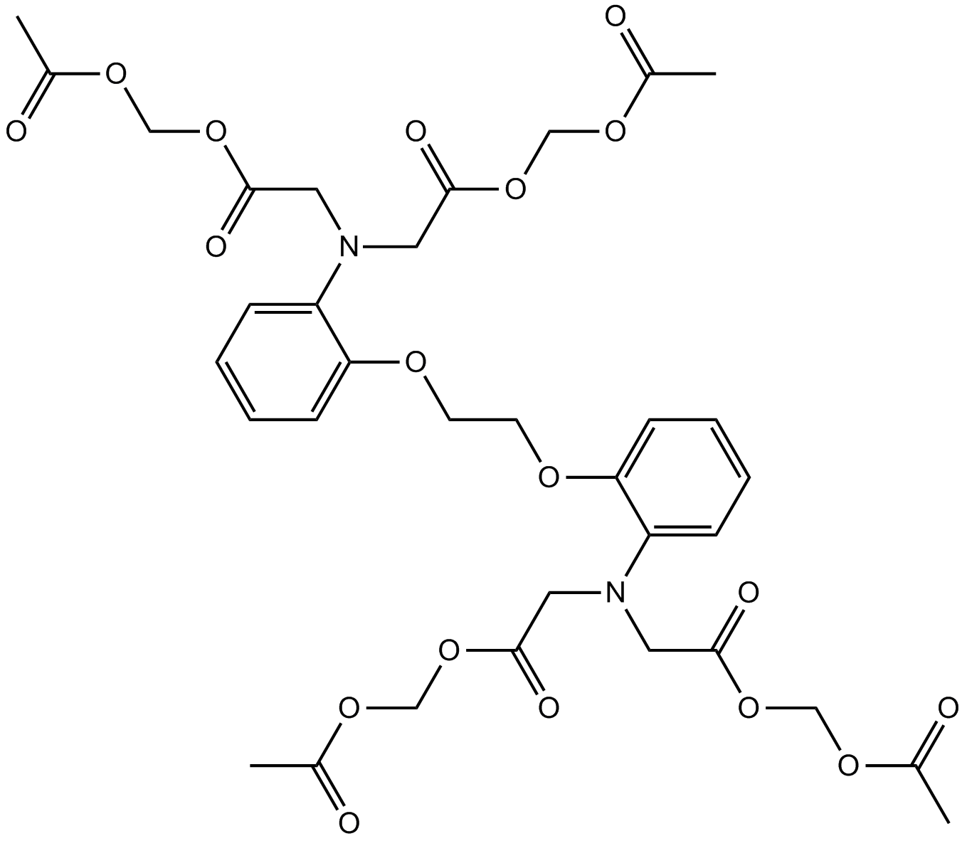
Cas No.: 126150-97-8
Sample solution is provided at 25 µL, 10mM.
BAPTA-AM is a cell membrane-permeable calcium ion chelator. BAPTA-AM directly inhibits hKv1.5, hERG, and hKv1.3 voltage-gated potassium channels (IC50=1.3, 1.45, 1.23µM). BAPTA-AM selectively chelates intracellular free Ca²⁺. BAPTA-AM inhibits cell damage caused by calcium overload. BAPTA-AM reduces intracellular reactive oxygen species. BAPTA-AM inhibits mitochondrial membrane potential collapse and cytochrome C release. BAPTA-AM reduces Caspase-8/9 activation. BAPTA-AM protects neurons from ischemic injury. BAPTA-AM can be used in studies related to excitotoxicity, ischemic brain injury, apoptosis, calcium signaling, arrhythmia, and immune cell function regulation[1-4].
In vitro, co-treatment of HeLa and SH-SY5Y cells with 10-50µM BAPTA-AM and 20µM Aβ42 for 24 hours inhibited Aβ42-induced cell death, suppressed the increase in caspase-3, -6, -8, -9 activities, and inhibited the rise in cathepsin L activity and cytosolic cathepsin L levels[5]. Co-treatment of SH-SY5Y cells with 10µM-30µM BAPTA-AM and 3µM MG132 for 24-48 hours inhibited MG132-induced superoxide anion generation and suppressed the MG132-induced increase in apoptotic cell proportion[6]. Co-treatment of mouse chondrocytes with 10µM BAPTA-AM and 100µM ferric ammonium citrate for 6-24 hours reduced intracellular iron concentration, inhibited ferric ammonium citrate-induced ROS generation, and prevented mitochondrial membrane potential collapse[7].
In vivo, C57BL/6 mice received intraperitoneal injection of 2.5mg/kg BAPTA-AM 0.5 hours before high tidal volume mechanical ventilation (20mL/kg). BAPTA-AM prevented high tidal volume-induced cytosolic calcium overload, improved alveolar septal thickening and pulmonary edema, and reduced lung wet-to-dry ratio, bronchoalveolar lavage fluid protein levels, and inflammatory cell infiltration numbers[8]. One week after destabilization of the medial meniscus (DMM) surgery, C57BL/6J mice received intra-articular injection of 1.5µg/kg-15µg/kg BAPTA-AM into the right knee joint once per week for 8 consecutive weeks. BAPTA-AM attenuated cartilage degeneration and reduced osteophyte formation and synovitis severity[9]. Swiss mice received intraperitoneal injection of 2.5-10mg/kg BAPTA-AM 30 minutes before intraperitoneal injection of 2.5g/kg ethanol. BAPTA-AM prevented ethanol-induced locomotor stimulation[10].
References:
[1] Li FJ, Tan KS, He CY. BAPTA-AM decreases cellular pH, inhibits acidocalcisome acidification and autophagy in amino acid-starved T. brucei. Mol Biochem Parasitol. 2017 Apr;213:26-29.
[2] Tymianski M, Charlton MP, Carlen PL, et al. Properties of neuroprotective cell-permeant Ca2+ chelators: effects on [Ca2+]i and glutamate neurotoxicity in vitro. J Neurophysiol. 1994;72(4):1973-1982.
[3] Wie MB, Koh JY, Won MH, et al. BAPTA/AM, an intracellular calcium chelator, induces delayed necrosis by lipoxygenase-mediated free radicals in mouse cortical cultures. Prog Neuropsychopharmacol Biol Psychiatry. 2001 Nov;25(8):1641-1659.
[4] Han KS, Kang HJ, Kim EY, et al. 1,2-bis(2-Aminophenoxy)ethane-N,N,N',N'-tetraacetic acid induces caspase-mediated apoptosis and reactive oxygen species-mediated necrosis in cultured cortical neurons. J Neurochem. 2001 Jul;78(2):230-239.
[5] Hossain MS, Ramasamy VS, Park IS. Involvement of calcium ions in amyloid-β-induced lamin fragmentation. BBA Mol Cell Res. 2023;1870:119383.
[6] Yamamuro-Tanabe A, Oshima Y, Iyama T, et al. Proteasome inhibitors induce apoptosis by superoxide anion generation via NADPH oxidase 5 in human neuroblastoma SH-SY5Y cells. J Pharmacol Sci. 2024;155(52-62).
[7] Jing X, Wang Q, Du T, et al. Calcium chelator BAPTA-AM protects against iron overload-induced chondrocyte mitochondrial dysfunction and cartilage degeneration. Int J Mol Med. 2021;48(4):196.
[8] Ye L, Zeng Q, Ling M, et al. Inhibition of IP3R/Ca2+ Dysregulation Protects Mice From Ventilator-Induced Lung Injury via Endoplasmic Reticulum and Mitochondrial Pathways. Front Immunol. 2021 Sep 15;12:729094.
[9] Sun K, Zhang X, Hou L, et al. TRPM2-mediated feed-forward loop promotes chondrocyte damage in osteoarthritis via calcium-cGAS-STING-NF-kB pathway. J Adv Res. 2025;75:213-227.
[10] Balino P, Monferrer L, Pastor R, et al. Intracellular calcium chelation with BAPTA-AM modulates ethanol-induced behavioral effects in mice. Exp Neurol. 2012 Apr;234(2):446-53.
| Cell experiment [1]: | |
|
Cell lines |
SH-SY5Y cells (human neuroblastoma cell line) |
|
Preparation Method |
SH-SY5Y cells were cultured in RPMI-1640 medium with 10% heat-inactivated FCS, 100μg/mL streptomycin, 100IU/mL penicillin, 1μg/mL fungizone at 37°C, 5% CO2. Cells were treated with 10μM or 30μM BAPTA-AM for 24h or 48h together with 3μM MG132. After treatment, intracellular superoxide anion was detected by DHE staining, apoptosis by Hoechst33342 nuclear morphology counting, and NOX5 involvement was probed by RT-PCR/Western blot alongside BAPTA-AM cohorts. |
|
Reaction Conditions |
10μM or 30μM; 24h or 48h |
|
Applications |
BAPTA-AM reduced the number of DHE-positive SH-SY5Y cells induced by MG132. BAPTA-AM reduced the number of Hoechst33342-positive apoptotic SH-SY5Y cells induced by MG132. BAPTA-AM suppressed MG132-induced superoxide anion generation via calcium chelation-mediated inhibition of NOX5 in SH-SY5Y cells. |
| Animal experiment [2]: | |
|
Animal models |
8-week-old male C57BL/6J wild-type mice subjected to destabilization of the medial meniscus (DMM) surgery |
|
Preparation Method |
One week after DMM surgery, mice received intra-articular injection of 10μL vehicle or 10μL BAPTA-AM (1.5μg/kg or 15μg/kg) into the right knee joint once per week for 8 consecutive weeks. Knee joints were collected after 8 weeks for Safranin O/Fast green and H&E staining, immunohistochemistry, micro-CT, and H&E synovitis scoring. |
|
Dosage form |
1.5μg/kg or 15μg/kg; intra-articular injection; once weekly for 8 weeks |
|
Applications |
BAPTA-AM attenuated cartilage degeneration (lower OARSI score, increased cartilage thickness). BAPTA-AM reduced catabolism markers MMP13 and ADAMTS5 in articular cartilage. BAPTA-AM reduced inflammation markers COX2 and iNOS in articular cartilage. BAPTA-AM increased anabolism markers COL2A1 and Aggrecan in articular cartilage. BAPTA-AM reduced osteophyte formation size. BAPTA-AM reduced synovitis severity score. |
References: [1] Yamamuro-Tanabe A, Oshima Y, Iyama T, et al. Proteasome inhibitors induce apoptosis by superoxide anion generation via NADPH oxidase 5 in human neuroblastoma SH-SY5Y cells. J Pharmacol Sci. 2024;155(1):52-62. [2] Sun K, Zhang X, Hou L, et al. TRPM2-mediated feed-forward loop promotes chondrocyte damage in osteoarthritis via calcium-cGAS-STING-NF-kB pathway. J Adv Res. 2025;75:213-227. | |
| Cas No. | 126150-97-8 | SDF | |
| Synonyms | BAPTA Acetoxymethyl ester | ||
| Canonical SMILES | O=C(CN(CC(OCOC(C)=O)=O)C1=CC=CC=C1OCCOC2=CC=CC=C2N(CC(OCOC(C)=O)=O)CC(OCOC(C)=O)=O)OCOC(C)=O | ||
| Formula | C34H40N2O18 | M.Wt | 764.68 |
| Solubility | ≥ 16.3mg/mL in DMSO with gentle warming | Storage | Desiccate at -20°C |
| Shipping Condition | Evaluation sample solution: shipped with blue ice. All other sizes available: with RT, or with Blue Ice upon request. | ||
| Prepare stock solution | |||

|
1 mg | 5 mg | 10 mg |
| 1 mM | 1.3077 mL | 6.5387 mL | 13.0774 mL |
| 5 mM | 261.5 μL | 1.3077 mL | 2.6155 mL |
| 10 mM | 130.8 μL | 653.9 μL | 1.3077 mL |
Step 1: Enter information below (Recommended: An additional animal making an allowance for loss during the experiment)
 g
μL
g
μL
Step 2: Enter the in vivo formulation (This is only the calculator, not formulation. Please contact us first if there is no in vivo formulation at the solubility Section.)
Calculation results:
Working concentration: mg/ml;
Method for preparing DMSO master liquid: mg drug pre-dissolved in μL DMSO ( Master liquid concentration mg/mL, Please contact us first if the concentration exceeds the DMSO solubility of the batch of drug. )
Method for preparing in vivo formulation: Take μL DMSO master liquid, next addμL PEG300, mix and clarify, next addμL Tween 80, mix and clarify, next add μL saline, mix and clarify.
Method for preparing in vivo formulation: Take μL DMSO master liquid, next add μL Corn oil, mix and clarify.
Note: 1. Please make sure the liquid is clear before adding the next solvent.
2. Be sure to add the solvent(s) in order. You must ensure that the solution obtained, in the previous addition, is a clear solution before proceeding to add the next solvent. Physical methods such as vortex, ultrasound or hot water bath can be used to aid dissolving.
3. All of the above co-solvents are available for purchase on the GlpBio website.
Quality Control & SDS
- View current batch:
- Purity: >99.00% Appearance: A solid
- COA (Certificate of Analysis)
- SDS (Safety Data Sheet)
- Datasheet
-
Related Biological Data

Excessive intracellular Ca2+ concentration activates calpain to degrade the microtubule of N2a cells.(D) SK-N-SH cells were transfected with Vector or CVS-M-FLAG for 36 h or transfected with CVS-M-FLAG for 12 h and treated with BAPTA-AM (25 µM) for 24 h; lysates were analyzed by WB.
N2a cells were transfected with Vector or CVS-M-FLAG for 12 h; then, cells were treated with DMSO, EGTA-AM, BAPTA-AM (Glpbio) (25 µM), Calpain inhibitor III, or Z-VAD-FMK for 24 h; lysates were analyzed by WB.
Mbio (2024). PMID: 38349129 IF: 6.3996 -
Related Biological Data

Different dihydropyridine-derived calcium channel blockers inhibit HTNV infection. (C) Huh7 cells were infected with HTNV upon treatment with BAPTA-AM, 24 hpi.
The calcium chelator BAPTA-AM was purchased from GlpBio (Montclair, CA, United States).
Front Pharmacol 13 (2022): 940178. PMID: 36105208 IF: 5.6 -
Related Biological Data

The PLC/IP3R/STIM-dependent Ca2+ channel inhibitors attenuate histone-induced apoptosis and defective bacterial phagocytosis in macrophages. A–D The percentage of apoptotic cells was quantified by Annexin V-FITC/PI staining.
To explore the effect of BAPTA-AM on histone-induced macrophage damage in CLP mice, mice were pretreated intraperitoneally with BAPTA-AM (GlpBio) (7.5 mg/kg) for 30 min and then subjected to CLP, followed by intravenous injection of histones (20 mg/kg).
Int Immunopharmacol 132 (2024): 111870. PMID: 38547771 IF: 5.5999 -
Related Biological Data

Isoflurance preconditioning was involved in intracellular Ca2+ of liver macrophages. C. The activation of NF-KB in KCs was examined by laser scanning confocal microscope (600×).
BAPTA-AM (GLPBIO, USA), a selective calcium chelator, was used to inhibit intracellular Ca2+.
Int Immunopharmacol 99 (2021): 107977. PMID: 34332342 IF: 4.93 -
Related Biological Data

The effects of BAPTA-AM on the IP3R-MCU calcium regulation axis in GC-1 spg cells.GC-1 spg cells were pretreated with BAPTA-AM before CdCl2 exposure. The mRNA expression levels in GC-1 spg cells were detected by RT-PCR.
However, BAPTA-AM (GlpBio) (10μM or 15μM) significantly increased the viability of the GC-1spg cells inhibited by CdCl2.
Toxicology (2023): 153448. PMID: 36731763 IF: 4.571 -
Related Biological Data

Activation of Piezo1 promoted chondrocytes senescence through Ca2+ accumulation. A and B. Representative fluorescence imaging of intracellular Ca2+, ROS and mean fluorescence intensity of chondrocytes after Yoda1 or BAPTA-AM treatment.
When primary chondrocytes proliferated to 80% density, they were intervened with 5 mM Yoda1 with or without 10 mM BAPTA-AM (GLPBio, GC13517) for 24 h, as in the previous study.
Biochem Bioph Res Co (2022). PMID: 35367826 IF: 3.5753
Average Rating: 5 (Based on Reviews and 20 reference(s) in Google Scholar.)