

Gambogenic acid |
Catalog No.GC38062
 One-Click Copy Product Info
|

Gambogenic acid, the main active component of gamboge, inhibits Aurora A kinase, with an IC50 value of 1.425μM.
Products are for research use only. Not for human use. We do not sell to patients.
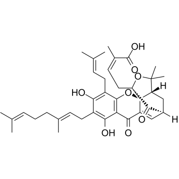
Cas No.: 173932-75-7
Sample solution is provided at 25 µL, 10mM.
_1-3.$$$39978408@51.jpg');)
_1-9.$$$38538795@65.jpg');)
_1-9.$$$39112701@51.jpg');)
_1-7.$$$37316672@70.jpg');)
_366-372.$$$36198801@70.jpg');)
.$$$38565142@65.jpg');)
_1867-1883.$$$33248023@67.jpg');)
_eads6055.$$$39977546@45.jpg');)
_eadm9805.$$$40080571@45.jpg');)
_eadj3020.$$$39666821@45.jpg');)
_1-20.$$$39757301@28.jpg');)
_1-24.$$$38898113@28.jpg');)
_273-287.$$$36806353@46.jpg');)
_1-16.$$$37156877@44.jpg');)
_1-19.$$$37460805@44.jpg');)
_1291-1307.$$$34518654@46.jpg');)
Gambogenic acid, the main active component of gamboge, inhibits Aurora A kinase, with an IC50 value of 1.425μM[1]. Gambogenic acid triggers endoplasmic reticulum (ER) stress-mediated apoptosis through the reactive oxygen species/IRE1α/c-Jun N-terminal kinase signalling pathway[2]. Gambogenic acid has been widely used as an anticancer agent to inhibit the growth of different cancer cells[3].
In vitro, Gambogenic acid treatment for 72h significantly inhibited the proliferation of HCC827, H1650, and HCC827 erlotinib-resistant (HCC827ER) cells with IC50 values of 1.510, 1.328, and 0.909μM, respectively[4]. Treatment of HepG2 cells with 3μM Gambogenic acid for 24h promoted apoptosis, accompanied by a decrease in Bcl-2 levels and an increase in Bax expression levels[5]. Treatment with 1μM Gambogenic acid for 48h inhibited the invasion and migration of A375 cells and induced ferroptosis in A375 cells treated with TGF-β1 (5ng/ml; 48h)[6].
In vivo, Gambogenic acid treatment via intraperitoneally injection at a dose of 2mg/kg/day for 21 days significantly reduced tumor volume and weight in HCT116 cell-xenograft mice, without affecting body weight[7]. Intraperitoneal injection of Gambogenic acid (10mg/kg/day) for 7 consecutive days in male Sprague-Dawley rats can prevent liver oxidative damage caused by acetaminophen (APAP) and alleviate liver histopathological changes caused by APAP[8].
References:
[1] Liu C, Xu J, Guo C, et al. Gambogenic acid induces endoplasmic reticulum stress in colorectal cancer via the aurora a pathway[J]. Frontiers in Cell and Developmental Biology, 2021, 9: 736350.
[2] Zhao Q, Zhong J, Bi Y, et al. Gambogenic acid induces Noxa-mediated apoptosis in colorectal cancer through ROS-dependent activation of IRE1α/JNK[J]. Phytomedicine, 2020, 78: 153306.
[3] Mi L, Xing Z, Zhang Y, et al. Unveiling gambogenic acid as a promising antitumor compound: a review[J]. Planta Medica, 2024, 90(05): 353-367.
[4] Xu L, Meng X, Xu N, et al. Gambogenic acid inhibits fibroblast growth factor receptor signaling pathway in erlotinib-resistant non-small-cell lung cancer and suppresses patient-derived xenograft growth[J]. Cell death & disease, 2018, 9(3): 262.
[5] Yan F, Wang M, Li J, et al. Gambogenic acid induced mitochondrial-dependent apoptosis and referred to phospho-Erk1/2 and phospho-p38 MAPK in human hepatoma HepG2 cells[J]. Environmental toxicology and pharmacology, 2012, 33(2): 181-190.
[6] Wang M, Li S, Wang Y, et al. Gambogenic acid induces ferroptosis in melanoma cells undergoing epithelial-to-mesenchymal transition[J]. Toxicology and applied pharmacology, 2020, 401: 115110.
[7] Zhao Q, Zhong J, Bi Y, et al. Gambogenic acid induces Noxa-mediated apoptosis in colorectal cancer through ROS-dependent activation of IRE1α/JNK[J]. Phytomedicine, 2020, 78: 153306.
[8] Ding Z, Li Y, Tang Z, et al. Role of gambogenic acid in regulating PI3K/Akt/NF-kβ signaling pathways in rat model of acute hepatotoxicity[J]. Bioscience, Biotechnology, and Biochemistry, 2021, 85(3): 520-527.
| Cell experiment [1]: | |
Cell lines | HepG2 cells |
Preparation Method | HepG2 cells were cultured in Dulbecco's modified Eagle's medium (DMEM) supplemented with 10% fetal bovine serum, 100units /ml penicillin, and 100μg/ml streptomycin. Cells were cultured at 37°C in a humidified atmosphere with 5% CO2. Subsequently, HepG2 cells in the logarithmic growth phase were collected and seeded in 96-well culture plates overnight at a density of approximately 6×10⁴ cells/ml. Cells were incubated with different concentrations of Gambogenic acid (0, 0.75, 1.5, 3, 6, and 12μM). At various time points of 24, 48, or 72h, 5.0mg/ml MTT was added, and cells were then incubated at 37°C for 4h and measured at a wavelength of 570nm. |
Reaction Conditions | 0, 0.75, 1.5, 3, 6, and 12μM; 24, 48, and 72h |
Applications | Gambogenic acid treatment reduced the percentage of viable HepG2 cells in a concentration- and time-dependent manner. |
| Animal experiment [2]: | |
Animal models | Female BALB/c-nu mice |
Preparation Method | Female BALB/c-nu mice (4 weeks of age) were maintained under specific pathogen free (SPF) conditions and provided with sterile food and water. HCT116 cells (1×10⁶ cells per mouse) were injected subcutaneously into the right abdomen of each mouse. When the tumor volume was approximately 100mm3, mice were randomly divided into a vehicle group, a 2mg/kg Gambogenic acid group, and a positive control 15mg/kg paclitaxel (PTX) group (n=6 mice in each group). Mice were intraperitoneally injected with Gambogenic acid daily and PTX three times a week. Tumor size was measured and calculated using the standard formula: 0.5×length×width2. The body weight of mice was measured at the same time. After 21 days, all mice were euthanized, and tumors were collected for analysis. |
Dosage form | 2mg/kg/day for 21 days; i.p. |
Applications | Gambogenic acid treatment significantly reduced tumor volume and weight in mice without affecting body weight. |
References: | |
| Cas No. | 173932-75-7 | SDF | |
| Canonical SMILES | O=C(O)/C(C)=C\C[C@@]1(C2=O)OC(C)(C)[C@]([C@]31OC4=C5C(O)=C(C/C=C(C)/CC/C=C(C)\C)C(O)=C4C/C=C(C)\C)([H])C[C@@]2([H])C=C3C5=O | ||
| Formula | C38H46O8 | M.Wt | 630.77 |
| Solubility | DMSO: 250 mg/mL (396.34 mM) | Storage | 4°C, protect from light |
| General tips | Please select the appropriate solvent to prepare the stock solution according to the
solubility of the product in different solvents; once the solution is prepared, please store it in
separate packages to avoid product failure caused by repeated freezing and thawing.Storage method
and period of the stock solution: When stored at -80°C, please use it within 6 months; when stored
at -20°C, please use it within 1 month. To increase solubility, heat the tube to 37°C and then oscillate in an ultrasonic bath for some time. |
||
| Shipping Condition | Evaluation sample solution: shipped with blue ice. All other sizes available: with RT, or with Blue Ice upon request. | ||
| Prepare stock solution | |||

|
1 mg | 5 mg | 10 mg |
| 1 mM | 1.5854 mL | 7.9268 mL | 15.8536 mL |
| 5 mM | 317.1 μL | 1.5854 mL | 3.1707 mL |
| 10 mM | 158.5 μL | 792.7 μL | 1.5854 mL |
Step 1: Enter information below (Recommended: An additional animal making an allowance for loss during the experiment)
 g
μL
g
μL
Step 2: Enter the in vivo formulation (This is only the calculator, not formulation. Please contact us first if there is no in vivo formulation at the solubility Section.)
Calculation results:
Working concentration: mg/ml;
Method for preparing DMSO master liquid: mg drug pre-dissolved in μL DMSO ( Master liquid concentration mg/mL, Please contact us first if the concentration exceeds the DMSO solubility of the batch of drug. )
Method for preparing in vivo formulation: Take μL DMSO master liquid, next addμL PEG300, mix and clarify, next addμL Tween 80, mix and clarify, next add μL saline, mix and clarify.
Method for preparing in vivo formulation: Take μL DMSO master liquid, next add μL Corn oil, mix and clarify.
Note: 1. Please make sure the liquid is clear before adding the next solvent.
2. Be sure to add the solvent(s) in order. You must ensure that the solution obtained, in the previous addition, is a clear solution before proceeding to add the next solvent. Physical methods such as vortex, ultrasound or hot water bath can be used to aid dissolving.
3. All of the above co-solvents are available for purchase on the GlpBio website.
Quality Control & SDS
- View current batch:
- Purity: >98.00% Appearance: A solid
- COA (Certificate of Analysis)
- SDS (Safety Data Sheet)
- Datasheet
Average Rating: 5 (Based on Reviews and 3 reference(s) in Google Scholar.)