

Palmitic acid (Synonyms: C16:0, Cetylic Acid) |
Catalog No.GN10676
 One-Click Copy Product Info
|

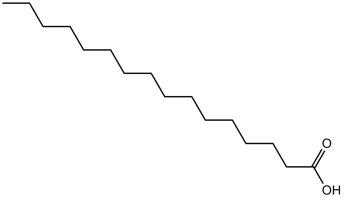
Palmitic acid is a 16-carbon long-chain saturated fatty acid that can post-translationally modify proteins.
Products are for research use only. Not for human use. We do not sell to patients.
Cas No.: 57-10-3
Sample solution is provided at 25 µL, 10mM.
Palmitic acid is a 16-carbon long-chain saturated fatty acid that can post-translationally modify proteins [1]. Palmitic acid can reduce the expressions of p-STAT3, p-JAK2, n-cadherin and vimentin, inhibit the nuclear localization of p-STAT3, and thereby suppress tumor cell metastasis and proliferation[2]. Palmitic acid has been widely used to enhance the signal transduction induced by TLR4 and to strengthen the inflammatory response[3].
In vitro, Palmitic acid treatment for 72 hours significantly inhibited the viability of Ishikawa cells and ECC-1 cells, with IC50 values of 348.2µM and 187.3µM, respectively[4]. Treatment with 300μM Palmitic acid for 24 hours significantly induced the production of reactive oxygen species (ROS) in HepG2 cells and led to a decrease in mitochondrial membrane potential[5]. 150μM of Palmitic acid treatment for 12 hours significantly promoted the maturation of monocyte-derived dendritic cells (MoDCs) and the secretion of IL-1β[6].
In vivo, Palmitic acid treatment via intraperitoneal injection at a dose of 50mg/kg/day for 18 days significantly inhibited the tumor growth in the SGC-7901 xenograft mouse model, without affecting body weight[7]. A single intraperitoneal injection of 30μmol (50μl) of Palmitic acid reduced the locomotor activity of mice within 2 hours, induced anxiety-like behaviors in mice, and simultaneously increased serotonin metabolism mediated by the amygdala[8].
References:
[1] Fatima S, Hu X, Gong R H, et al. Palmitic acid is an intracellular signaling molecule involved in disease development[J]. Cellular and Molecular Life Sciences, 2019, 76(13): 2547-2557.
[2] Wang X, Zhang C, Bao N. Molecular mechanism of palmitic acid and its derivatives in tumor progression[J]. Frontiers in oncology, 2023, 13: 1224125.
[3] Korbecki J, Bajdak-Rusinek K. The effect of palmitic acid on inflammatory response in macrophages: an overview of molecular mechanisms[J]. Inflammation Research, 2019, 68(11): 915-932.
[4] Zhao Z, Wang J, Kong W, et al. Palmitic acid exerts anti-tumorigenic activities by modulating cellular stress and lipid droplet formation in endometrial cancer[J]. Biomolecules, 2024, 14(5): 601.
[5] Alnahdi A, John A, Raza H. Augmentation of glucotoxicity, oxidative stress, apoptosis and mitochondrial dysfunction in HepG2 cells by palmitic acid[J]. Nutrients, 2019, 11(9): 1979.
[6] Nicholas D A, Zhang K, Hung C, et al. Palmitic acid is a toll-like receptor 4 ligand that induces human dendritic cell secretion of IL-1β[J]. PloS one, 2017, 12(5): e0176793.
[7] Yu X, Peng W, Wang Y, et al. Palmitic acid inhibits the growth and metastasis of gastric cancer by blocking the STAT3 signaling pathway[J]. Cancers, 2023, 15(2): 388.
[8] Moon M L, Joesting J J, Lawson M A, et al. The saturated fatty acid, palmitic acid, induces anxiety-like behavior in mice[J]. Metabolism, 2014, 63(9): 1131-1140.
| Cell experiment [1]: | |
Cell lines | Ishikawa cells |
Preparation Method | Preparation of Palmitic acid working solution: Add 78μl of anhydrous ethanol to 1mg Palmitic acid and dissolve to 50mM stock solution (ultrasonic heating for 3min; 37°C); Take an appropriate amount of Fatty acid free BSA Add DMEM culture medium to prepare a 10% BSA solution (100mg/ml). Take 5μl of Palmitic acid stock solution (50mM), add 100μl of BSA solution (10%), mix well (ultrasonic heating for 3min; 37°C), add 895μl of DMEM culture medium, mix well (ultrasonic heating for 3min; 37°C) to prepare 0.25mM Palmitic acid working solution. Ishikawa cells were cultured in DMEM/F-12 medium enriched with 10% fetal bovine serum (FBS) and 1% antimycotic-antibiotic at 37°C in 5% CO2/atmosphere. Cells were seeded at a density of 2×10⁴ cells/ml in the 96-well plates and allowed to adhere in a 5% CO2 incubator at 37°C for 24h. The cells were treated with different concentrations of Palmitic acid (0, 0.1, 1, 10, 50, 100, and 250μM) for 96h. The cell viability was evaluated. |
Reaction Conditions | 0, 0.1, 1, 10, 50, 100, and 250μM; 96h |
Applications | Palmitic acid treatment significantly inhibited cell viability of Ishikawa cells in a dose-dependent manner. |
| Animal experiment [2]: | |
Animal models | Male BALB/c nude mice |
Preparation Method | Male BALB/c nude mice (6-week-old; 18-22g) were housed in a room with controlled temperature (21-23°C), humidity (55%-60%) and lighting (12h light/dark cycle) and were supplied with water ad libitum. Approximately 5×10⁶ SGC-7901 cells were injected into the flanks of the nude mice to establish a human gastric cancer syngeneic s.c. xenograft model. When the cancer volume reached approximately 50mm3, Palmitic acid was administered via i.p. injections once daily at the designed doses (50mg/kg or vehicle) for 18 days. Cancer growth and mouse body weights were measured every other day throughout the experiment. Cancer volumes (mm3) were calculated as 0.5×length (mm)×width2(mm). |
Dosage form | 50mg/kg/day; 18 days; i.p. |
Applications | Palmitic acid treatment significantly inhibited the tumor growth in the SGC-7901 xenograft mouse model, without affecting body weight. |
References: | |
| Cas No. | 57-10-3 | SDF | |
| Synonyms | C16:0, Cetylic Acid | ||
| Chemical Name | hexadecanoic acid | ||
| Canonical SMILES | CCCCCCCCCCCCCCCC(=O)O | ||
| Formula | C16H32O2 | M.Wt | 256.42 |
| Solubility | ≥ 51mg/mL in DMSO; ≥ 51mg/mL in Ethanol | Storage | Store at -20°C,protect from light |
| Shipping Condition | Evaluation sample solution: shipped with blue ice. All other sizes available: with RT, or with Blue Ice upon request. | ||
| Prepare stock solution | |||

|
1 mg | 5 mg | 10 mg |
| 1 mM | 3.8999 mL | 19.4993 mL | 38.9985 mL |
| 5 mM | 780 μL | 3.8999 mL | 7.7997 mL |
| 10 mM | 390 μL | 1.9499 mL | 3.8999 mL |
Step 1: Enter information below (Recommended: An additional animal making an allowance for loss during the experiment)
 g
μL
g
μL
Step 2: Enter the in vivo formulation (This is only the calculator, not formulation. Please contact us first if there is no in vivo formulation at the solubility Section.)
Calculation results:
Working concentration: mg/ml;
Method for preparing DMSO master liquid: mg drug pre-dissolved in μL DMSO ( Master liquid concentration mg/mL, Please contact us first if the concentration exceeds the DMSO solubility of the batch of drug. )
Method for preparing in vivo formulation: Take μL DMSO master liquid, next addμL PEG300, mix and clarify, next addμL Tween 80, mix and clarify, next add μL saline, mix and clarify.
Method for preparing in vivo formulation: Take μL DMSO master liquid, next add μL Corn oil, mix and clarify.
Note: 1. Please make sure the liquid is clear before adding the next solvent.
2. Be sure to add the solvent(s) in order. You must ensure that the solution obtained, in the previous addition, is a clear solution before proceeding to add the next solvent. Physical methods such as vortex, ultrasound or hot water bath can be used to aid dissolving.
3. All of the above co-solvents are available for purchase on the GlpBio website.
Quality Control & SDS
- View current batch:
- Purity: >98.00% Appearance: A solid
- COA (Certificate of Analysis)
- SDS (Safety Data Sheet)
- Datasheet
Average Rating: 5 (Based on Reviews and 30 reference(s) in Google Scholar.)