

Quinolinic acid (Synonyms: NSC 13127, NSC 18836, NSC 403247) |
Catalog No.GC14547
 One-Click Copy Product Info
|

Quinolinic acid, a neuroactive metabolite of the kynurenine pathway, is an endogenous neurotoxin.
Products are for research use only. Not for human use. We do not sell to patients.
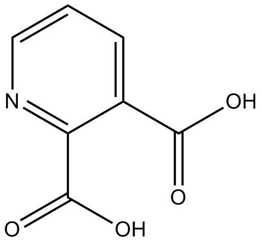
Cas No.: 89-00-9
Sample solution is provided at 25 µL, 10mM.
_1-3.$$$39978408@51.jpg');)
_1-9.$$$38538795@65.jpg');)
_1-9.$$$39112701@51.jpg');)
_1-7.$$$37316672@70.jpg');)
_366-372.$$$36198801@70.jpg');)
.$$$38565142@65.jpg');)
_1867-1883.$$$33248023@67.jpg');)
_eads6055.$$$39977546@45.jpg');)
_eadm9805.$$$40080571@45.jpg');)
_eadj3020.$$$39666821@45.jpg');)
_1-20.$$$39757301@28.jpg');)
_1-24.$$$38898113@28.jpg');)
_273-287.$$$36806353@46.jpg');)
_1-16.$$$37156877@44.jpg');)
_1-19.$$$37460805@44.jpg');)
_1291-1307.$$$34518654@46.jpg');)
Quinolinic acid, a neuroactive metabolite of the kynurenine pathway, is an endogenous neurotoxin[1]. Quinolinic acid increases glutamate release, induces the generation of reactive oxygen species (ROS), activates nitric oxide synthase, produces excessive NO, and subsequently leads to calcium influx and neuronal apoptosis[2]. Quinolinic acid has been widely used to induce lipoperoxidation and cytoskeletal destabilisation[3].
In vitro, Quinolinic acid treatment for 48 hours significantly inhibited cell viability of B16-F10, RAW 264.7, and HaCaT cells, with the IC50 values of 8.23mM, 68.08mM, and 20.07mM, respectively[4]. Quinolinic acid treatment (350nM) for 24 hours resulted in a significant increase in the levels of IL-1β and Vimentin in human astrocytes, and a decrease in the activity of glutamine synthetase (GS)[5]. Treatment with 30mM Quinolinic acid for 24 hours significantly induced cell death in N18D3 cells, resulting in nuclear condensation and fragmentation of DNA[6].
In vivo, a single injection of Quinolinic acid (30nmol; 0.1µl) into the medial prefrontal cortex (mPFC) of mice resulted in spatial cognitive impairment in the mice within 10 days, and reversed hippocampal synaptic plasticity[7]. Oral administration of Quinolinic acid at 30mg/kg/day for 14 days significantly increased the plasma PAI-1 level in rats and promoted thrombosis[8].
References:
[1] Lugo-Huitrón R, Ugalde Muñiz P, Pineda B, et al. Quinolinic acid: an endogenous neurotoxin with multiple targets[J]. Oxidative medicine and cellular longevity, 2013, 2013(1): 104024.
[2] La Cruz V P D, Carrillo-Mora P, Santamaría A. Quinolinic acid, an endogenous molecule combining excitotoxicity, oxidative stress and other toxic mechanisms[J]. International Journal of Tryptophan Research, 2012, 5: IJTR. S8158.
[3] Hestad K, Alexander J, Rootwelt H, et al. The role of tryptophan dysmetabolism and quinolinic acid in depressive and neurodegenerative diseases[J]. Biomolecules, 2022, 12(7): 998.
[4] Basson C, Serem J C, Hlophe Y N, et al. An in vitro investigation of L‐kynurenine, quinolinic acid, and kynurenic acid on B16 F10 melanoma cell cytotoxicity and morphology[J]. Cell Biochemistry and Function, 2023, 41(7): 912-922.
[5] Ting K K, Brew B J, Guillemin G J. Effect of quinolinic acid on human astrocytes morphology and functions: implications in Alzheimer's disease[J]. Journal of neuroinflammation, 2009, 6(1): 36.
[6] Jang S, Jeong H S, Park J S, et al. Neuroprotective effects of (−)-epigallocatechin-3-gallate against quinolinic acid-induced excitotoxicity via PI3K pathway and NO inhibition[J]. Brain Research, 2010, 1313: 25-33.
[7] Latif-Hernandez A, Shah D, Ahmed T, et al. Quinolinic acid injection in mouse medial prefrontal cortex affects reversal learning abilities, cortical connectivity and hippocampal synaptic plasticity[J]. Scientific reports, 2016, 6(1): 36489.
[8] Leszczyńska A, Misztal T, Marcińczyk N, et al. Effect of quinolinic acid–a uremic toxin from tryptophan metabolism–on hemostatic profile in rat and mouse thrombosis models[J]. Advances in medical sciences, 2019, 64(2): 370-380.
| Cell experiment [1]: | |
Cell lines | B16-F10 cells |
Preparation Method | B16-F10 cells were cultured in DMEM medium supplemented with 10% fetal bovine serum, 100U/ml penicillin, and 100μg/ml streptomycin at 37°C with 5% CO2 and 95% saturated atmospheric humidity. Cells were seeded into 96-well microplates at a density of 5×10³ cells/ml for 24h. Various concentrations of Quinolinic acid (0, 5, 10, 20, 40, 60, 80, and 100mM) were added to each well. At the end of the incubation period (48h at 37°C), cell viability was analyzed. |
Reaction Conditions | 0, 5, 10, 20, 40, 60, 80, and 100mM; 48h |
Applications | Quinolinic acid treatment significantly inhibited the cell viability of B16-F10 cells in a dose-dependent manner. |
| Animal experiment [2]: | |
Animal models | Male Wistar rats |
Preparation Method | Male Wistar rats (6 weeks old; 190-200g) were grouped in individually ventilated cages (IVC) and maintained under specific pathogen-free (SPF) conditions in an environment-controlled room (12h light/ 12h dark cycle, 21±2°C, 55% relative humidity) and had access to sterilized tap water and standard chow ad libitum. Wistar rats were randomly assigned to the vehicle control group, comprising rats receiving pure drinking water, and to the Quinolinic acid group, comprising rats receiving Quinolinic acid dissolved in drinking water in a dose of 30mg/kg/day for 14 days. One day before the induction of arterial thrombosis, the rats underwent monitoring of blood pressure (BP) using the tail-cuff method. On the day of the experiment, the rats were anesthetized with pentobarbital (50mg/kg) and subjected to electrical arterial injury. The formed thrombus was dissected, and blood samples were collected into anticoagulant 3.13% trisodium citrate for analysis. |
Dosage form | 30mg/kg/day; 14 days; p.o. |
Applications | Quinolinic acid treatment increased the plasma PAI-1 level and promoted thrombosis in rats. |
References: | |
| Cas No. | 89-00-9 | SDF | |
| Synonyms | NSC 13127, NSC 18836, NSC 403247 | ||
| Chemical Name | pyridine-2,3-dicarboxylic acid | ||
| Canonical SMILES | OC(C1=CC=CN=C1C(O)=O)=O | ||
| Formula | C7H5NO4 | M.Wt | 167.12 |
| Solubility | ≥ 7.8mg/mL in DMSO | Storage | Store at RT |
| General tips | Please select the appropriate solvent to prepare the stock solution according to the
solubility of the product in different solvents; once the solution is prepared, please store it in
separate packages to avoid product failure caused by repeated freezing and thawing.Storage method
and period of the stock solution: When stored at -80°C, please use it within 6 months; when stored
at -20°C, please use it within 1 month. To increase solubility, heat the tube to 37°C and then oscillate in an ultrasonic bath for some time. |
||
| Shipping Condition | Evaluation sample solution: shipped with blue ice. All other sizes available: with RT, or with Blue Ice upon request. | ||
| Prepare stock solution | |||

|
1 mg | 5 mg | 10 mg |
| 1 mM | 5.9837 mL | 29.9186 mL | 59.8372 mL |
| 5 mM | 1.1967 mL | 5.9837 mL | 11.9674 mL |
| 10 mM | 598.4 μL | 2.9919 mL | 5.9837 mL |
Step 1: Enter information below (Recommended: An additional animal making an allowance for loss during the experiment)
 g
μL
g
μL
Step 2: Enter the in vivo formulation (This is only the calculator, not formulation. Please contact us first if there is no in vivo formulation at the solubility Section.)
Calculation results:
Working concentration: mg/ml;
Method for preparing DMSO master liquid: mg drug pre-dissolved in μL DMSO ( Master liquid concentration mg/mL, Please contact us first if the concentration exceeds the DMSO solubility of the batch of drug. )
Method for preparing in vivo formulation: Take μL DMSO master liquid, next addμL PEG300, mix and clarify, next addμL Tween 80, mix and clarify, next add μL saline, mix and clarify.
Method for preparing in vivo formulation: Take μL DMSO master liquid, next add μL Corn oil, mix and clarify.
Note: 1. Please make sure the liquid is clear before adding the next solvent.
2. Be sure to add the solvent(s) in order. You must ensure that the solution obtained, in the previous addition, is a clear solution before proceeding to add the next solvent. Physical methods such as vortex, ultrasound or hot water bath can be used to aid dissolving.
3. All of the above co-solvents are available for purchase on the GlpBio website.
Quality Control & SDS
- View current batch:
- Purity: >99.00% Appearance: A solid
- COA (Certificate of Analysis)
- SDS (Safety Data Sheet)
- Datasheet
Average Rating: 5 (Based on Reviews and 17 reference(s) in Google Scholar.)