

Thapsigargin |
Catalog No.GC11482
 One-Click Copy Product Info
|

Thapsigargin is a sesquiterpene lactone isolated from Thapsia garganica and acts as an irreversible sarco/endoplasmic reticulum Ca²⁺-ATPase (SERCA) inhibitor.
Products are for research use only. Not for human use. We do not sell to patients.
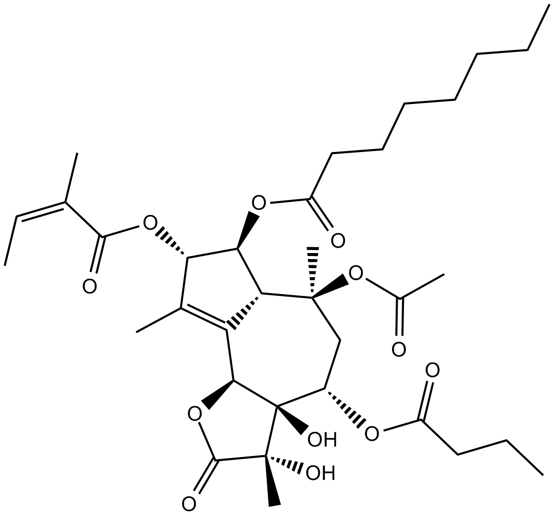
Cas No.: 67526-95-8
Sample solution is provided at 25 µL, 10mM.
Thapsigargin is a sesquiterpene lactone isolated from Thapsia garganica and acts as an irreversible sarco/endoplasmic reticulum Ca²⁺-ATPase (SERCA) inhibitor. Thapsigargin depletes intracellular calcium stores by inhibiting the SERCA, thereby activating the unfolded protein response (UPR) and ATF3/ATF4 pathways. This process induces apoptosis in tumor cells or triggers a broad-spectrum antiviral state in host cells. Meanwhile, calcium dyshomeostasis activates multiple stress signals to amplify inflammatory and autophagic responses. Thapsigargin can be used in research related to cancer therapy and broad-spectrum antiviral applications including COVID-19[1-4].
In vitro, treatment of HT29, HCT116, and SW1116 colorectal cancer cells with 0.1μM or 10μM Thapsigargin for 24 hours induced cell death[5]. Treatment of β-TC-6 pancreatic β cells with 0.1μM Thapsigargin for 24 hours induced apoptosis[6]. Treatment of MH7A human rheumatoid arthritis synovial cells with 0.001μM, 0.1μM, or 1μM Thapsigargin for 2 or 4 days inhibited cell proliferation and induced apoptosis in a time- and dose-dependent manner[7].
In vivo, male Swiss albino mice (10-week-old) with AOM/DSS-induced colitis received weekly intraperitoneal injections of 1mg/kg Thapsigargin for 5 consecutive weeks. Thapsigargin alleviated body weight loss and disease activity index scores, reduced colon weight-to-length ratio, decreased colonic tumor numbers and aberrant crypt foci formation, and improved colonic epithelial ultrastructure and tissue architecture[8]. Male BALB/c nude mice subcutaneously inoculated with SW-13 human adrenocortical carcinoma cells received daily intraperitoneal injections of 1mg/kg Thapsigargin for 15 days. Thapsigargin inhibited xenograft tumor growth, reduced tumor volume and weight, and did not affect mouse body weight[9].
References:
[1] Geiszt M, Káldi K, Szeberényi JB, et al. Thapsigargin inhibits Ca2+ entry into human neutrophil granulocytes. Biochem J. 1995 Jan 15;305 ( Pt 2)(Pt 2):525-8.
[2] Wang H, Jia XZ, Sui CJ, et al. Effects of thapsigargin on the proliferation and survival of human rheumatoid arthritis synovial cells. ScientificWorldJournal. 2014 Feb 9;2014:605416.
[3] Garavito-Aguilar ZV, Recio-Pinto E, Corrales AV, et al. Differential thapsigargin-sensitivities and interaction of Ca2+ stores in human SH-SY5Y neuroblastoma cells. Brain Res. 2004 Jun 18;1011(2):177-86.
[4] Abdullahi A, Stanojcic M, Parousis A, et al. Modeling Acute ER Stress in Vivo and in Vitro. Shock. 2017 Apr;47(4):506-513.
[5] Jiang X, Li D, Wang G, et al. Thapsigargin promotes colorectal cancer cell migration through upregulation of lncRNA MALAT1. Oncol Rep. 2020;43(4):1245-1255.
[6] Mwangi S, Anitha M, Mallikarjun C, et al. Glial Cell Line-Derived Neurotrophic Factor increases beta cell mass and improves glucose tolerance. Gastroenterology. 2008 Mar;134(3):727-737.
[7] Wang H, Jia X, Sui C, et al. Effects of Thapsigargin on the Proliferation and Survival of Human Rheumatoid Arthritis Synovial Cells. The Scientific World Journal. 2014;2014:605416.
[8] Baruah S, Bharali M K, Akhtara N, et al. Thapsigargin enhanced chemotherapeutic sensitivity of irinotecan in the inflammation-induced colorectal cancer model in mice. Sci Rep. 2026;16:4551.
[9] Wu L, Huang X, Kuang Y, et al. Thapsigargin induces apoptosis in adrenocortical carcinoma by activating endoplasmic reticulum stress and the JNK signaling pathway: an in vitro and in vivo study. Drug Des Devel Ther. 2019;13:2787-2798.
| Cell experiment [1]: | |
|
Cell lines |
β-TC-6 cells (mouse pancreatic beta cell line) |
|
Preparation Method |
β-TC-6 cells were cultured in Dulbecco's Modified Eagles' Medium (DMEM) supplemented with 15% fetal bovine serum. Cells were serum-deprived for 48 hours and then treated with Thapsigargin at 0.1μM for 24 hours. |
|
Reaction Conditions |
0.1μM; 24h |
|
Applications |
Thapsigargin induced apoptosis in β-TC-6 cells, increased the content of cleaved caspase-3. |
| Animal experiment [2]: | |
|
Animal models |
Male BALB/c nude mice (4-5 weeks old, 20-22g); subcutaneous adrenocortical carcinoma xenograft models were established by injecting 2×10⁶ SW-13 human adrenocortical carcinoma cells in 200 μL medium into the left shoulder of each mouse |
|
Preparation Method |
When the subcutaneous tumors grew to approximately 0.2×0.2cm² in size, mice were intraperitoneally injected with Thapsigargin (1mg/kg) or vehicle (0.5% methylcellulose) daily. Mouse body weights and tumor sizes were recorded regularly, and xenograft tumor tissues were harvested after 15 consecutive days. |
|
Dosage form |
1mg/kg; i.p.; daily for 15 days |
|
Applications |
Thapsigargin inhibited the growth of SW-13 xenograft tumors, with reduced tumor volume and weight, and no significant change in mouse body weight relative to the vehicle group. In xenograft tumor tissues, p-JNK, p-ERK, p-MAPK, p-PERK, IRE1 and GRP78 protein levels were upregulated, while total JNK, ERK, PERK and MAPK protein levels remained unchanged. |
|
References: [1] Mwangi S, Anitha M, Mallikarjun C, et al. Glial Cell Line-Derived Neurotrophic Factor increases beta cell mass and improves glucose tolerance. Gastroenterology. 2008 Mar;134(3):727-737. [2] Wu L, Huang X, Kuang Y, et al. Thapsigargin induces apoptosis in adrenocortical carcinoma by activating endoplasmic reticulum stress and the JNK signaling pathway: an in vitro and in vivo study. Drug Des Devel Ther. 2019;13:2787-2798. |
|
| Cas No. | 67526-95-8 | SDF | |
| Chemical Name | (3S,3aR,4S,6S,6aR,7S,8S,9bS)-6-acetoxy-4-(butyryloxy)-3,3a-dihydroxy-3,6,9-trimethyl-8-(((Z)-2-methylbut-2-enoyl)oxy)-2-oxo-2,3,3a,4,5,6,6a,7,8,9b-decahydroazuleno[4,5-b]furan-7-yl octanoate | ||
| Canonical SMILES | O[C@@]([C@H]1OC(CCC)=O)([C@]2(C)O)[C@H](C([C@H]([C@@H]3OC(CCCCCCC)=O)[C@@](C1)(C)OC(C)=O)=C(C)[C@@H]3OC(/C(C)=C\C)=O)OC2=O | ||
| Formula | C34H50O12 | M.Wt | 650.76 |
| Solubility | 30mg/ml in ethanol, DMSO, DMF | Storage | Store at -20°C, sealed storage, away from moisture and light |
| General tips | Please select the appropriate solvent to prepare the stock solution according to the
solubility of the product in different solvents; once the solution is prepared, please store it in
separate packages to avoid product failure caused by repeated freezing and thawing.Storage method
and period of the stock solution: When stored at -80°C, please use it within 6 months; when stored
at -20°C, please use it within 1 month. To increase solubility, heat the tube to 37°C and then oscillate in an ultrasonic bath for some time. |
||
| Shipping Condition | Evaluation sample solution: shipped with blue ice. All other sizes available: with RT, or with Blue Ice upon request. | ||
| Prepare stock solution | |||

|
1 mg | 5 mg | 10 mg |
| 1 mM | 1.5367 mL | 7.6833 mL | 15.3666 mL |
| 5 mM | 307.3 μL | 1.5367 mL | 3.0733 mL |
| 10 mM | 153.7 μL | 768.3 μL | 1.5367 mL |
Step 1: Enter information below (Recommended: An additional animal making an allowance for loss during the experiment)
 g
μL
g
μL
Step 2: Enter the in vivo formulation (This is only the calculator, not formulation. Please contact us first if there is no in vivo formulation at the solubility Section.)
Calculation results:
Working concentration: mg/ml;
Method for preparing DMSO master liquid: mg drug pre-dissolved in μL DMSO ( Master liquid concentration mg/mL, Please contact us first if the concentration exceeds the DMSO solubility of the batch of drug. )
Method for preparing in vivo formulation: Take μL DMSO master liquid, next addμL PEG300, mix and clarify, next addμL Tween 80, mix and clarify, next add μL saline, mix and clarify.
Method for preparing in vivo formulation: Take μL DMSO master liquid, next add μL Corn oil, mix and clarify.
Note: 1. Please make sure the liquid is clear before adding the next solvent.
2. Be sure to add the solvent(s) in order. You must ensure that the solution obtained, in the previous addition, is a clear solution before proceeding to add the next solvent. Physical methods such as vortex, ultrasound or hot water bath can be used to aid dissolving.
3. All of the above co-solvents are available for purchase on the GlpBio website.
Quality Control & SDS
- View current batch:
- Purity: >99.50% Appearance: A solid
- COA (Certificate of Analysis)
- SDS (Safety Data Sheet)
- Datasheet
-
Related Biological Data

RHBDF1 is critical for maintaining BiP protein stability in breast cancer cells.c, d BiP levels in MCF-7 cells (MT or R1KO) treated with Thapsigargin (TG, 10 μM).
When the cells were further treated with thapsigargin (TG)(GlpBio), an ER stress inducer, the BiP protein level progressively increased in the control cells; however, this increase was significantly slower in R1KO cells
Acta Pharmacol Sin (2023): 1-13. PMID: 37798352 IF: 8.1996 -
Related Biological Data

SPC alleviates AngII-induced cardiac hypertrophy by inhibiting CaM. c, d, g, h Surface area of CMs was determined by TRITC Phalloidin staining and data analysis.
To explore whether SPC inhibits Ang II-induced cardiac hypertrophy through its interactions with CaM, we conducted an in vitro experiment by treating cells with siCaM and TG(GlpBio) to decrease or increase CaM levels, respectively.
Acta Pharmacol Sin (2023): 1-15. PMID: 37833535 IF: 8.1996 -
Related Biological Data

DAPA inhibited ER stress in Ang II-induced cardiomyocytes. a, b The protein levels of Bcl-2, Bax, and cleaved caspase 3 in each group were detected.
To confirm whether the DAPA-induced inhibition of ERS attenuates Ang II-induced apoptosis, we used TG to activate ERS and measured the levels of apoptosis biomarkers, including Bcl-2,cleaved caspase 3, and Bax.
Acta Pharmacol Sin (2021): 1-12. PMID: 34853445 IF: 6.152 -
Related Biological Data

TG and 3-MA treatment partially restores SEEC function after BMS30940 suppression of FABP4 (a, b) Expression levels of key proteins in the endoplasmic reticulum stress signaling pathway after combined treatment with hormones, BMS309403, and TG.
In the case of TG (1 μM)(Glpbio) or 3-MA (5 mM) treatment groups, TG and 3-MA were added 1 h prior to the addition of IFN-τ. The total treatment duration for all cells was 24 h.
J Reprod Develop (2023). PMID: 37779094 IF: 1.7998
Average Rating: 5 (Based on Reviews and 25 reference(s) in Google Scholar.)