Apoptosis
As one of the cellular death mechanisms, apoptosis, also known as programmed cell death, can be defined as the process of a proper death of any cell under certain or necessary conditions. Apoptosis is controlled by the interactions between several molecules and responsible for the elimination of unwanted cells from the body.
Many biochemical events and a series of morphological changes occur at the early stage and increasingly continue till the end of apoptosis process. Morphological event cascade including cytoplasmic filament aggregation, nuclear condensation, cellular fragmentation, and plasma membrane blebbing finally results in the formation of apoptotic bodies. Several biochemical changes such as protein modifications/degradations, DNA and chromatin deteriorations, and synthesis of cell surface markers form morphological process during apoptosis.
Apoptosis can be stimulated by two different pathways: (1) intrinsic pathway (or mitochondria pathway) that mainly occurs via release of cytochrome c from the mitochondria and (2) extrinsic pathway when Fas death receptor is activated by a signal coming from the outside of the cell.
Different gene families such as caspases, inhibitor of apoptosis proteins, B cell lymphoma (Bcl)-2 family, tumor necrosis factor (TNF) receptor gene superfamily, or p53 gene are involved and/or collaborate in the process of apoptosis.
Caspase family comprises conserved cysteine aspartic-specific proteases, and members of caspase family are considerably crucial in the regulation of apoptosis. There are 14 different caspases in mammals, and they are basically classified as the initiators including caspase-2, -8, -9, and -10; and the effectors including caspase-3, -6, -7, and -14; and also the cytokine activators including caspase-1, -4, -5, -11, -12, and -13. In vertebrates, caspase-dependent apoptosis occurs through two main interconnected pathways which are intrinsic and extrinsic pathways. The intrinsic or mitochondrial apoptosis pathway can be activated through various cellular stresses that lead to cytochrome c release from the mitochondria and the formation of the apoptosome, comprised of APAF1, cytochrome c, ATP, and caspase-9, resulting in the activation of caspase-9. Active caspase-9 then initiates apoptosis by cleaving and thereby activating executioner caspases. The extrinsic apoptosis pathway is activated through the binding of a ligand to a death receptor, which in turn leads, with the help of the adapter proteins (FADD/TRADD), to recruitment, dimerization, and activation of caspase-8 (or 10). Active caspase-8 (or 10) then either initiates apoptosis directly by cleaving and thereby activating executioner caspase (-3, -6, -7), or activates the intrinsic apoptotic pathway through cleavage of BID to induce efficient cell death. In a heat shock-induced death, caspase-2 induces apoptosis via cleavage of Bid.
Bcl-2 family members are divided into three subfamilies including (i) pro-survival subfamily members (Bcl-2, Bcl-xl, Bcl-W, MCL1, and BFL1/A1), (ii) BH3-only subfamily members (Bad, Bim, Noxa, and Puma9), and (iii) pro-apoptotic mediator subfamily members (Bax and Bak). Following activation of the intrinsic pathway by cellular stress, pro‑apoptotic BCL‑2 homology 3 (BH3)‑only proteins inhibit the anti‑apoptotic proteins Bcl‑2, Bcl-xl, Bcl‑W and MCL1. The subsequent activation and oligomerization of the Bak and Bax result in mitochondrial outer membrane permeabilization (MOMP). This results in the release of cytochrome c and SMAC from the mitochondria. Cytochrome c forms a complex with caspase-9 and APAF1, which leads to the activation of caspase-9. Caspase-9 then activates caspase-3 and caspase-7, resulting in cell death. Inhibition of this process by anti‑apoptotic Bcl‑2 proteins occurs via sequestration of pro‑apoptotic proteins through binding to their BH3 motifs.
One of the most important ways of triggering apoptosis is mediated through death receptors (DRs), which are classified in TNF superfamily. There exist six DRs: DR1 (also called TNFR1); DR2 (also called Fas); DR3, to which VEGI binds; DR4 and DR5, to which TRAIL binds; and DR6, no ligand has yet been identified that binds to DR6. The induction of apoptosis by TNF ligands is initiated by binding to their specific DRs, such as TNFα/TNFR1, FasL /Fas (CD95, DR2), TRAIL (Apo2L)/DR4 (TRAIL-R1) or DR5 (TRAIL-R2). When TNF-α binds to TNFR1, it recruits a protein called TNFR-associated death domain (TRADD) through its death domain (DD). TRADD then recruits a protein called Fas-associated protein with death domain (FADD), which then sequentially activates caspase-8 and caspase-3, and thus apoptosis. Alternatively, TNF-α can activate mitochondria to sequentially release ROS, cytochrome c, and Bax, leading to activation of caspase-9 and caspase-3 and thus apoptosis. Some of the miRNAs can inhibit apoptosis by targeting the death-receptor pathway including miR-21, miR-24, and miR-200c.
p53 has the ability to activate intrinsic and extrinsic pathways of apoptosis by inducing transcription of several proteins like Puma, Bid, Bax, TRAIL-R2, and CD95.
Some inhibitors of apoptosis proteins (IAPs) can inhibit apoptosis indirectly (such as cIAP1/BIRC2, cIAP2/BIRC3) or inhibit caspase directly, such as XIAP/BIRC4 (inhibits caspase-3, -7, -9), and Bruce/BIRC6 (inhibits caspase-3, -6, -7, -8, -9).
Any alterations or abnormalities occurring in apoptotic processes contribute to development of human diseases and malignancies especially cancer.
References:
1.Yağmur Kiraz, Aysun Adan, Melis Kartal Yandim, et al. Major apoptotic mechanisms and genes involved in apoptosis[J]. Tumor Biology, 2016, 37(7):8471.
2.Aggarwal B B, Gupta S C, Kim J H. Historical perspectives on tumor necrosis factor and its superfamily: 25 years later, a golden journey.[J]. Blood, 2012, 119(3):651.
3.Ashkenazi A, Fairbrother W J, Leverson J D, et al. From basic apoptosis discoveries to advanced selective BCL-2 family inhibitors[J]. Nature Reviews Drug Discovery, 2017.
4.McIlwain D R, Berger T, Mak T W. Caspase functions in cell death and disease[J]. Cold Spring Harbor perspectives in biology, 2013, 5(4): a008656.
5.Ola M S, Nawaz M, Ahsan H. Role of Bcl-2 family proteins and caspases in the regulation of apoptosis[J]. Molecular and cellular biochemistry, 2011, 351(1-2): 41-58.
What is Apoptosis? The Apoptotic Pathways and the Caspase Cascade
Targets for Apoptosis
- Pyroptosis(15)
- Caspase(77)
- 14.3.3 Proteins(3)
- Apoptosis Inducers(71)
- Bax(15)
- Bcl-2 Family(136)
- Bcl-xL(13)
- c-RET(15)
- IAP(32)
- KEAP1-Nrf2(73)
- MDM2(21)
- p53(137)
- PC-PLC(6)
- PKD(8)
- RasGAP (Ras- P21)(2)
- Survivin(8)
- Thymidylate Synthase(12)
- TNF-α(141)
- Other Apoptosis(1144)
- Apoptosis Detection(0)
- Caspase Substrate(0)
- APC(6)
- PD-1/PD-L1 interaction(60)
- ASK1(4)
- PAR4(2)
- RIP kinase(47)
- FKBP(22)
Products for Apoptosis
- Cat.No. 상품명 정보
-
GC43274
Citromycetin
시트로마이세틴은 호주 해양 유래 및 육상 Penicillium spp의 방향족 폴리케타이드 화합물입니다.

-
GC63393
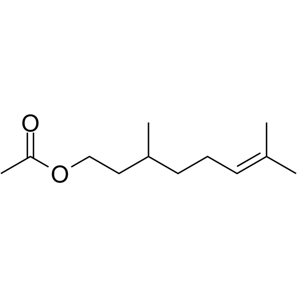
Citronellyl acetate
시트로넬릴 아세테이트는 항통각수용 활성을 갖는 식물의 2차 대사의 모노테르펜 생성물입니다.
-
GC52367
Citrullinated Vimentin (G146R) (R144 + R146) (139-159)-biotin Peptide
A biotinylated and citrullinated mutant vimentin peptide

-
GC52370
Citrullinated Vimentin (R144) (139-159)-biotin Peptide
A biotinylated and citrullinated vimentin peptide

-
GN10219
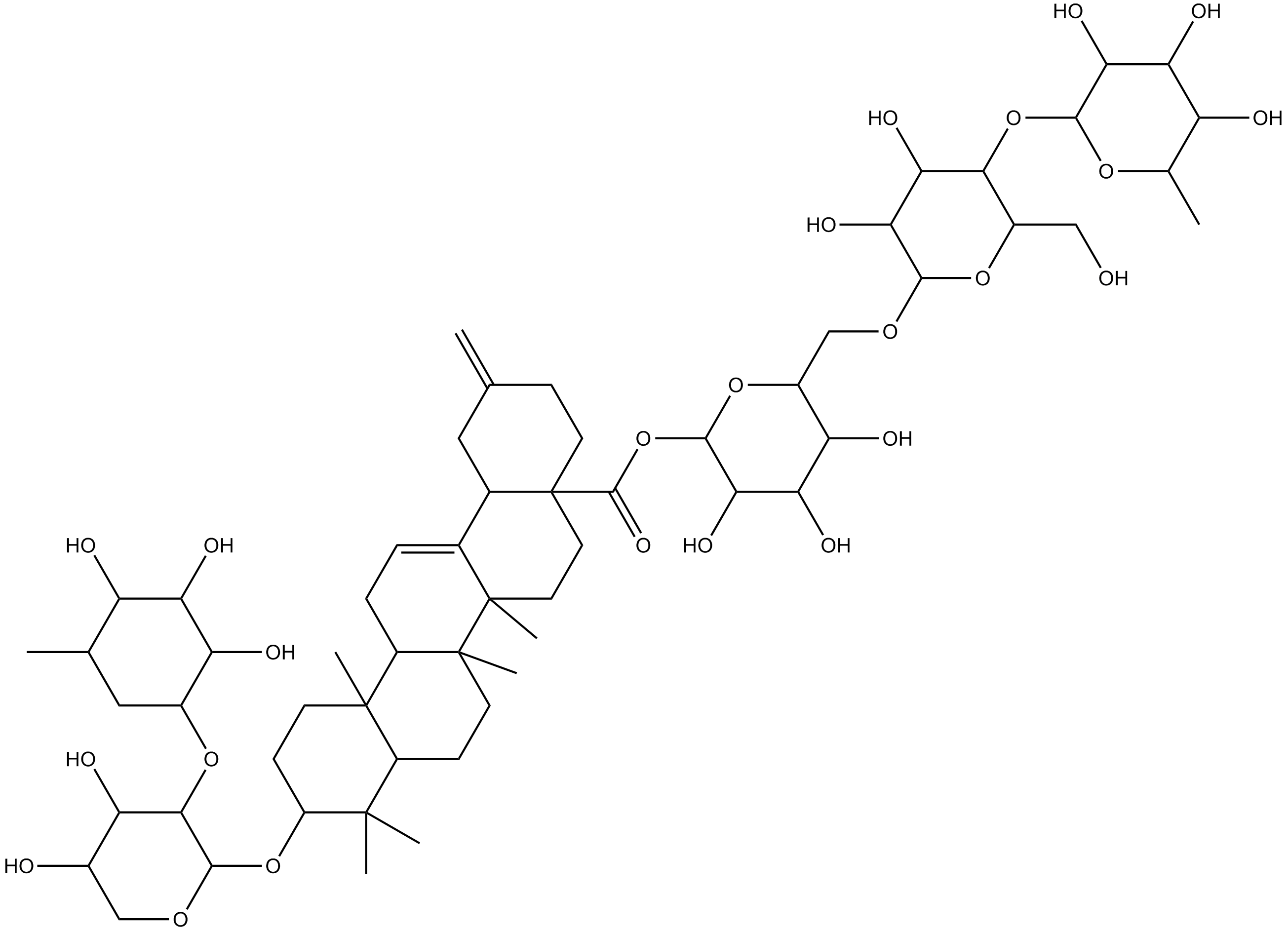
Ciwujianoside-B
-
GC64649
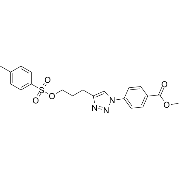
Cjoc42
Cjoc42는 간키린에 결합할 수 있는 화합물이다. Cjoc42는 용량 의존적 방식으로 간키린 활성을 억제합니다. Cjoc42는 일반적으로 다량의 간키린과 관련된 p53 단백질 수준의 감소를 방지합니다. Cjoc42는 p53 의존성 전사와 DNA 손상에 대한 민감성을 회복시킵니다.
-
GC39485
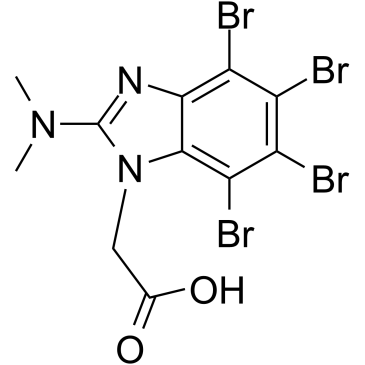
CK2/ERK8-IN-1
CK2/ERK8-IN-1은 IC50이 0.50μM인 이중 카제인 키나제 2(CK2)(Ki 0.25μM) 및 ERK8(MAPK15, ERK7) 억제제입니다. CK2/ERK8-IN-1은 또한 각각 8.65μM, 15.25μM 및 11.9μM의 Kis로 PIM1, HIPK2(호메오도메인 상호작용 단백질 키나제 2) 및 DYRK1A에 결합합니다. CK2/ERK8-IN-1은 세포자멸사 효능이 있습니다.
-
GC49556
Cl-Necrostatin-1
A RIPK1 inhibitor

-
GC47098
CL2-SN-38 (dichloroacetic acid salt)
An antibody-drug conjugate containing SN-38

-
GC60714
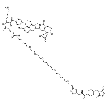
CL2A-SN-38
CL2A-SN-38은 강력한 DNA 토포이소머라제 I 억제제 SN-38과 링커 CL2A로 구성된 약물-링커 접합체로 항체 약물 접합체(ADC)를 만듭니다.
-
GC52469
CL2A-SN-38 (dichloroacetic acid salt)
An antibody-drug conjugate containing SN-38

-
GC10509
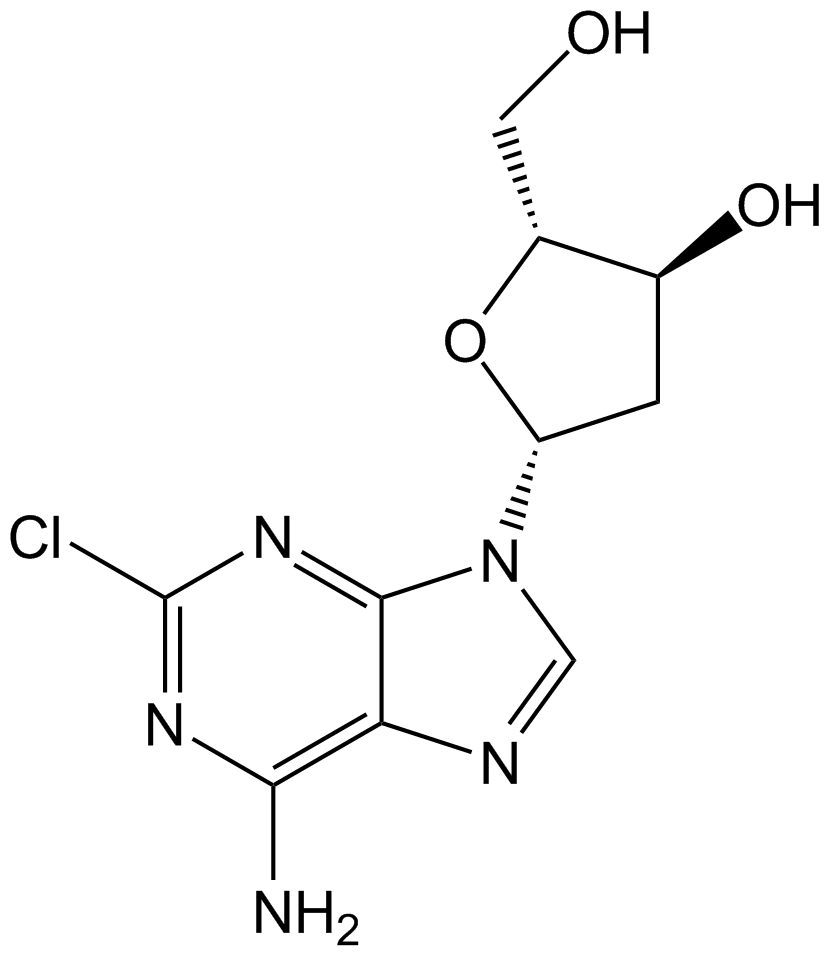
Cladribine
퓨린 뉴클레오사이드 유사체인 클라드리빈(2-클로로-2'-데옥시아데노신)은 경구 활성 아데노신 디아미나제 억제제입니다.
-
GC60111
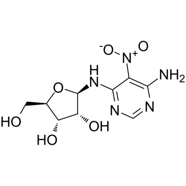
Clitocine
버섯에서 분리된 아데노신 뉴클레오사이드 유사체인 클리토신은 강력하고 효과적인 해독제입니다. 클리토신은 넌센스 돌연변이의 억제인자 역할을 하며 p53 넌센스 돌연변이 대립유전자를 보유한 세포에서 p53 단백질의 생산을 유도할 수 있습니다. 클리토신은 Mcl-1을 표적으로 하여 다제내성 인간 암세포에서 세포자멸사를 유도할 수 있습니다. 항암 활성.
-
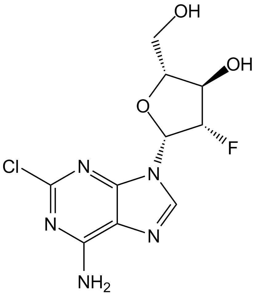
GC15219
Clofarabine
암 연구를 위한 뉴클레오시드 유사체인 클로파라빈은 조절 소단위의 알로스테릭 부위에 결합하여 리보뉴클레오티드 환원효소(IC50=65nM)의 강력한 억제제입니다.
-
GC10813
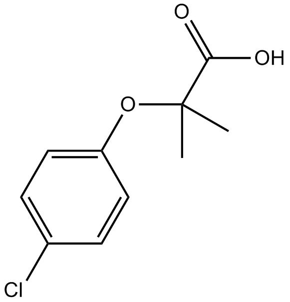
Clofibric Acid
지질 조절제인 Clofibrate, Etofibrate 및 Etofyllinclofibrate의 약학적 활성 대사물질인 Clofibric acid(Chlorofibrinic acid)는 PPARα 작용제로서 저지방혈증 효과를 나타냅니다.
-
GC32587
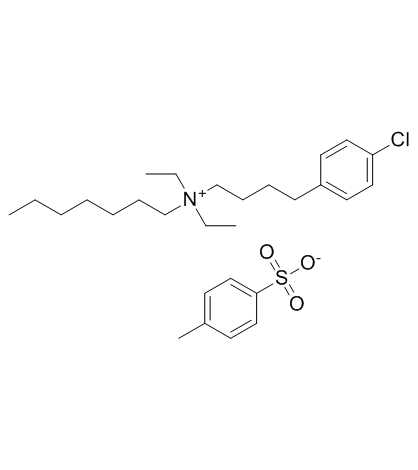
Clofilium tosylate
칼륨 채널 차단제인 클로필리움 토실레이트는 Bcl-2-비민감성 카스파제-3 활성화를 통해 인간 전골수구성 백혈병(HL-60) 세포의 세포자멸사를 유도합니다.
-
GC47105
Clonostachydiol
Clonostachydiol은 Clonostachys cylindrospora(균주 FH-A 6607) 곰팡이의 구충성 거대 디올라이드입니다.

-
GC12367
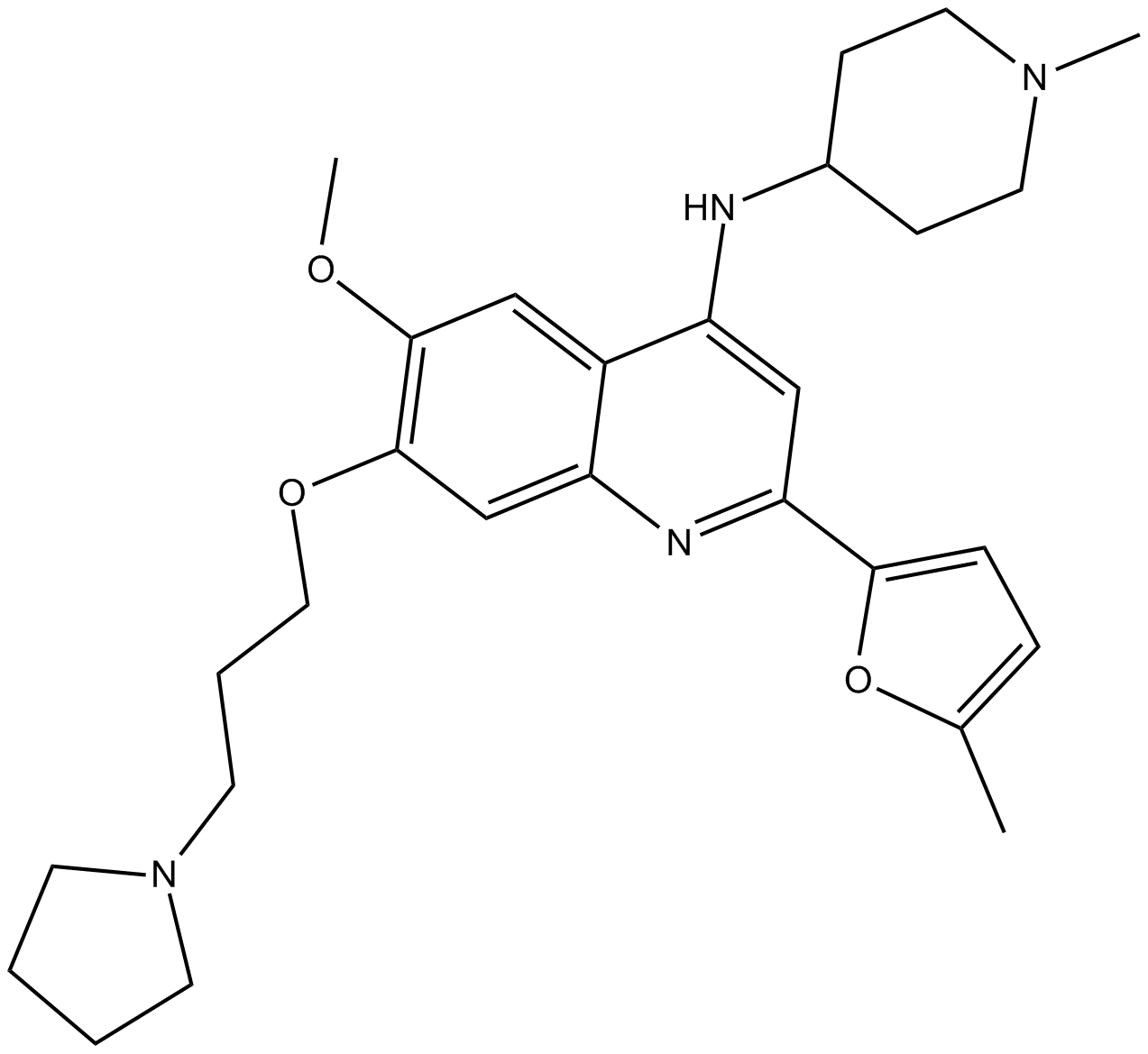
CM-272
CM-272는 항종양 활성을 가진 동급 최초의 강력하고 선택적인 기질 경쟁적이고 가역적인 이중 G9a/DNA 메틸트랜스퍼라제(DNMT) 억제제입니다. CM-272는 각각 8nM, 382nM, 85nM, 1200nM 및 2nM의 IC50으로 G9a, DNMT1, DNMT3A, DNMT3B 및 GLP를 억제합니다. CM-272는 세포 증식을 억제하고 세포 사멸을 촉진하여 IFN-자극 유전자 및 면역원성 세포 사멸을 유도합니다.
-
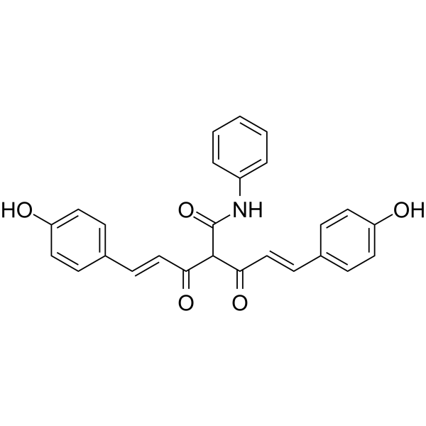
GC62347
CMC2.24
경구 활성 트리카르보닐메탄 제제인 CMC2.24(TRB-N0224)는 Ras 활성화 및 이의 다운스트림 이펙터 ERK1/2 경로를 억제함으로써 마우스의 췌장 종양에 효과적입니다.
-
GC61567
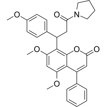
CMLD-2
HuR-ARE 상호작용의 억제제인 CMLD-2는 HuR 단백질에 경쟁적으로 결합하여 아데닌-우리딘 풍부 요소(ARE) 함유 mRNA(Ki=350 nM)와의 상호작용을 방해합니다. CMLD-2는 결장암, 췌장암, 갑상선암 및 폐암 세포주와 같은 다양한 암세포에서 세포자멸사를 유도하여 항종양 활성을 나타냅니다. Hu 항원 R(HuR)은 RNA 결합 단백질로서 표적 mRNA의 안정성 및 번역을 조절할 수 있습니다.
-
GC49096
Cobaltic Protoporphyrin IX (chloride)
An inducer of HO-1 activity

-
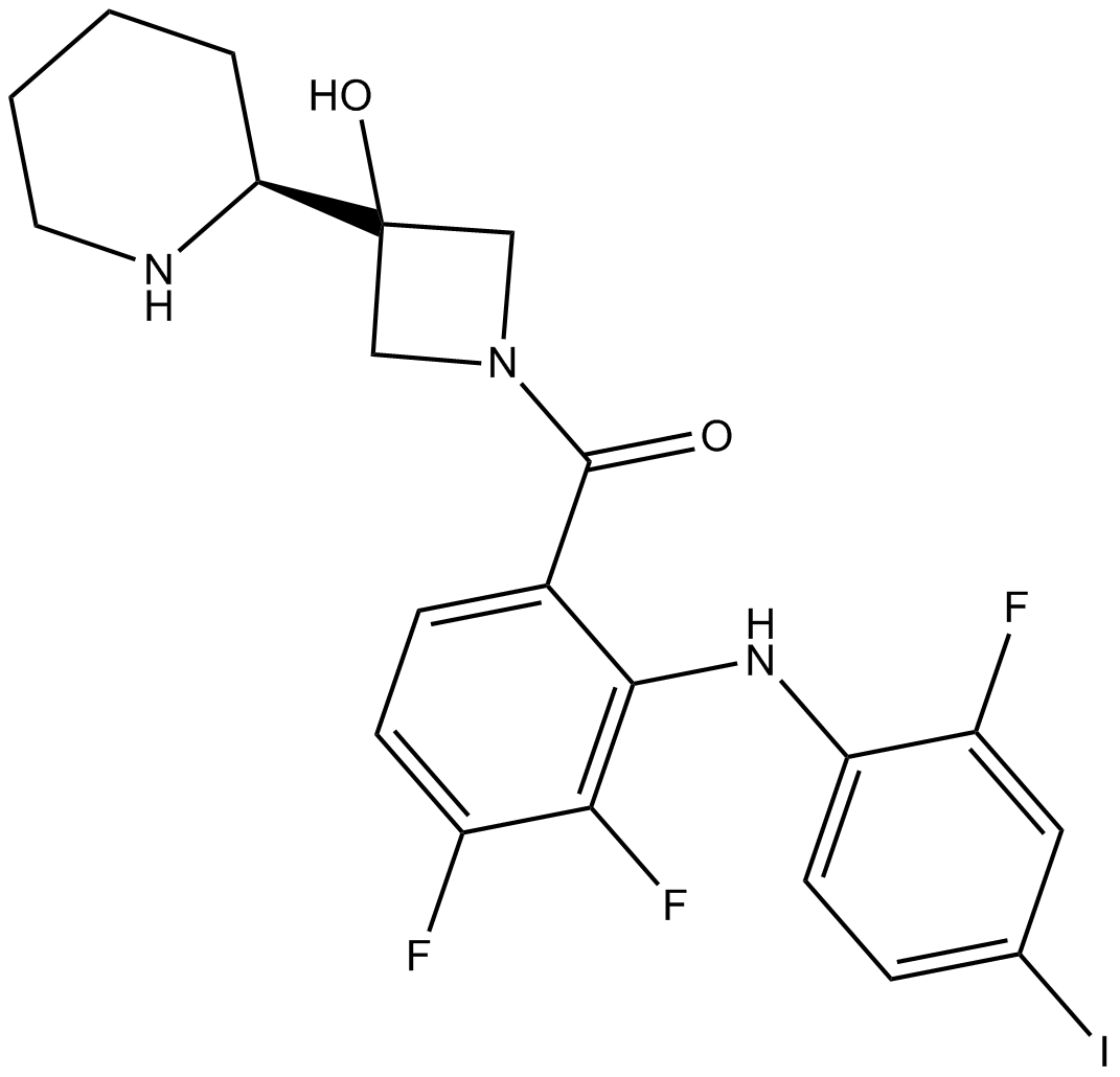
GC10033
Cobimetinib
A potent, orally available MEK1 inhibitor
-
GC43297
Coenzyme Q2
Coenzyme Q10 is a component of the electron transport chain and participates in aerobic cellular respiration, generating energy in the form of ATP.

-
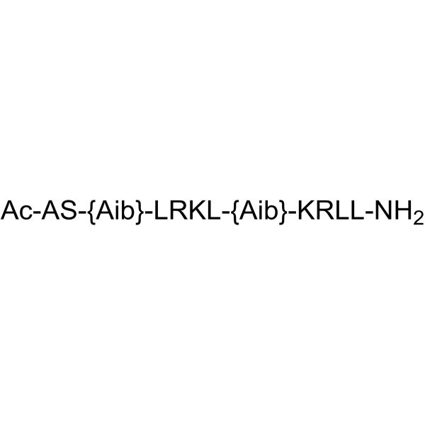
GC62192
COG1410
COG1410은 아포지단백 E 유래 펩타이드입니다.
-
GC40664
Colcemid
콜세미드는 포유류 세포나 난자에서 각각 G2/M 단계의 유사분열 정지 또는 소포 파열(GVBD) 단계의 준비 분열 정지를 유도하는 세포골격 억제제입니다.

-
GN10123
Columbianadin
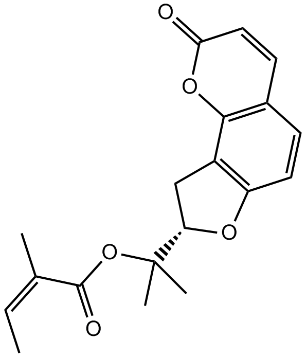
-
GC49454
Complex 3
A fluorescent copper complex with anticancer activity

-
GC18572
Concanavalin A
콘카나마이신 A는 Streptomyces diastatochromogenes에서 분리된 매크로라이드 항생물질인 콘카나마이신 가족에 속하며, 고도로 활성화되고 선택적으로 세포질 프로톤-ATPase (v-[H+]ATPase)를 억제하는 항생제입니다.

-
GC18832
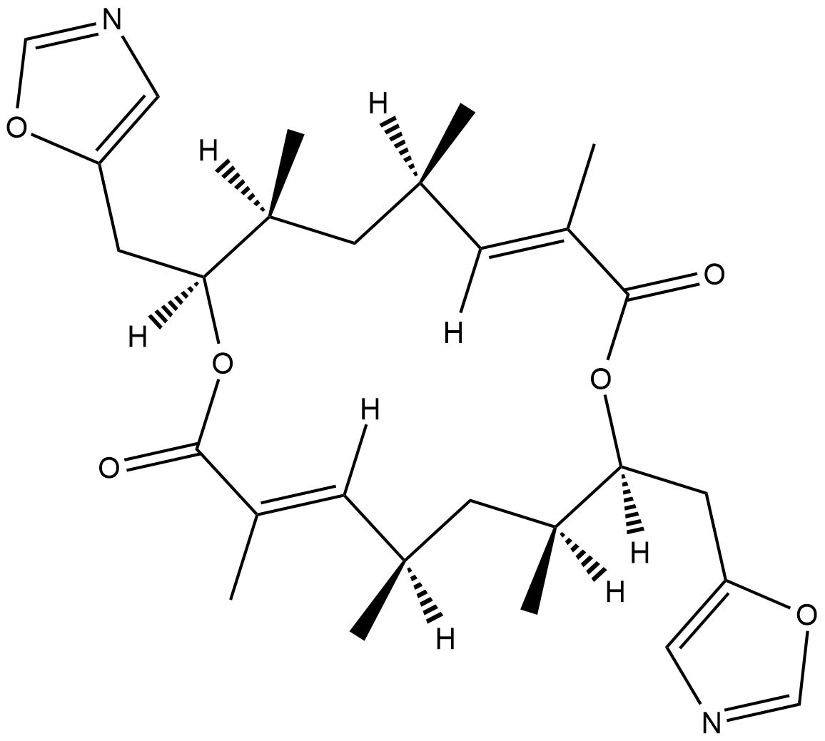
Conglobatin
Conglobatin is a dimeric macrolide dilactone originally isolated from S.
-
GC48483
Conglobatin B
A bacterial metabolite

-
GC48497
Conglobatin C1
A bacterial metabolite

-
GC38376
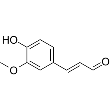
Coniferaldehyde
Coniferaldehyde(Ferulaldehyde)는 heme oxygenase-1(HO-1)의 효과적인 유도제입니다.
-
GC63379
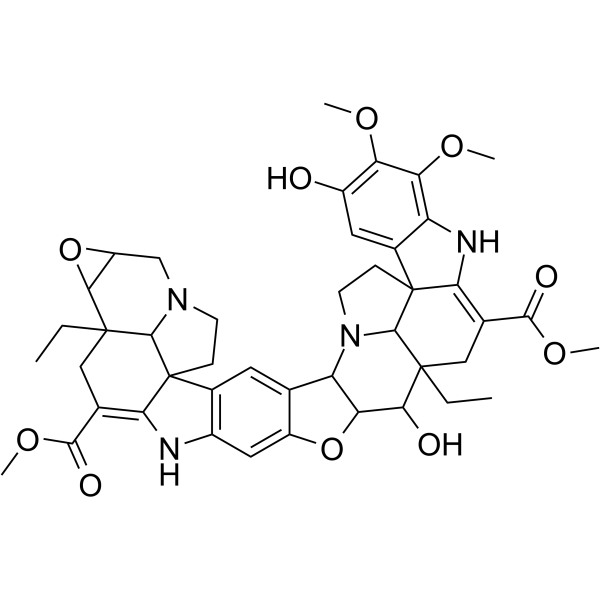
Conophylline
Conophylline은 열대 식물 Ervatamia microphylla의 잎에서 추출한 vinca alkaloid입니다.
-
GC16772
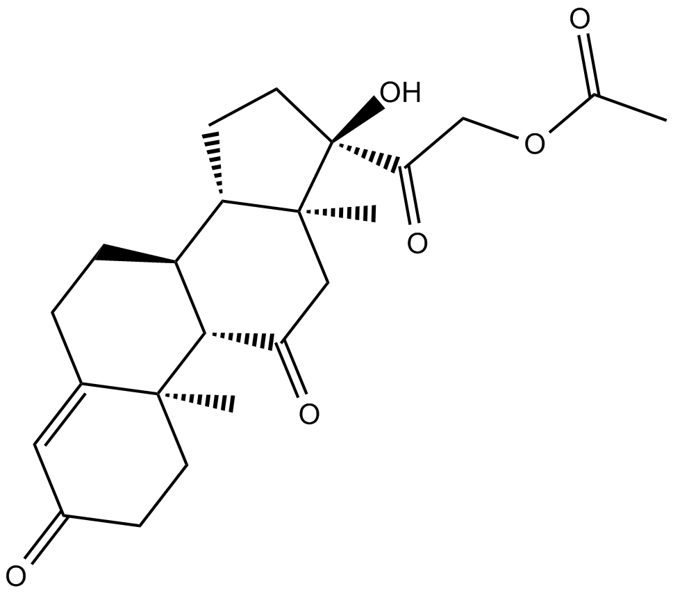
Cortisone acetate
코르티손 아세테이트(Cortisone 21-acetate), 코르티솔(글루코코르티코이드)의 산화된 대사 산물.
-
GC16116
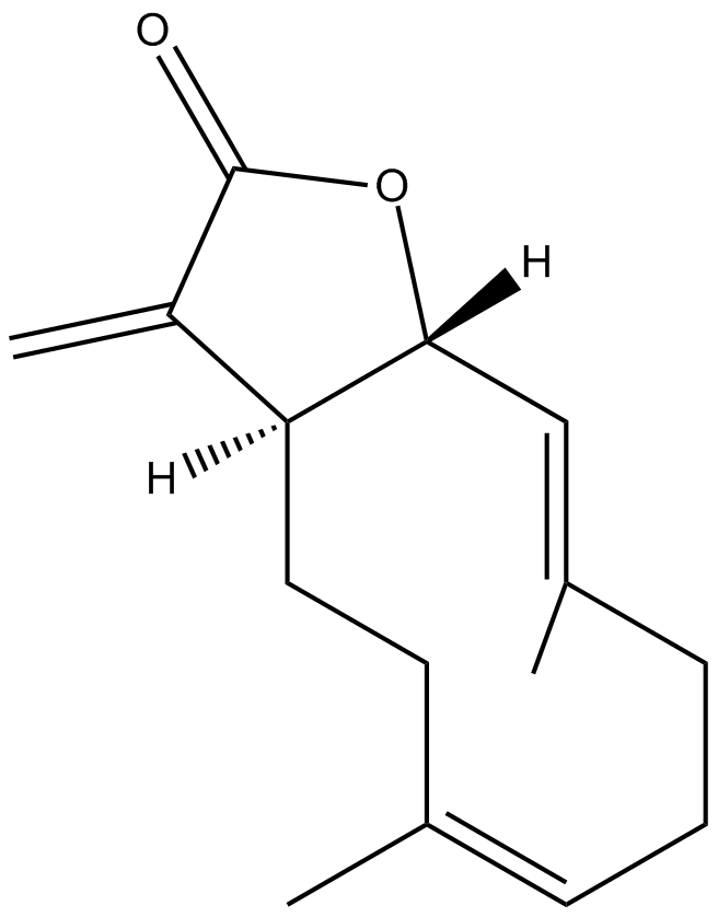
Costunolide
A natural sesquiterpene lactone
-
GC15225
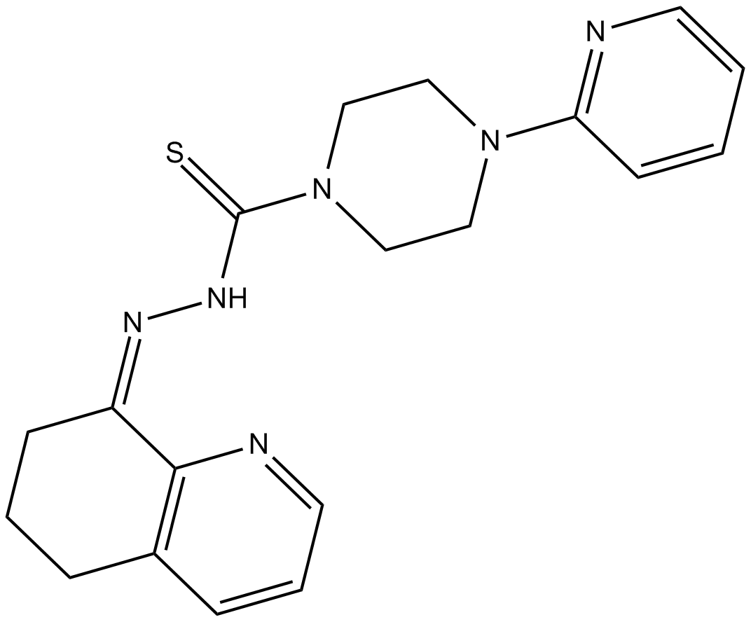
COTI-2
독성이 낮은 항암제 COTI-2는 p53 돌연변이 형태의 경구용 3세대 활성제이다. COTI-2는 돌연변이 p53을 재활성화하고 PI3K/AKT/mTOR 경로를 억제함으로써 작용합니다. COTI-2는 여러 인간 종양 세포주에서 세포자멸사를 유도합니다. COTI-2는 p53-의존 및 -독립 기전을 통해 HNSCC에서 항종양 활성을 나타낸다. COTI-2는 돌연변이 p53을 야생형 형태로 변환합니다.
-
GC15840
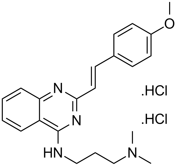
CP 31398 dihydrochloride
A p53 stabilizing agent
-
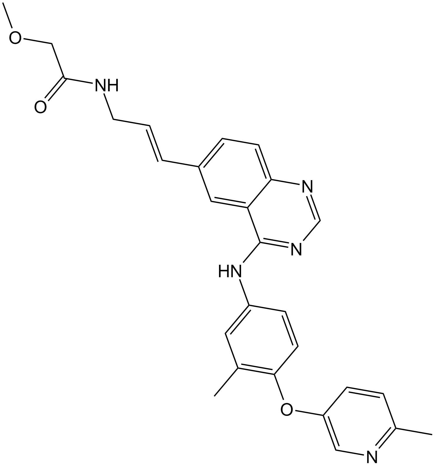
GC13091
CP-724714
CP-724714는 IC50이 10nM인 강력한 선택적 경구 활성 ErbB2(HER2) 티로신 키나제 억제제입니다. CP-724714는 EGFR 키나제에 대해 현저한 선택성을 나타냅니다(IC50=6400nM). CP-724714는 손상되지 않은 세포에서 ErbB2 수용체 자가인산화를 강력하게 억제합니다. 항종양 활동.
-
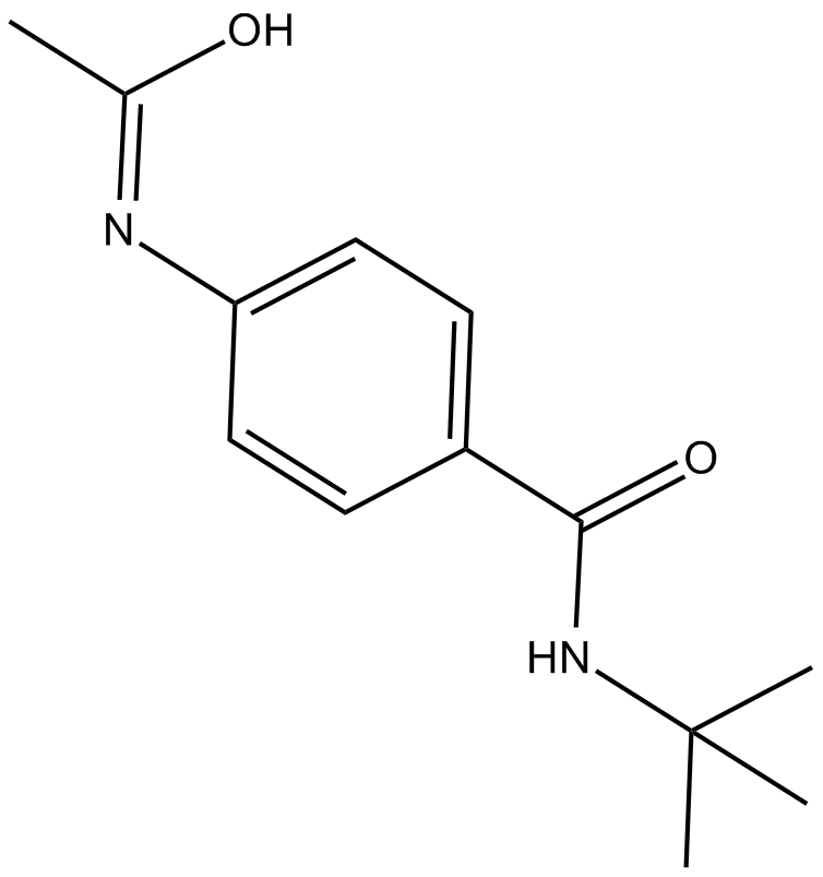
GC14500
CPI-1189
CPI-1189는 항산화 및 신경 보호 특성을 가진 TNF-α 방출 억제제입니다.
-
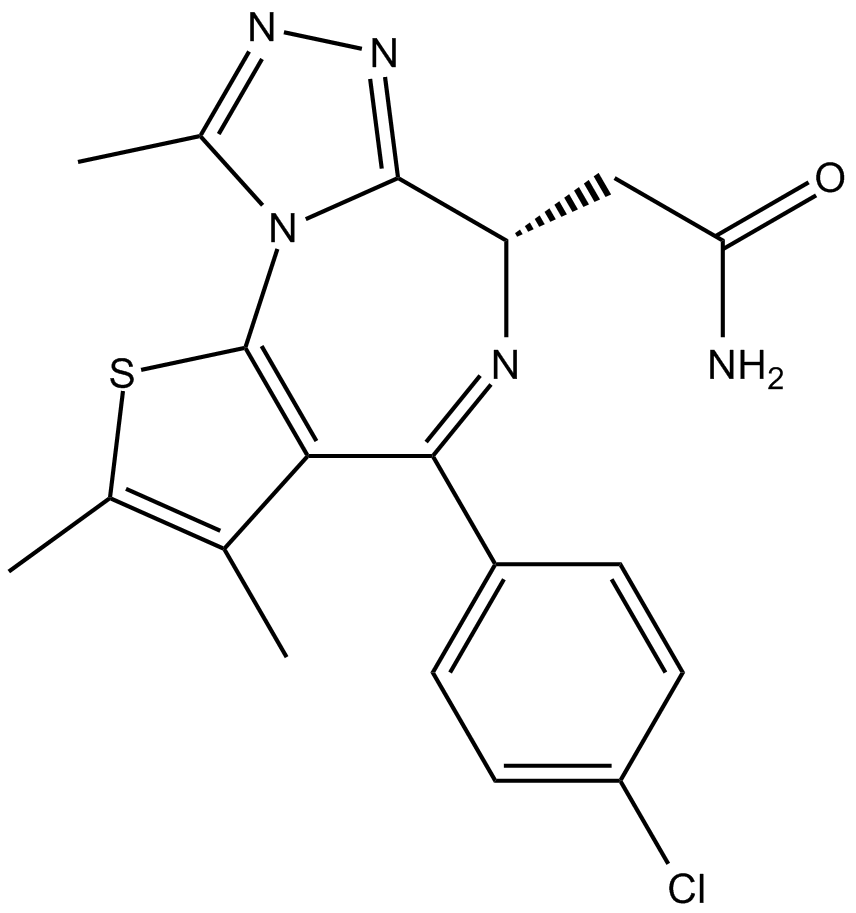
GC14699
CPI-203
CPI-203은 IC50 값이 약 37nM(BRD4 α-스크린 분석)인 BET 브로모도메인의 새롭고 강력한 선택적 세포 투과성 억제제입니다.
-
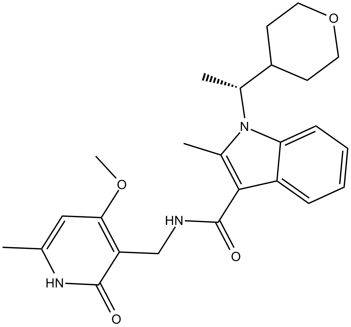
GC10021
CPI-360
EZH2 inhibitor
-
GC14921
CPI-613
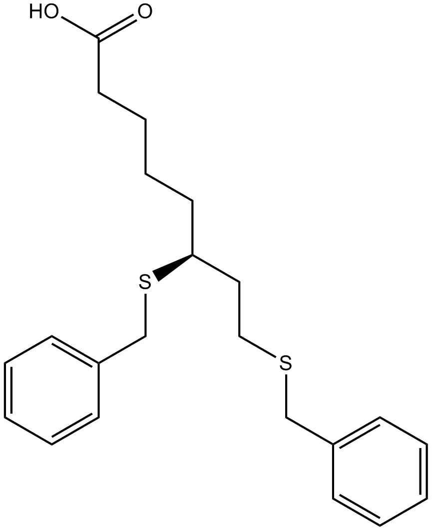
An inhibitor of α-ketoglutarate dehydrogenase
-
GC39365
CPTH2
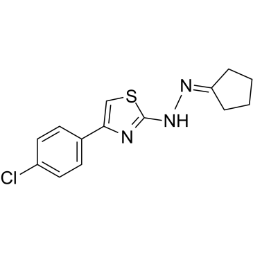
CPTH2는 강력한 히스톤 아세틸트랜스퍼라제(HAT) 억제제입니다. CPTH2는 Gcn5에 의한 히스톤 H3의 아세틸화를 선택적으로 억제합니다. CPTH2는 아폽토시스를 유도하고 아세틸트랜스퍼라제 p300(KAT3B)의 억제를 통해 투명 세포 신암(ccRCC) 세포주의 침습성을 감소시킵니다.
-
GC35747
Crebanine
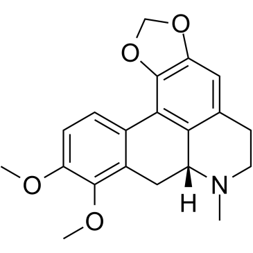
Stephania venosa의 알칼로이드인 Crebanine은 인간 암세포에서 G1 정지 및 세포자멸사를 유도합니다.
-
GC34543
cRIPGBM
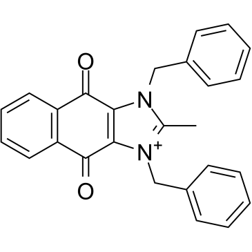
cRIPGBM은 수용체-상호작용 단백질 키나제 2(RIPK2)에 결합하여 GBM 암 줄기 세포(CSC)에서 세포 유형 선택적 세포자멸사 유도제인 RIPGBM의 세포자멸사 유도 유도체이며 GBM-1 세포에서 EC50이 68nM입니다.
-
GC13838
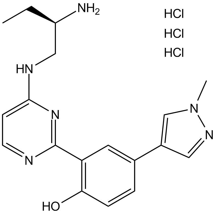
CRT 0066101
PKD inhibitor
-
GC35750
CRT0066101 dihydrochloride
CRT0066101 디히드로클로라이드는 PKD1, 2 및 3에 대해 각각 IC50 값이 1, 2.5 및 2nM인 강력하고 특이적인 PKD 억제제입니다.
-
GC45414
CRT0066854
CRT0066854는 강력하고 선택적인 비정형 PKC 동종효소 억제제입니다.

-
GC14355
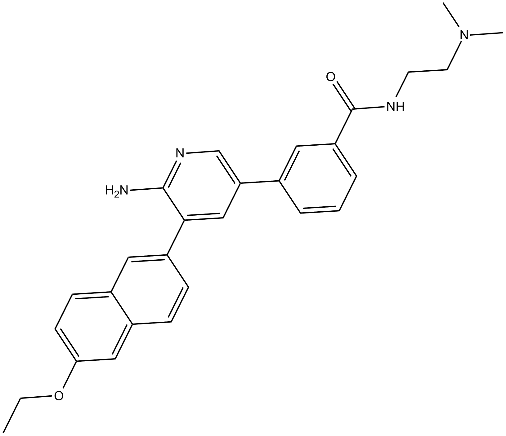
CRT5
피라진 벤즈아미드인 CRT5는 VEGF로 처리된 내피 세포에서 PKD의 세 가지 동형 모두에 대한 강력하고 선택적 억제제입니다(각각 PKD1, PKD2 및 PKD3에 대해 IC50s = 1, 2 및 1.5 nM).
-
GC32911
CTX1
CTX1은 HdmX 매개 p53 억제를 극복하는 p53 활성제입니다. CTX1은 마우스 급성 골수성 백혈병(AML) 모델 시스템에서 강력한 항암 활성을 나타냅니다.
-
GN10535
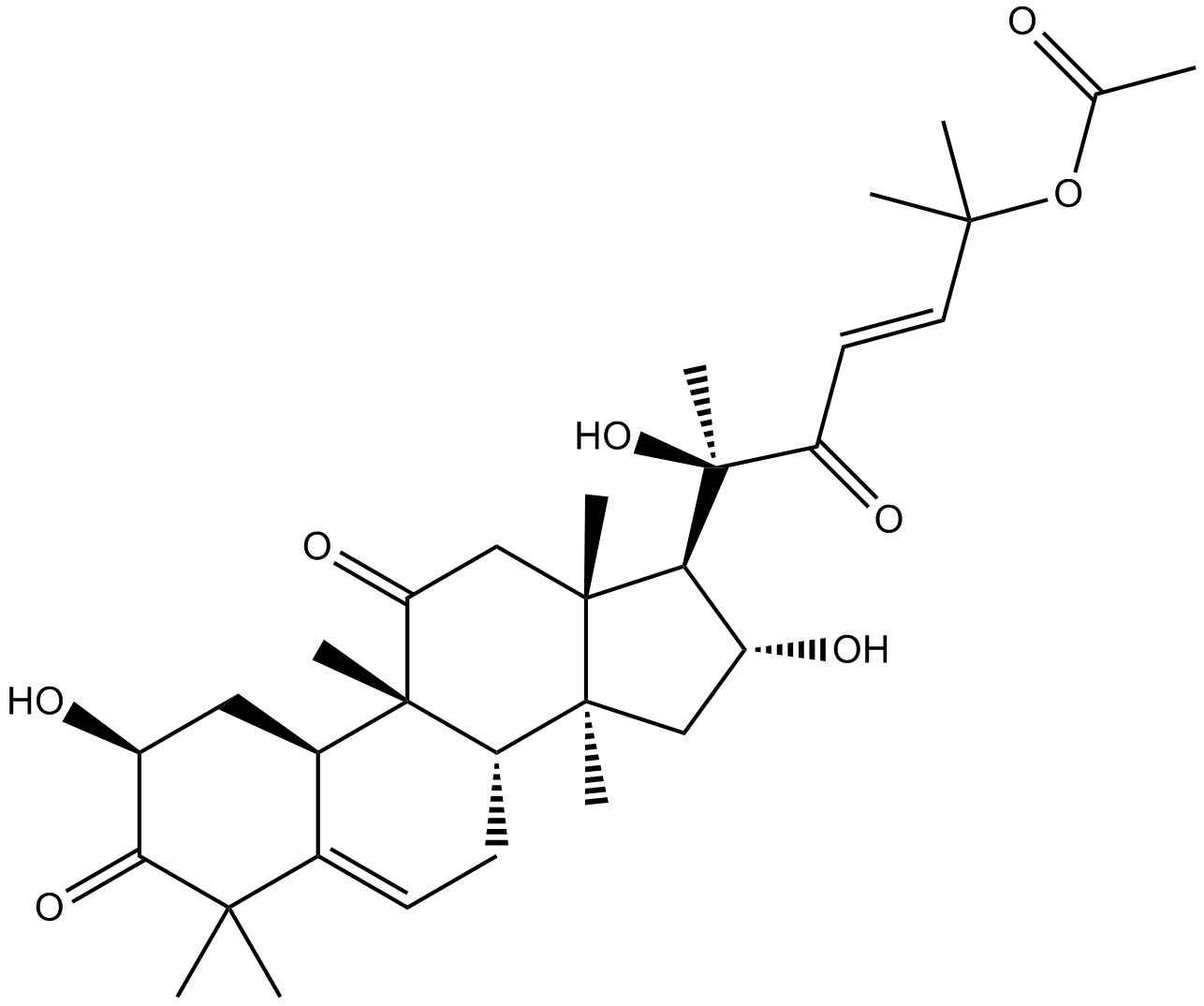
Cucurbitacin B
-
GC35758
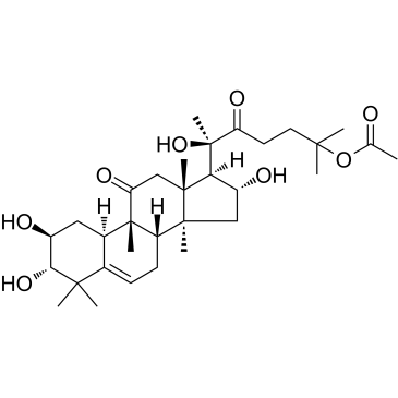
Cucurbitacin IIa
Cucurbitacin IIa는 Hemsleya amalils Diels에서 분리된 트리테르펜으로 암세포의 세포자살을 유도하고 서바이빈의 발현을 감소시키며 phospho-Histone H3를 감소시키고 암세포에서 절단된 PARP를 증가시킵니다.
-
GN10788
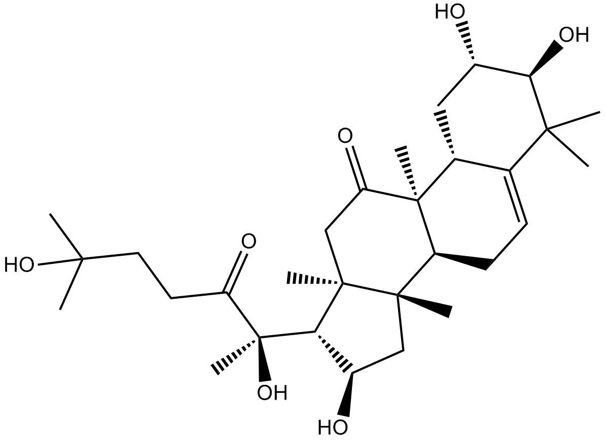
Cucurbitacin IIb
-
GC32781
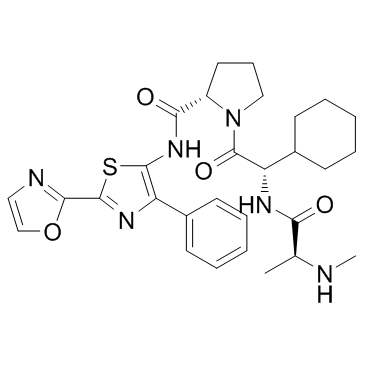
CUDC-427 (GDC-0917)
CUDC-427(GDC-0917)은 다양한 암 치료에 사용되는 강력한 2세대 범선택적 IAP 길항제입니다.
-
GC12115
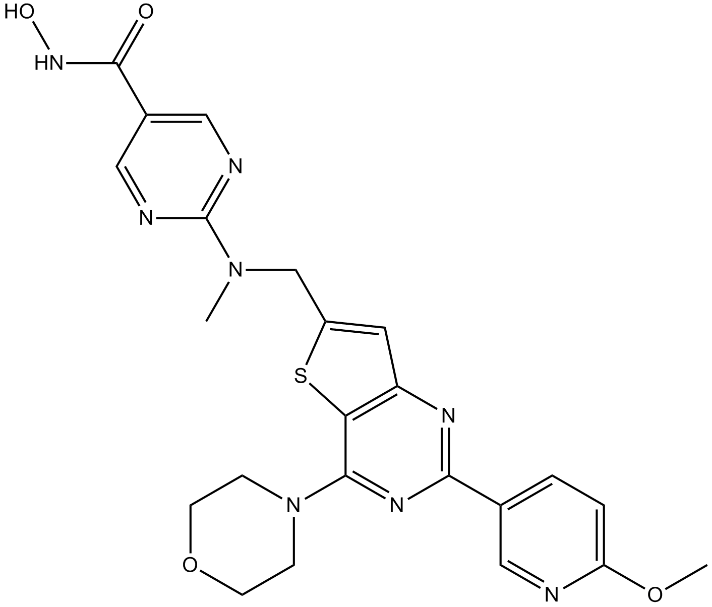
CUDC-907
A dual inhibitor of HDACs and PI3Ks
-
GC11217
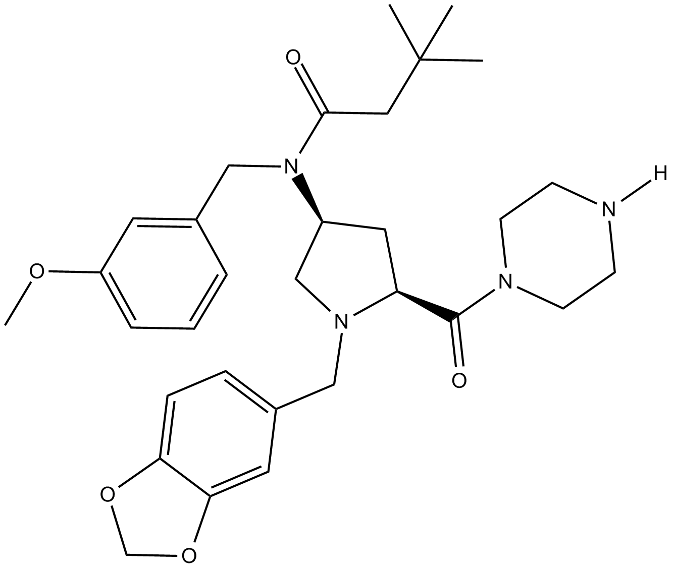
CUR 61414
CUR 61414는 새롭고 강력한 세포 투과성 고슴도치 신호 전달 경로 억제제(IC50 \u003d100-200nM)입니다.
-
GC14787
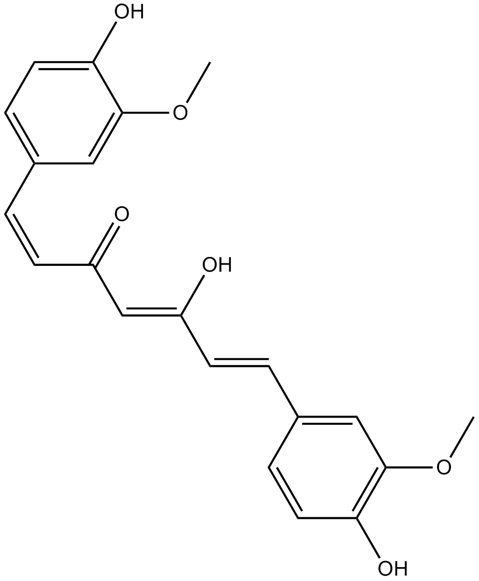
Curcumin
다양한 생물학적 활동을 가진 노란색 색소
-
GC40226
Curcumin-d6
커큐민 D6(디페룰로일메탄 D6)은 커큐민(강황 황색)이라고 표시된 중수소입니다.

-
GN10521
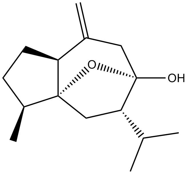
Curcumol
-
GC66356
Cusatuzumab
Cusatuzumab은 인간 αCD70 단클론 항체입니다. 821d96072c2d58d8970e76f526b0f6b8Cusatuzumab은 향상된 항체 의존성 세포로 세포 독성 활성을 나타냅니다. 821d96072c2d58d8970e76f526b0f6b8Cusatuzumab은 백혈병 줄기 세포(LSC)를 감소시키고 골수 분화 및 세포 사멸과 관련된 유전자 서명을 유발합니다. 821d96072c2d58d8970e76f526b0f6b8Cusatuzumab은 급성 골수성 백혈병(AML) 연구에 잠재력이 있습니다.821d96072c2d58d8970e76f526b0f6b8

-
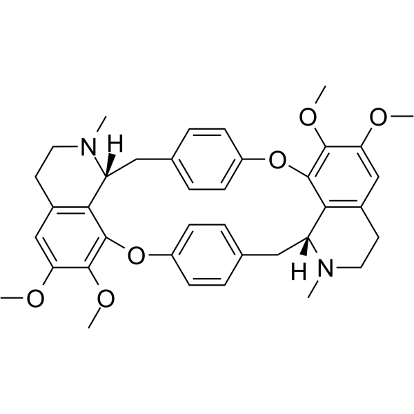
GC63967
Cycleanine
Cycleanine은 강력한 혈관 선택적 칼슘 길항제입니다.
-
GC49716
Cyclo(RGDyK) (trifluoroacetate salt)
A cyclic peptide ligand of αVβ3 integrin

-
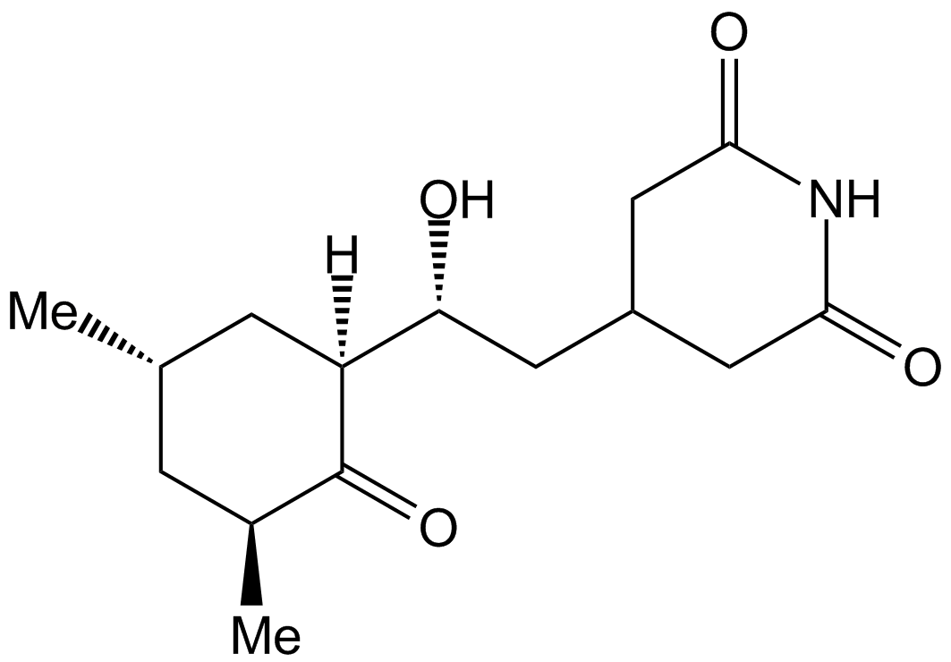
GC17198
Cycloheximide
Cycloheximide is an antibiotic that inhibits protein synthesis at the translation level, acting exclusively on cytoplasmic (80s) ribosomes of eukaryotes.
-
GC43346
Cyclopamine-KAAD
고슴도치 신호 억제제인 Cyclopamine-KAAD는 부드러운 길항제입니다.

-
GC47148
Cyclophosphamide-d4
Cyclophosphamide-d4는 Cyclophosphamide로 표시된 중수소입니다. 사이클로포스파미드는 면역억제제인 항종양 활성을 갖는 질소 머스타드와 화학적으로 관련된 합성 알킬화제입니다.

-
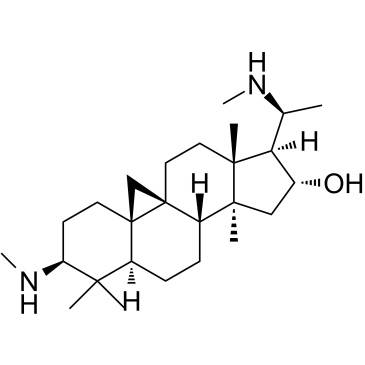
GC38419
Cyclovirobuxine D
Cyclovirobuxine D(CVB-D)는 중국 전통 의학 Buxus microphylla의 주요 활성 성분입니다.
-
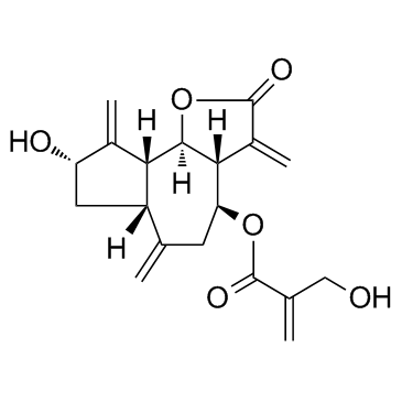
GC33330
Cynaropicrin
시나로피크린은 쥐와 인간 대식세포에 대해 각각 8.24 및 3.18μM의 IC50으로 종양 괴사 인자(TNF-α) 방출을 억제할 수 있는 세스퀴테르펜 락톤입니다. Cynaropicrin은 또한 연골 분해 인자(MMP13)의 증가를 억제하고 NF-κB 신호를 억제합니다.
-
GC65565
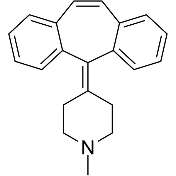
Cyproheptadine
Cyproheptadine은 강력한 경구 활성 5-HT2A 수용체 길항제로 항우울제 및 항 세로토닌 효과가 있습니다.
-
GC33779
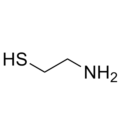
Cysteamine (β-Mercaptoethylamine)
-
GC13502
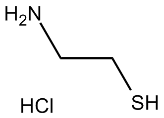
Cysteamine HCl
Cysteamine HCl(2-Aminoethanethiol hydrochloride)은 신증성 시스틴증 치료를 위한 경구 활성제이자 항산화제입니다.
-
GC17050
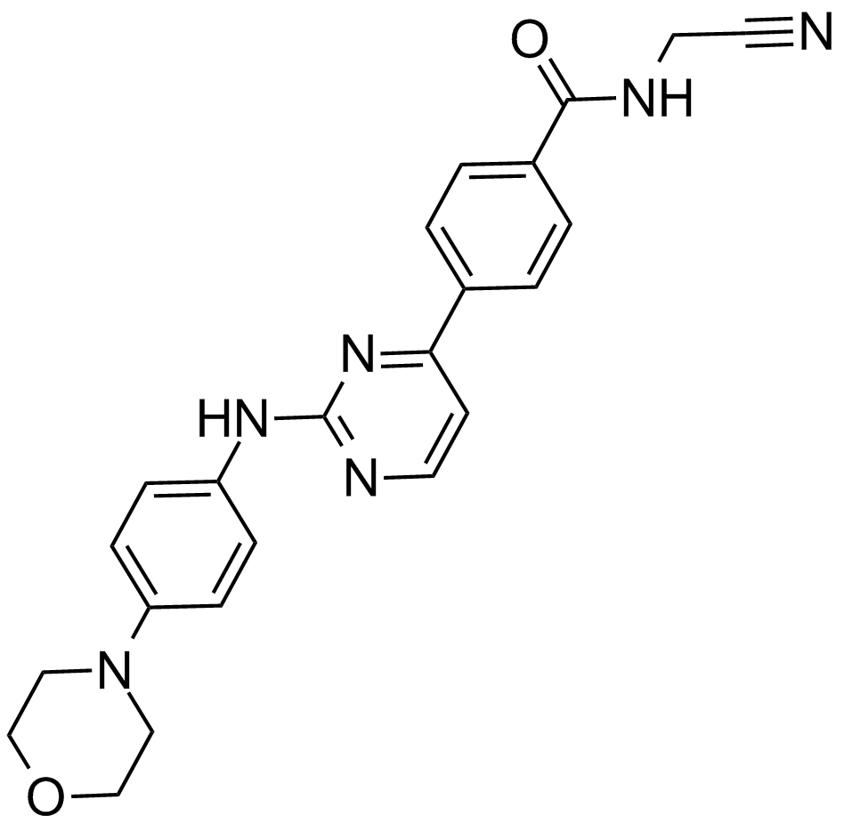
CYT387
A potent inhibitor of JAK1 and JAK2
-
GC11383
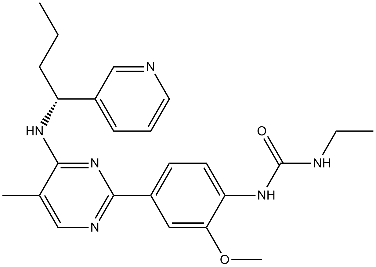
CYT997 (Lexibulin)
CYT997(Lexibulin)(CYT-997)은 암 세포주에서 IC50이 10-100nM인 강력한 경구 활성 튜불린 중합 억제제입니다. 시험관내 및 생체내에서 강력한 세포독성 및 혈관 교란 활성을 갖는다. CYT997(Lexibulin)은 세포 사멸을 유도하고 GC 세포에서 미토콘드리아 ROS 생성을 유도합니다.
-
GC13070
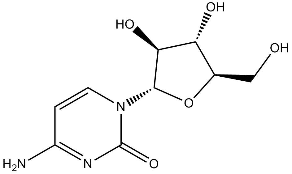
Cytarabine
세포독성제, DNA 합성 차단
-
GC43356
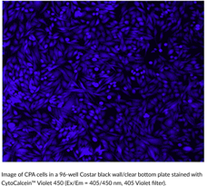
CytoCalcein™ Violet 450
CytoCalcein? Violet 450 is a fluorogenic dye used to assess cell viability.
-
GC43357
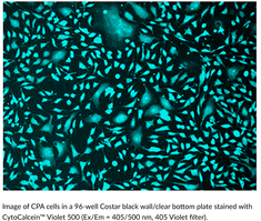
CytoCalcein™ Violet 500
CytoCalcein? Violet 500 is a fluorogenic dye used to assess cell viability.
-
GC43361
Cytostatin (sodium salt)
Cytostatin is a natural antitumor inhibitor of cell adhesion to extracellular matrix, blocking adhesion of B16 melanoma cells to laminin and collagen type IV in vitro (IC50s = 1.3 and 1.4 μg/ml, respectively) and B16 cells metastatic activity in mice.

-
GC43368
D,L-1′-Acetoxychavicol Acetate
D,L-1′-Acetoxychavicol acetate is a natural compound first isolated from the rhizomes of ginger-like plants.

-
GC66824
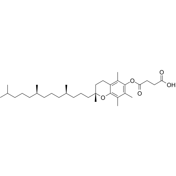
D-α-Tocopherol Succinate
D-α-토코페롤 석시네이트(비타민 E 석시네이트)는 항산화제인 토코페롤 및 비타민 E의 염 형태입니다. D-α-토코페롤 석시네이트는 시스플라틴 유도 세포 독성을 억제합니다. 821d96072c2d58d8970e76f526b0f6b8D-α-토코페롤 석시네이트는 암 연구에 사용할 수 있습니다.821d96072c2d58d8970e76f526b0f6b8
-
GC12256
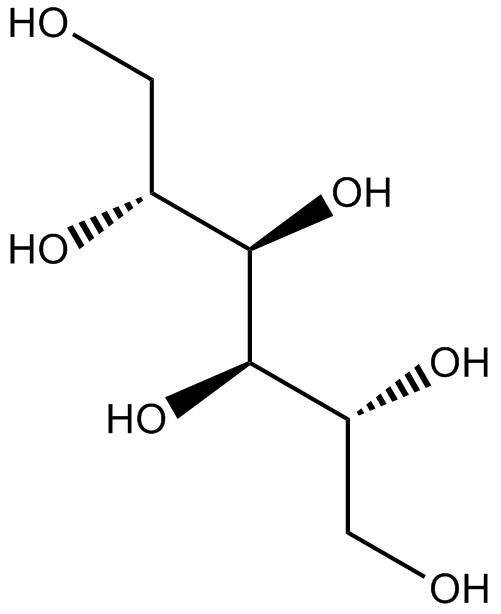
D-Mannitol
D-만니톨은 삼투성 이뇨제이자 약한 신장 혈관 확장제입니다.
-
GC60796
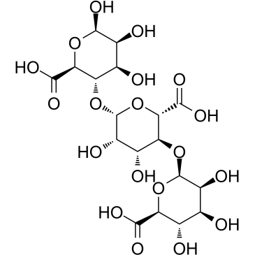
D-Trimannuronic acid
D-트리만누론산, 알지네이트 올리고머는 해조류에서 추출됩니다.
-
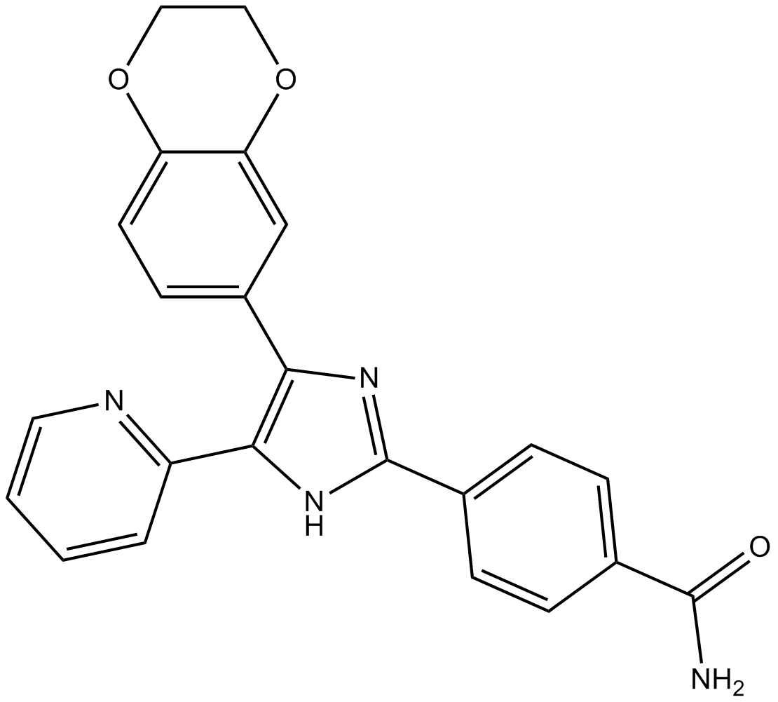
GC13202
D4476
Inhibitor of CK1 and ALK5
-
GC17851
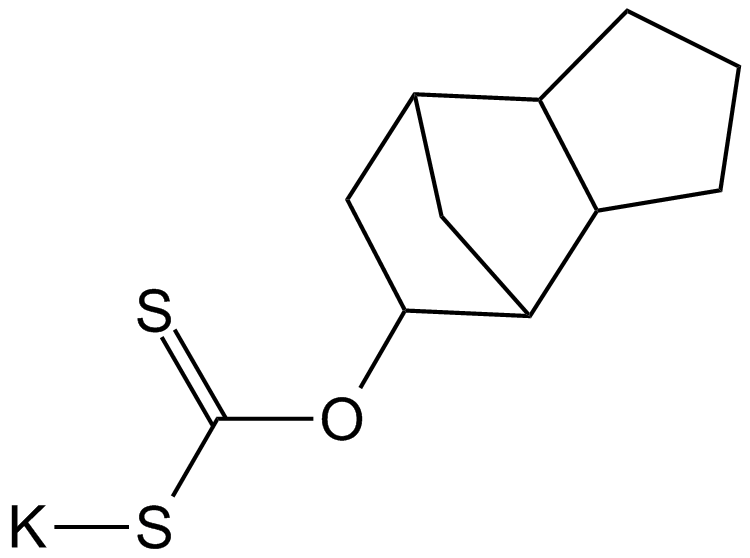
D609
A competitive inhibitor of PC-specific PLC
-
GC50296
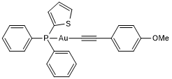
D9
D9는 2.8nM의 EC50을 갖는 티오레독신 환원효소(TrxR)의 강력하고 선택적인 억제제입니다. D9는 시험관내 및 생체내 모두에서 종양 증식을 억제하는 능력이 있습니다.
-
GC18421
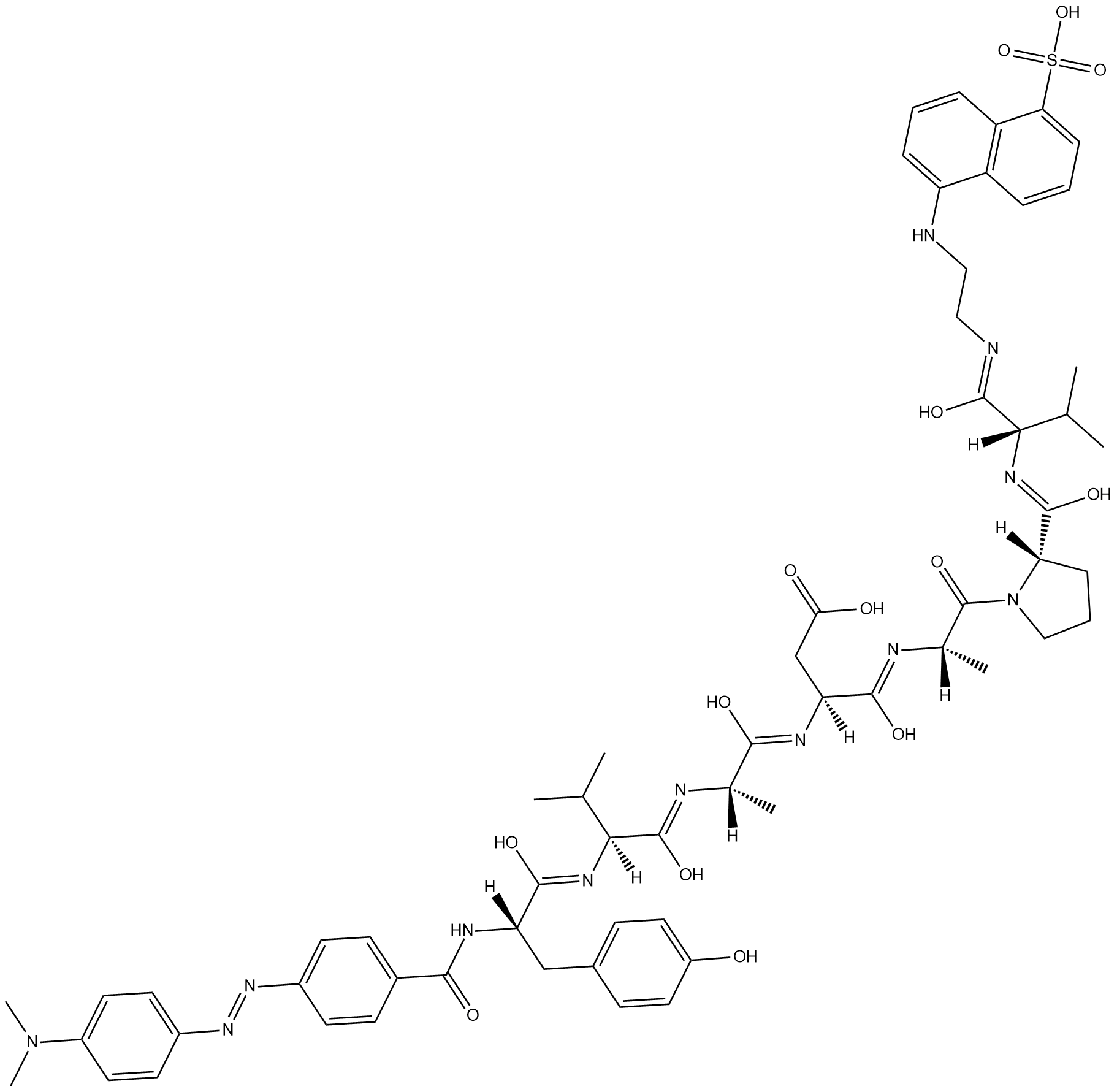
Dabcyl-YVADAPV-EDANS
Dabcyl-YVADAPV-EDANS is a fluorogenic substrate for caspase-1.
-
GC47166
Dabrafenib-d9
다브라페닙-d9(GSK2118436A-d9)는 다브라페닙으로 표지된 중수소입니다. 다브라페닙(GSK2118436A)은 C-Raf 및 B-RafV600E에 대해 IC50이 각각 5nM 및 0.6nM인 Raf의 ATP 경쟁적 억제제입니다.

-
GC14485
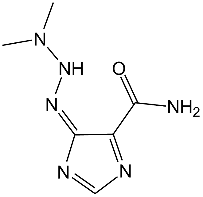
Dacarbazine
Dacarbazine은 세포주기 비특이적 항종양 알킬화제입니다. Dacarbazine은 각각 50 및 10μg/ml의 IC50 값으로 T 및 B 림프모구 반응을 억제합니다. Dacarbazine은 전이성 악성 흑색종의 연구에 사용할 수 있습니다.
-
GC47167
Dacarbazine-d6
Dacarbazine-d6(Imidazole Carboxamide-d6)은 Dacarbazine으로 표시된 중수소입니다. Dacarbazine(DTIC-Dome; DTIC)은 항종양제입니다. 흑색종에 대한 상당한 활성이 있습니다.

-
GC68305
Dacetuzumab

-
GC10225
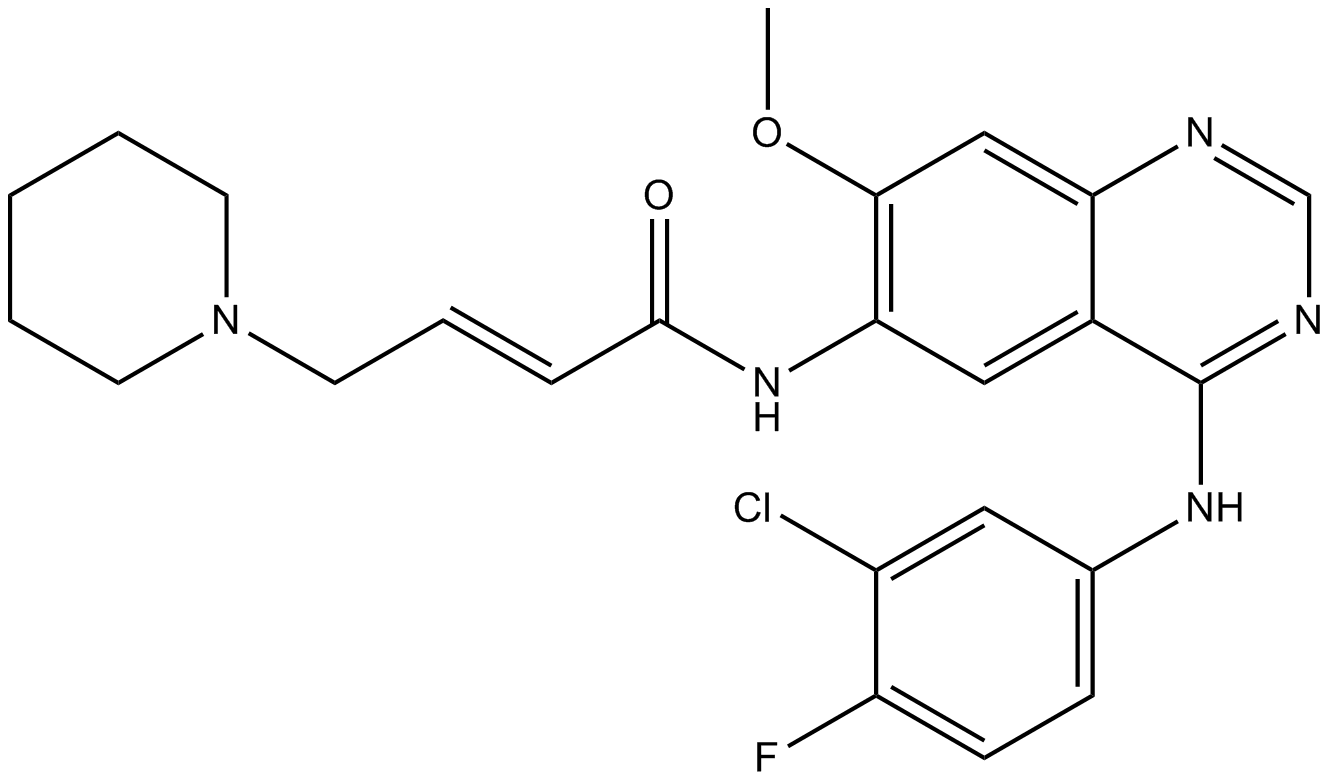
Dacomitinib (PF299804, PF299)
다코미티닙(PF299804, PF299)(PF-00299804)은 EGFR, ERBB2 및 ERBB4에 대해 각각 IC50이 6nM, 45.7nM 및 73.7nM인 키나제의 ERBB 계열에 대한 특이적이고 비가역적인 억제제입니다.
-
GC15211
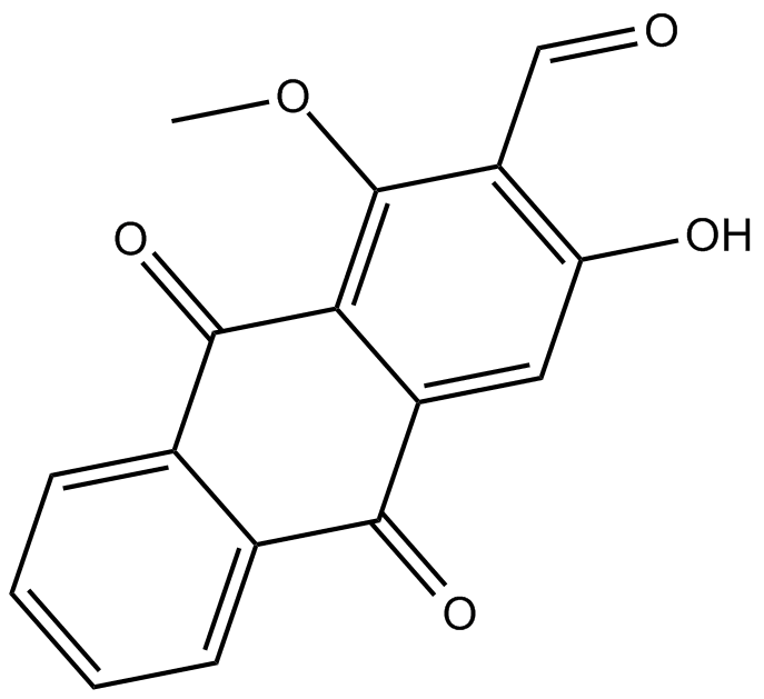
Damnacanthal
Damnacanthal은 Morinda citrifolia의 뿌리에서 분리된 안트라퀴논입니다.
-
GN10318
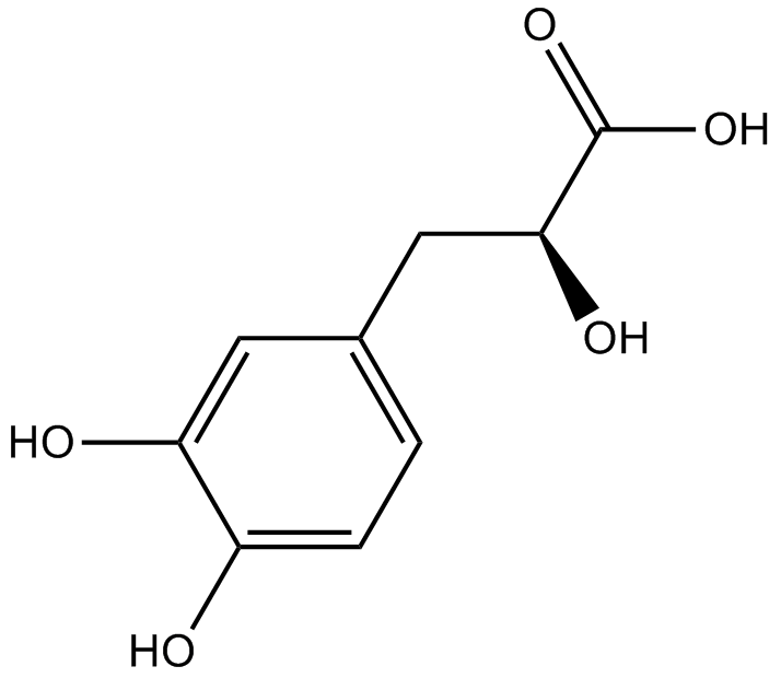
Danshensu
-
GC34010
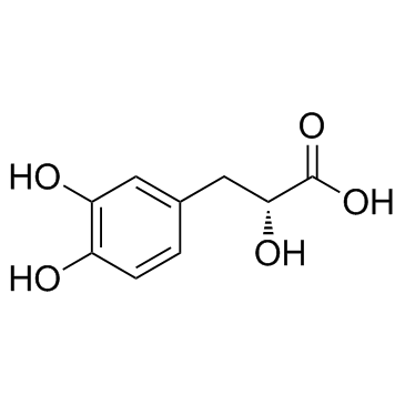
Danshensu (Dan shen suan A)
Danshensu, an active ingredient of Salvia miltiorrhiza, shows wide cardiovascular benefit by activating Nrf2 signaling pathway.
-
GC15217
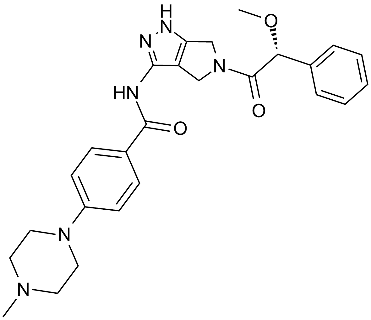
Danusertib (PHA-739358)
A pan-Aurora kinase and Abl inhibitor
-
GC17650
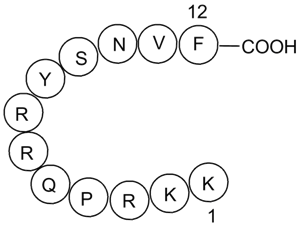
DAPK Substrate Peptide
A DAPK1 peptide substrate
-
GC49883
DAPK Substrate Peptide (trifluoroacetate salt)
A DAPK1 peptide substrate

-
GC12942
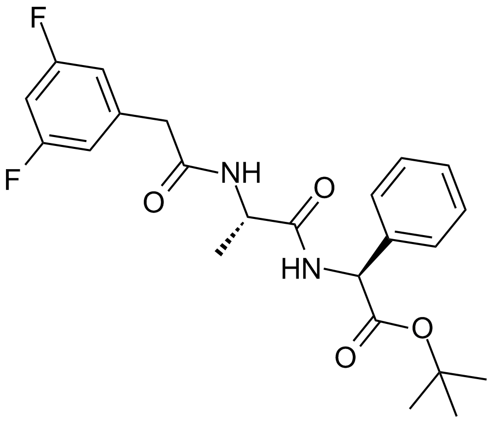
DAPT (GSI-IX)
γ-시크레타아제 억제제
-
GC43379
Darinaparsin
유기 비소인 다리나파르신(ZIO-101)은 미토콘드리아를 표적으로 하는 약제입니다. Darinaparsin은 암세포에서 apoptosis를 유도하고 항암효과가 있다.

-
GC15568
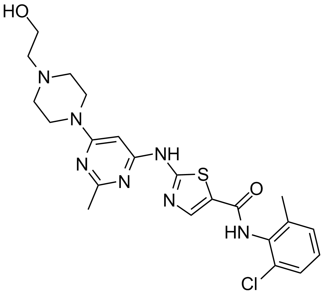
Dasatinib (BMS-354825)
Abl과 Src의 억제제
-
GC35812
Dasatinib hydrochloride
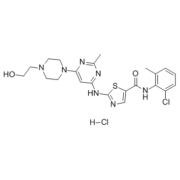
Dasatinib(BMS-354825) 염산염은 강력한 항종양 활성을 가진 매우 강력한 ATP 경쟁적 경구 활성 이중 Src/Bcr-Abl 억제제입니다. Kis는 Src 및 Bcr-Abl에 대해 각각 16pM 및 30pM입니다. Dasatinib 염산염은 각각 <1.0 nM 및 0.5 nM의 IC50으로 Bcr-Abl 및 Src를 억제합니다. Dasatinib 염산염은 또한 apoptosis와 autophagy를 유도합니다.
-
GC10354
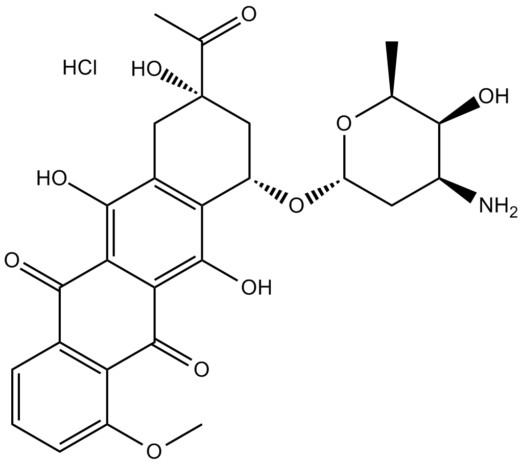
Daunorubicin HCl
Daunorubicin(Daunomycin) 염산염은 강력한 항종양 활성을 가진 토포이소머라제 II 억제제입니다.
