Apoptosis
As one of the cellular death mechanisms, apoptosis, also known as programmed cell death, can be defined as the process of a proper death of any cell under certain or necessary conditions. Apoptosis is controlled by the interactions between several molecules and responsible for the elimination of unwanted cells from the body.
Many biochemical events and a series of morphological changes occur at the early stage and increasingly continue till the end of apoptosis process. Morphological event cascade including cytoplasmic filament aggregation, nuclear condensation, cellular fragmentation, and plasma membrane blebbing finally results in the formation of apoptotic bodies. Several biochemical changes such as protein modifications/degradations, DNA and chromatin deteriorations, and synthesis of cell surface markers form morphological process during apoptosis.
Apoptosis can be stimulated by two different pathways: (1) intrinsic pathway (or mitochondria pathway) that mainly occurs via release of cytochrome c from the mitochondria and (2) extrinsic pathway when Fas death receptor is activated by a signal coming from the outside of the cell.
Different gene families such as caspases, inhibitor of apoptosis proteins, B cell lymphoma (Bcl)-2 family, tumor necrosis factor (TNF) receptor gene superfamily, or p53 gene are involved and/or collaborate in the process of apoptosis.
Caspase family comprises conserved cysteine aspartic-specific proteases, and members of caspase family are considerably crucial in the regulation of apoptosis. There are 14 different caspases in mammals, and they are basically classified as the initiators including caspase-2, -8, -9, and -10; and the effectors including caspase-3, -6, -7, and -14; and also the cytokine activators including caspase-1, -4, -5, -11, -12, and -13. In vertebrates, caspase-dependent apoptosis occurs through two main interconnected pathways which are intrinsic and extrinsic pathways. The intrinsic or mitochondrial apoptosis pathway can be activated through various cellular stresses that lead to cytochrome c release from the mitochondria and the formation of the apoptosome, comprised of APAF1, cytochrome c, ATP, and caspase-9, resulting in the activation of caspase-9. Active caspase-9 then initiates apoptosis by cleaving and thereby activating executioner caspases. The extrinsic apoptosis pathway is activated through the binding of a ligand to a death receptor, which in turn leads, with the help of the adapter proteins (FADD/TRADD), to recruitment, dimerization, and activation of caspase-8 (or 10). Active caspase-8 (or 10) then either initiates apoptosis directly by cleaving and thereby activating executioner caspase (-3, -6, -7), or activates the intrinsic apoptotic pathway through cleavage of BID to induce efficient cell death. In a heat shock-induced death, caspase-2 induces apoptosis via cleavage of Bid.
Bcl-2 family members are divided into three subfamilies including (i) pro-survival subfamily members (Bcl-2, Bcl-xl, Bcl-W, MCL1, and BFL1/A1), (ii) BH3-only subfamily members (Bad, Bim, Noxa, and Puma9), and (iii) pro-apoptotic mediator subfamily members (Bax and Bak). Following activation of the intrinsic pathway by cellular stress, pro‑apoptotic BCL‑2 homology 3 (BH3)‑only proteins inhibit the anti‑apoptotic proteins Bcl‑2, Bcl-xl, Bcl‑W and MCL1. The subsequent activation and oligomerization of the Bak and Bax result in mitochondrial outer membrane permeabilization (MOMP). This results in the release of cytochrome c and SMAC from the mitochondria. Cytochrome c forms a complex with caspase-9 and APAF1, which leads to the activation of caspase-9. Caspase-9 then activates caspase-3 and caspase-7, resulting in cell death. Inhibition of this process by anti‑apoptotic Bcl‑2 proteins occurs via sequestration of pro‑apoptotic proteins through binding to their BH3 motifs.
One of the most important ways of triggering apoptosis is mediated through death receptors (DRs), which are classified in TNF superfamily. There exist six DRs: DR1 (also called TNFR1); DR2 (also called Fas); DR3, to which VEGI binds; DR4 and DR5, to which TRAIL binds; and DR6, no ligand has yet been identified that binds to DR6. The induction of apoptosis by TNF ligands is initiated by binding to their specific DRs, such as TNFα/TNFR1, FasL /Fas (CD95, DR2), TRAIL (Apo2L)/DR4 (TRAIL-R1) or DR5 (TRAIL-R2). When TNF-α binds to TNFR1, it recruits a protein called TNFR-associated death domain (TRADD) through its death domain (DD). TRADD then recruits a protein called Fas-associated protein with death domain (FADD), which then sequentially activates caspase-8 and caspase-3, and thus apoptosis. Alternatively, TNF-α can activate mitochondria to sequentially release ROS, cytochrome c, and Bax, leading to activation of caspase-9 and caspase-3 and thus apoptosis. Some of the miRNAs can inhibit apoptosis by targeting the death-receptor pathway including miR-21, miR-24, and miR-200c.
p53 has the ability to activate intrinsic and extrinsic pathways of apoptosis by inducing transcription of several proteins like Puma, Bid, Bax, TRAIL-R2, and CD95.
Some inhibitors of apoptosis proteins (IAPs) can inhibit apoptosis indirectly (such as cIAP1/BIRC2, cIAP2/BIRC3) or inhibit caspase directly, such as XIAP/BIRC4 (inhibits caspase-3, -7, -9), and Bruce/BIRC6 (inhibits caspase-3, -6, -7, -8, -9).
Any alterations or abnormalities occurring in apoptotic processes contribute to development of human diseases and malignancies especially cancer.
References:
1.Yağmur Kiraz, Aysun Adan, Melis Kartal Yandim, et al. Major apoptotic mechanisms and genes involved in apoptosis[J]. Tumor Biology, 2016, 37(7):8471.
2.Aggarwal B B, Gupta S C, Kim J H. Historical perspectives on tumor necrosis factor and its superfamily: 25 years later, a golden journey.[J]. Blood, 2012, 119(3):651.
3.Ashkenazi A, Fairbrother W J, Leverson J D, et al. From basic apoptosis discoveries to advanced selective BCL-2 family inhibitors[J]. Nature Reviews Drug Discovery, 2017.
4.McIlwain D R, Berger T, Mak T W. Caspase functions in cell death and disease[J]. Cold Spring Harbor perspectives in biology, 2013, 5(4): a008656.
5.Ola M S, Nawaz M, Ahsan H. Role of Bcl-2 family proteins and caspases in the regulation of apoptosis[J]. Molecular and cellular biochemistry, 2011, 351(1-2): 41-58.
対象は Apoptosis
- Pyroptosis(15)
- Caspase(77)
- 14.3.3 Proteins(3)
- Apoptosis Inducers(71)
- Bax(15)
- Bcl-2 Family(136)
- Bcl-xL(13)
- c-RET(15)
- IAP(32)
- KEAP1-Nrf2(73)
- MDM2(21)
- p53(137)
- PC-PLC(6)
- PKD(8)
- RasGAP (Ras- P21)(2)
- Survivin(8)
- Thymidylate Synthase(12)
- TNF-α(141)
- Other Apoptosis(1145)
- Apoptosis Detection(0)
- Caspase Substrate(0)
- APC(6)
- PD-1/PD-L1 interaction(60)
- ASK1(4)
- PAR4(2)
- RIP kinase(47)
- FKBP(22)
製品は Apoptosis
- Cat.No. 商品名 インフォメーション
-
GC43274
Citromycetin
シトロミセチンは、オーストラリアの海洋由来および陸生のペニシリウム種に由来する芳香族ポリケチド化合物です 。

-
GC63393
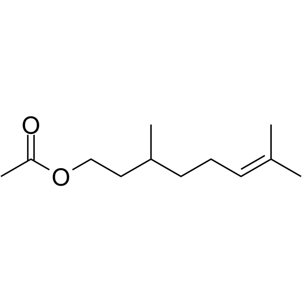
Citronellyl acetate
酢酸シトロネリルは、植物の二次代謝のモノテルペン生成物であり、抗侵害受容活性があります。
-
GC52367
Citrullinated Vimentin (G146R) (R144 + R146) (139-159)-biotin Peptide
バイオチニル化されたシトルリン化変異型ビメンチンペプチド

-
GC52370
Citrullinated Vimentin (R144) (139-159)-biotin Peptide
バイオチニル化されたシトルリン化ビメンチンペプチド

-
GN10219
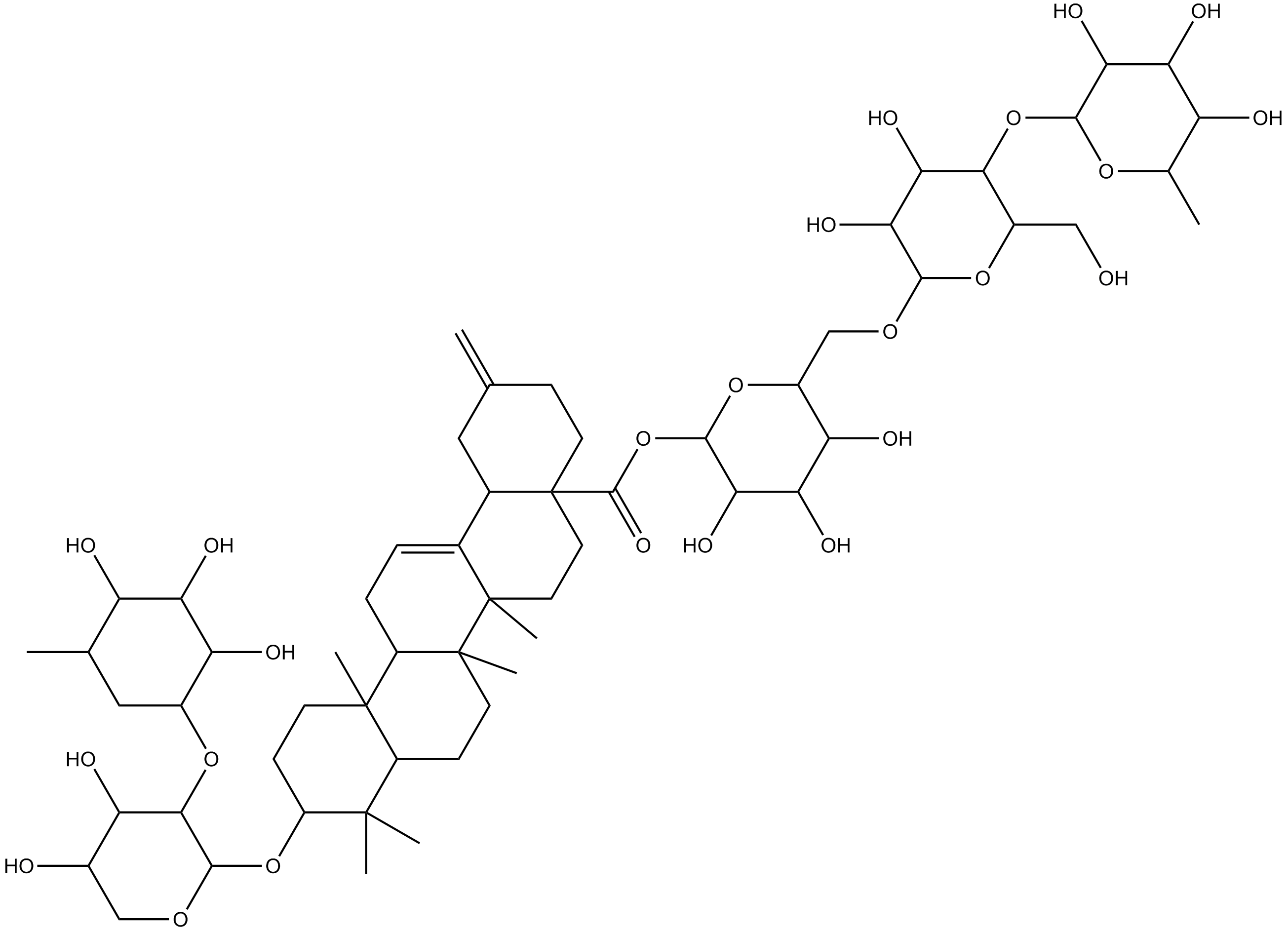
Ciwujianoside-B
-
GC64649
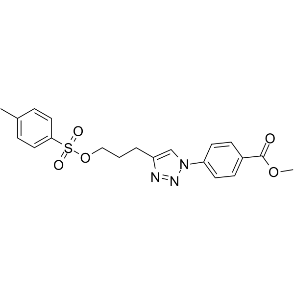
Cjoc42
Cjoc42はガンキリンに結合する化合物です。 Cjoc42 は用量依存的にガンキリン活性を阻害します。 Cjoc42 は、通常大量のガンキリンに関連する p53 タンパク質レベルの低下を防ぎます。 Cjoc42 は、p53 依存性の転写と DNA 損傷に対する感受性を回復します。
-
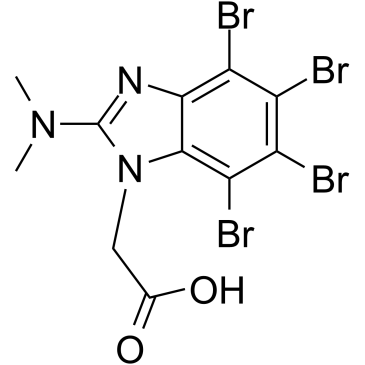
GC39485
CK2/ERK8-IN-1
CK2/ERK8-IN-1 は、デュアル カゼイン キナーゼ 2 (CK2) (Ki 0.25 μM) および ERK8 (MAPK15、ERK7) 阻害剤であり、IC50 は 0.50 μM です。 CK2/ERK8-IN-1 は、PIM1、HIPK2 (ホメオドメイン相互作用タンパク質キナーゼ 2)、DYRK1A にもそれぞれ 8.65 μM、15.25 μM、11.9 μM の Kis で結合します。 CK2/ERK8-IN-1 にはアポトーシス促進効果があります。
-
GC49556
Cl-Necrostatin-1
RIPK1阻害剤

-
GC47098
CL2-SN-38 (dichloroacetic acid salt)
SN-38を含む抗体薬物複合体

-
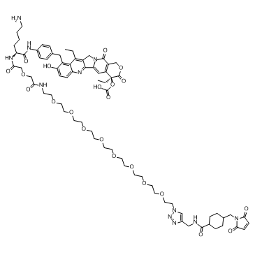
GC60714
CL2A-SN-38
CL2A-SN-38 は、強力な DNA トポイソメラーゼ I 阻害剤である SN-38 と、抗体薬物複合体 (ADC) を作成するためのリンカー CL2A で構成される薬物-リンカー複合体です。
-
GC52469
CL2A-SN-38 (dichloroacetic acid salt)
SN-38を含む抗体薬物複合体

-
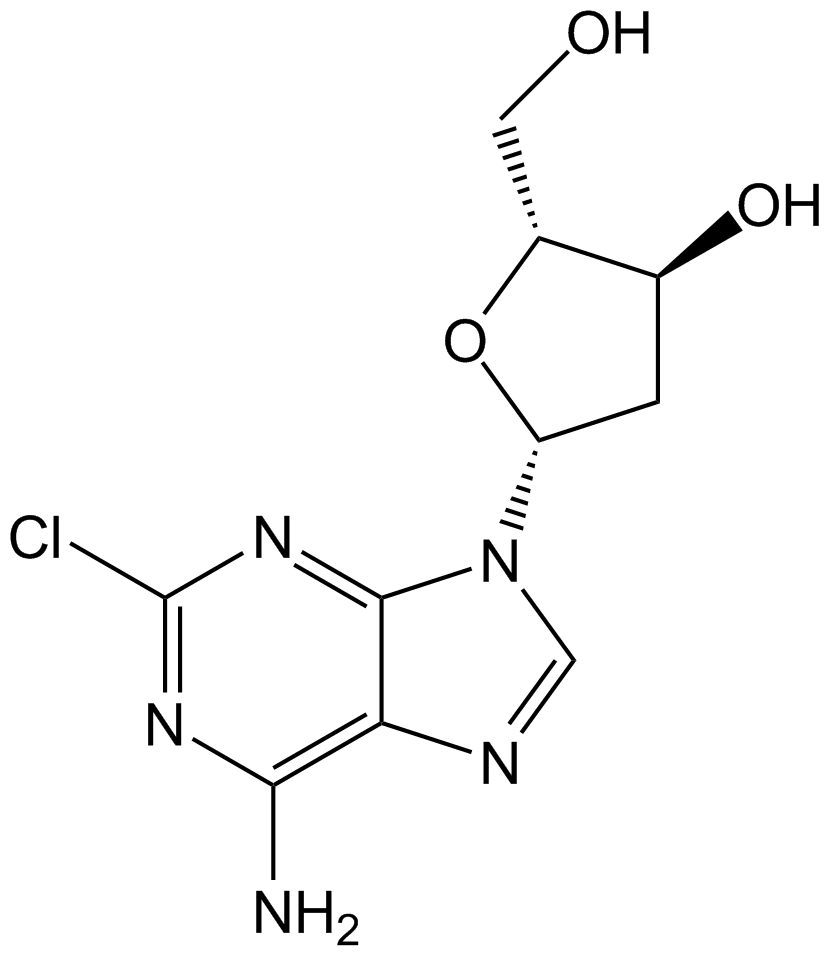
GC10509
Cladribine
プリンヌクレオシド類似体であるクラドリビン(2-クロロ-2'-デオキシアデノシン)は、経口活性アデノシンデアミナーゼ阻害剤です。
-
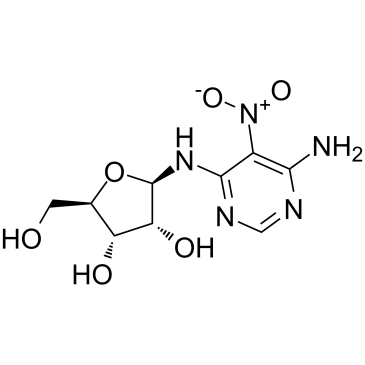
GC60111
Clitocine
キノコから分離されたアデノシン ヌクレオシド アナログであるクリトシンは、強力で効果的なリードスルー エージェントです。クリトシンはナンセンス変異の抑制因子として作用し、p53 ナンセンス変異対立遺伝子を保有する細胞で p53 タンパク質の産生を誘導することができます。クリトシンは、Mcl-1 を標的とすることにより、多剤耐性ヒト癌細胞のアポトーシスを誘導できます。抗がん作用。
-
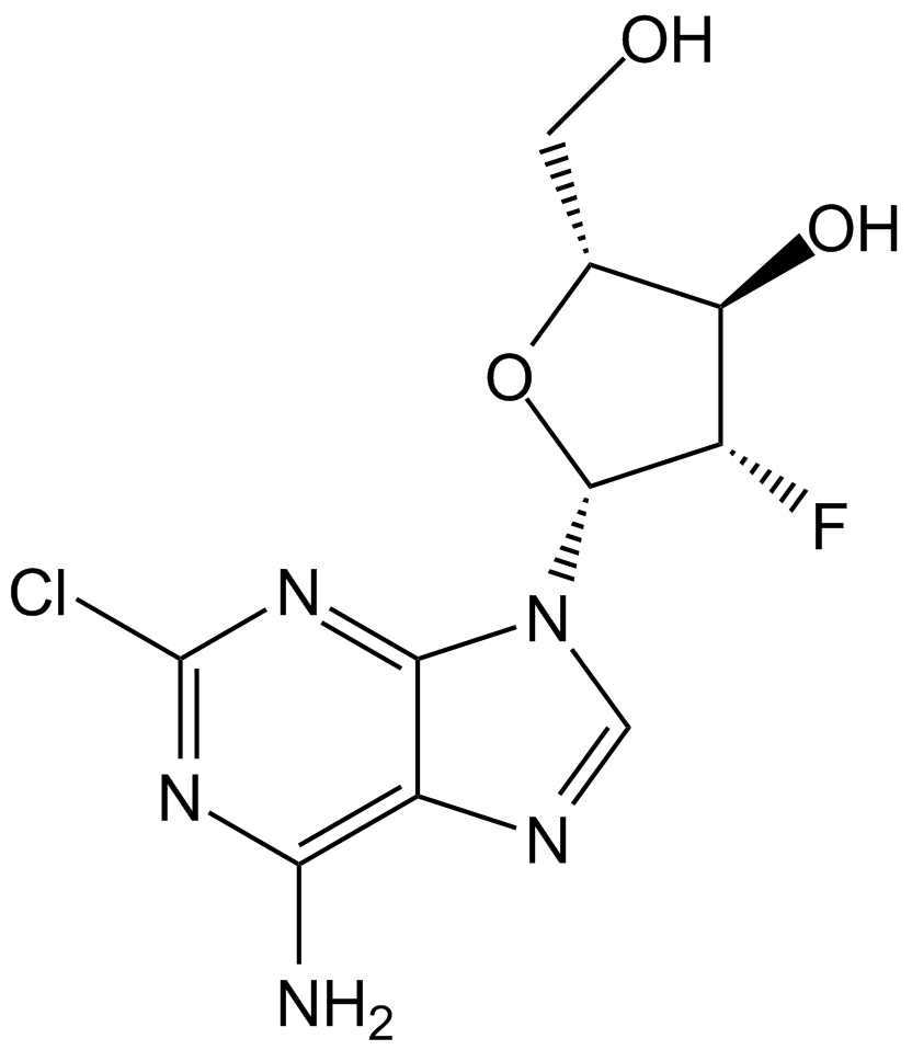
GC15219
Clofarabine
がん研究用のヌクレオシド類似体であるクロファラビンは、調節サブユニットのアロステリック部位に結合することにより、リボヌクレオチドレダクターゼの強力な阻害剤 (IC50=65 nM) です 。
-
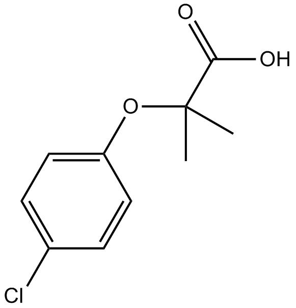
GC10813
Clofibric Acid
クロフィブリン酸 (クロロフィブリン酸) は、脂質調節物質であるクロフィブラート、エトフィブラート、およびエトフィリンクロフィブラートの薬学的に活性な代謝産物であり、脂質低下作用を示す PPARα アゴニストです。
-
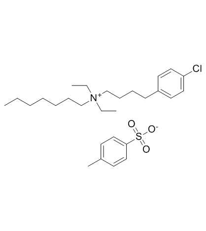
GC32587
Clofilium tosylate
カリウム チャネル遮断薬であるトシル酸クロフィリウムは、カスパーゼ 3 の Bcl-2 非感受性活性化を介して、ヒト前骨髄球性白血病 (HL-60) 細胞のアポトーシスを誘導します。
-
GC47105
Clonostachydiol
クロノスタキジオールは、真菌 Clonostachys cylindrospora (菌株 FH-A 6607) 由来の駆虫性マクロジオリドです 。

-
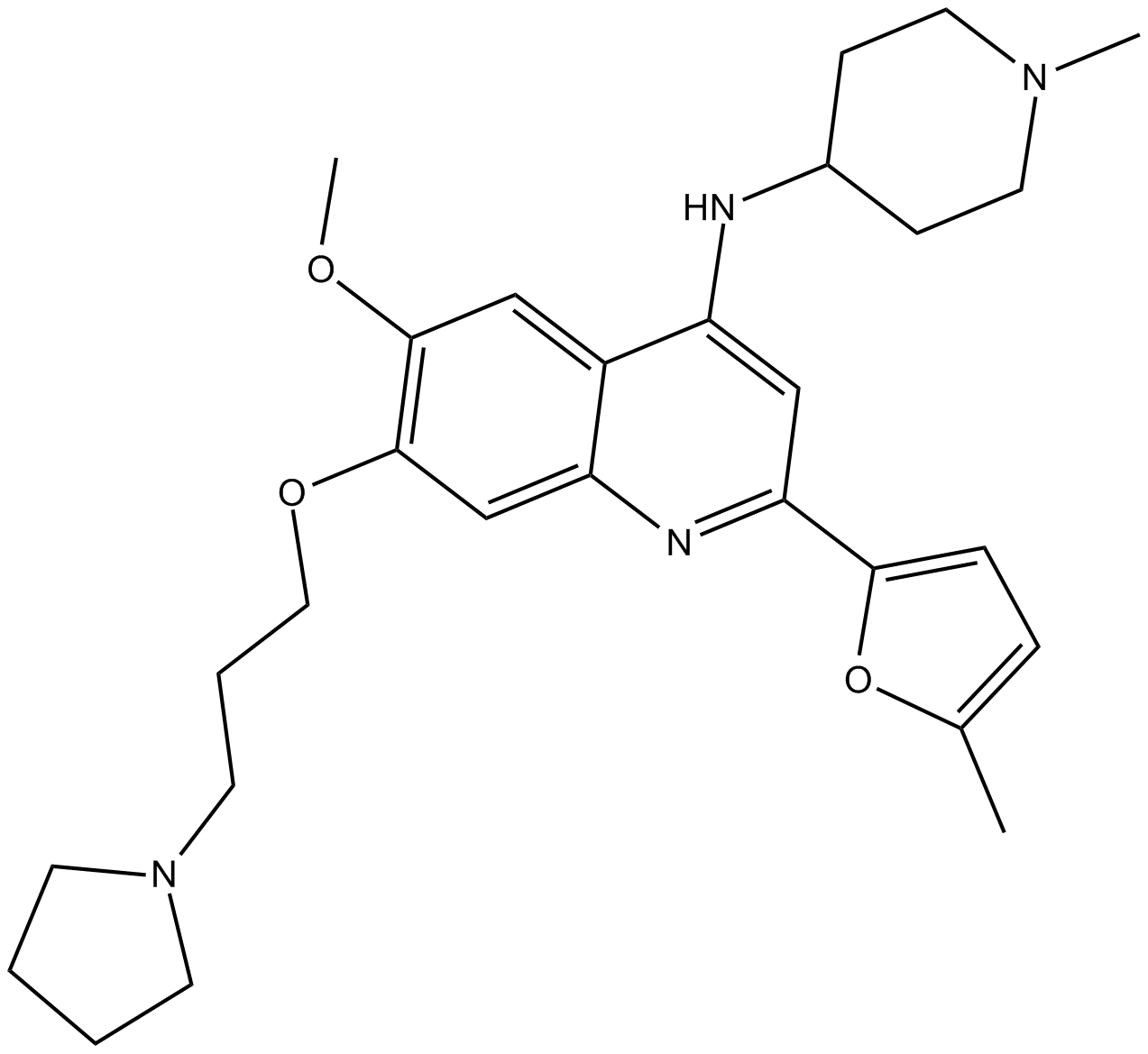
GC12367
CM-272
CM-272 は、抗腫瘍活性を備えたクラス初の強力な選択的基質競合性可逆デュアル G9a/DNA メチルトランスフェラーゼ (DNMT) 阻害剤です。 CM-272 は、G9a、DNMT1、DNMT3A、DNMT3B、および GLP を、それぞれ 8 nM、382 nM、85 nM、1200 nM、および 2 nM の IC50 で阻害します。 CM-272 は細胞増殖を阻害し、アポトーシスを促進し、IFN 刺激遺伝子と免疫原性細胞死を誘導します。
-
GC62347
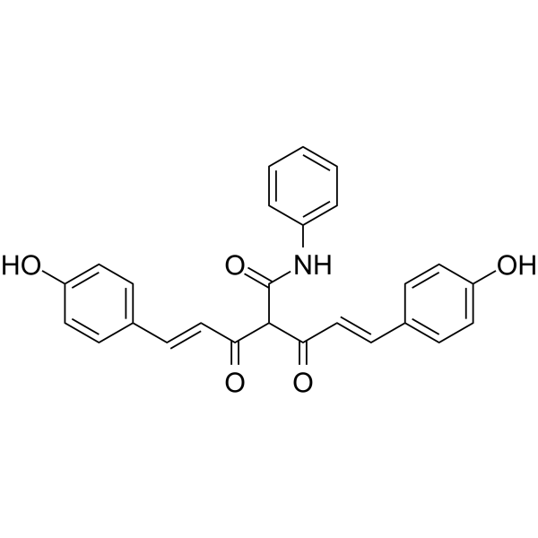
CMC2.24
CMC2.24 (TRB-N0224) は、経口で活性なトリカルボニルメタン剤であり、Ras 活性化とその下流のエフェクター ERK1/2 経路を阻害することにより、マウスの膵臓腫瘍に対して効果的です。
-
GC61567
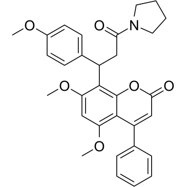
CMLD-2
HuR-ARE 相互作用の阻害剤である CMLD-2 は、競合的に HuR タンパク質に結合し、アデニン-ウリジン リッチ エレメント (ARE) を含む mRNA (Ki=350 nM) との相互作用を破壊します。 CMLD-2 誘導アポトーシスは、結腸、膵臓、甲状腺、および肺癌細胞株などのさまざまな癌細胞で抗腫瘍活性を示します。 Hu 抗原 R (HuR) は RNA 結合タンパク質であり、標的 mRNA の安定性と翻訳を調節できます。
-
GC49096
Cobaltic Protoporphyrin IX (chloride)
HO-1活性の誘導剤

-
GC10033
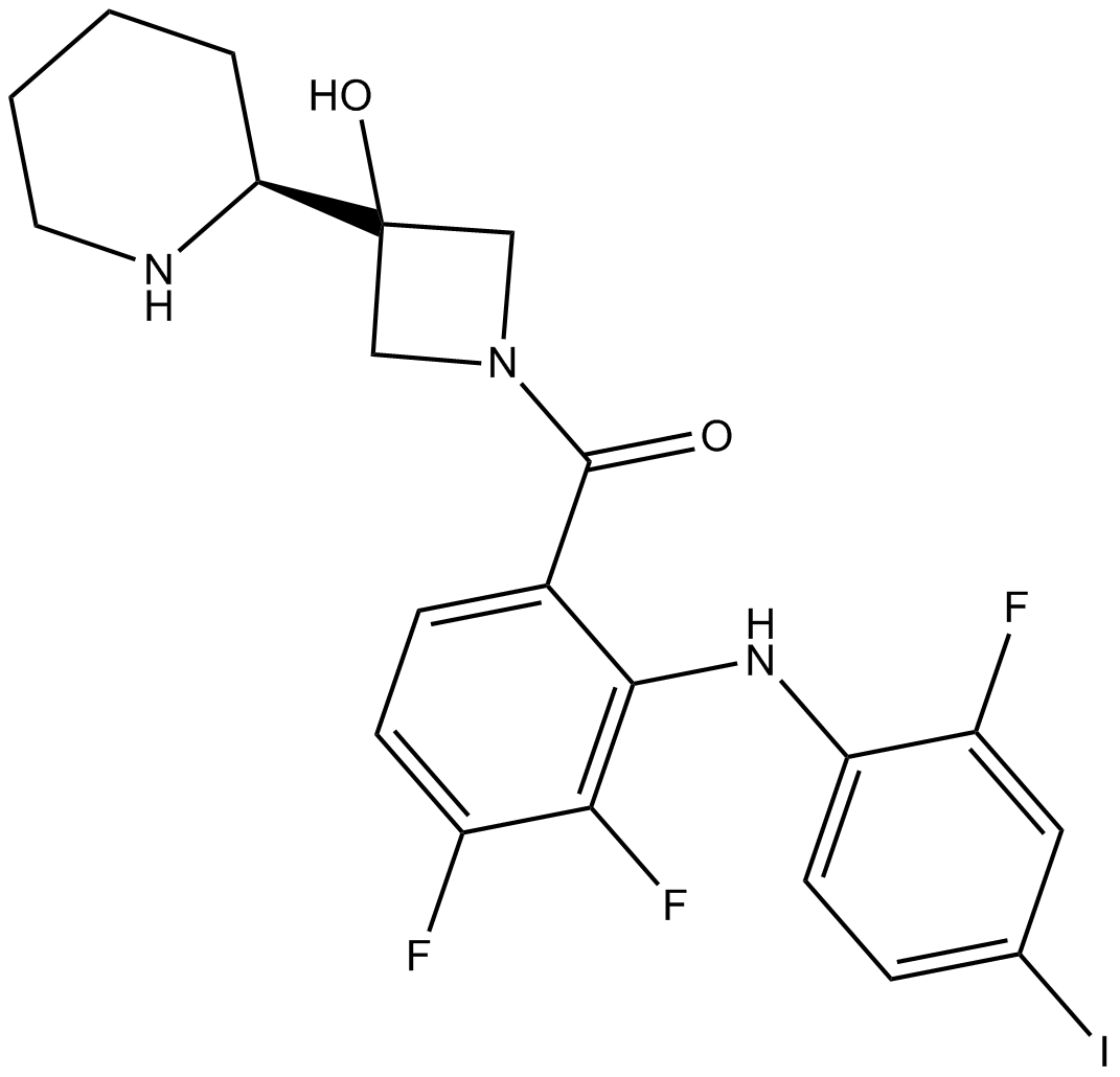
Cobimetinib
口服可能な強力なMEK1阻害剤
-
GC43297
Coenzyme Q2
補酵素Q10は電子伝達系の成分であり、有酸素細胞呼吸に参加し、ATPという形でエネルギーを生成します。

-
GC62192
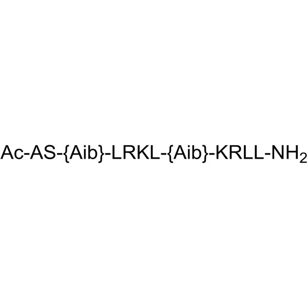
COG1410
COG1410 は、アポリポタンパク質 E 由来のペプチドです。
-
GC40664
Colcemid
Colcemidは、哺乳類の細胞または卵母細胞において、G2/M期の有糸分裂停止または小胞破裂(GVBD)期の減数分裂停止を誘導する細胞骨格阻害剤である。

-
GN10123
Columbianadin
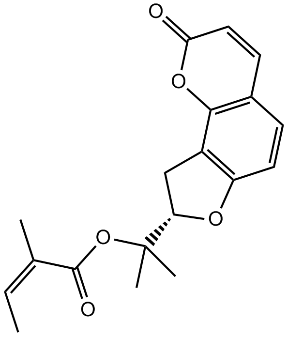
-
GC49454
Complex 3
抗がん活性を持つ蛍光銅錯体

-
GC18572
Concanavalin A
コンカナマイシンAは、高い活性と選択的なバキュオールプロトン-ATPase(v-[H+]ATPase)阻害剤である、Streptomyces diastatochromogenesから分離されたマクロライド系抗生物質の一種であるコンカナマイシンに属しています。

-
GC18832
Conglobatin
コングロバチンは、元々Sから単離された二量体のマクロライドジラクトンです。
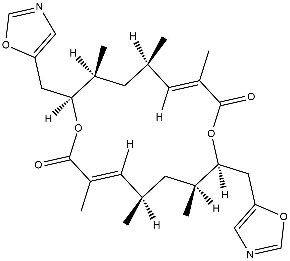
-
GC48483
Conglobatin B
細菌代謝産物

-
GC48497
Conglobatin C1
細菌代謝産物

-
GC38376
Coniferaldehyde
コニフェルアルデヒド (フェルルアルデヒド) は、ヘムオキシゲナーゼ-1 (HO-1) の効果的なインデューサーです。
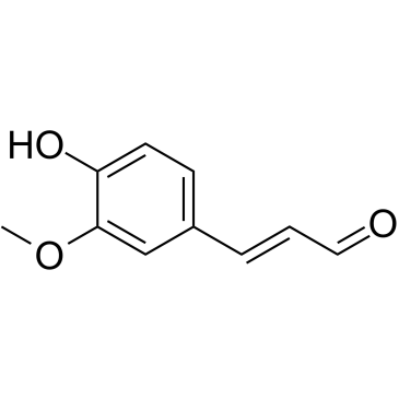
-
GC63379
Conophylline
コノフィリンは、熱帯植物エルバタミア・ミクロフィラの葉から抽出されるビンカアルカロイドです。
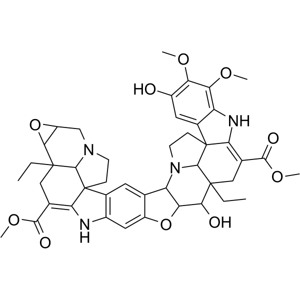
-
GC16772
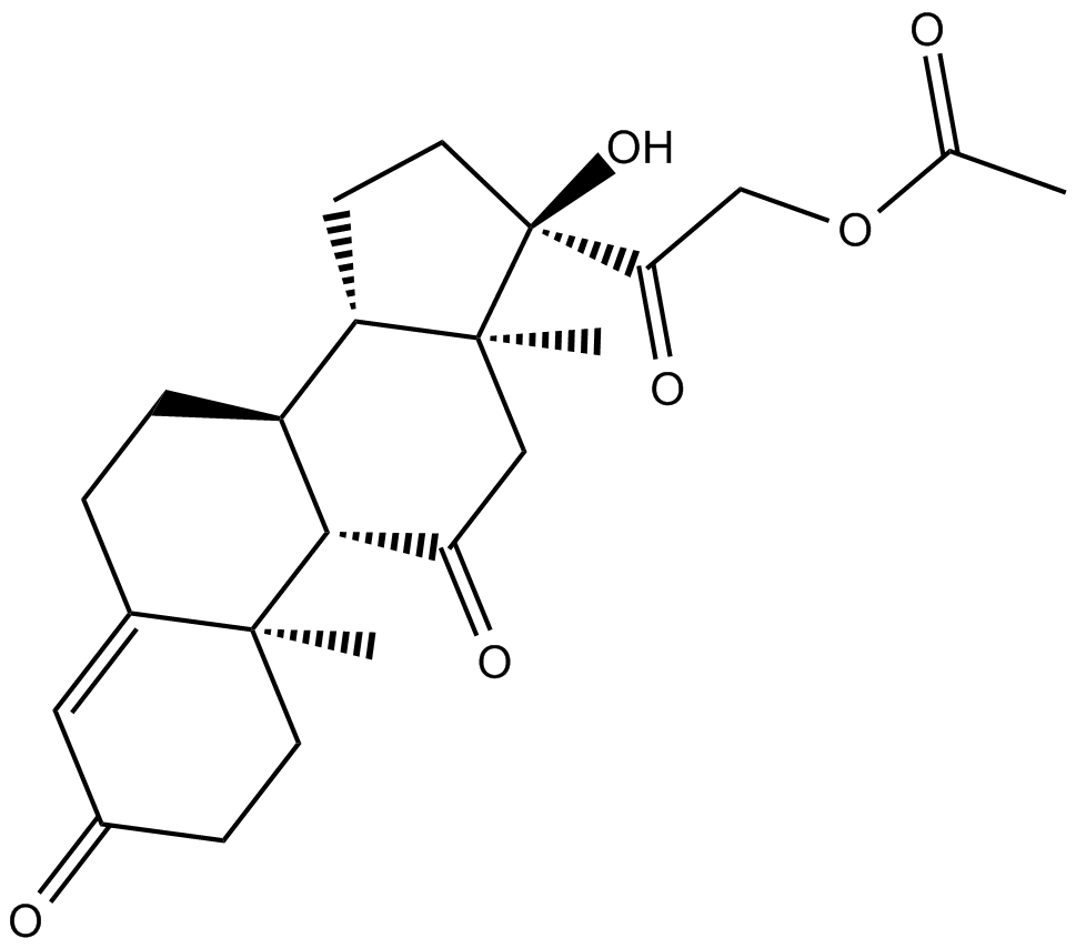
Cortisone acetate
酢酸コルチゾン (コルチゾン 21-アセテート)、コルチゾール (グルココルチコイド) の酸化代謝産物。
-
GC16116
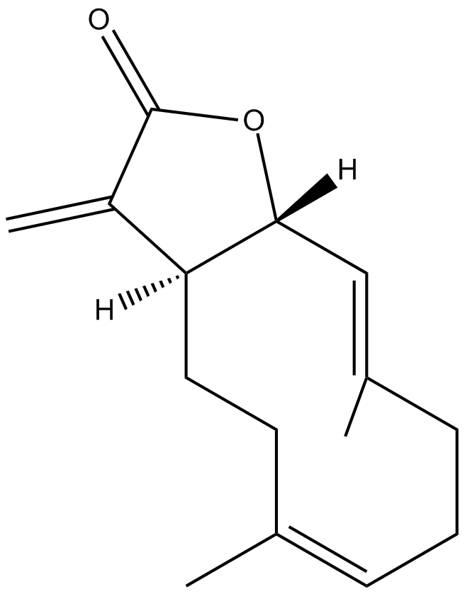
Costunolide
天然セスキテルペンラクトン
-
GC15225
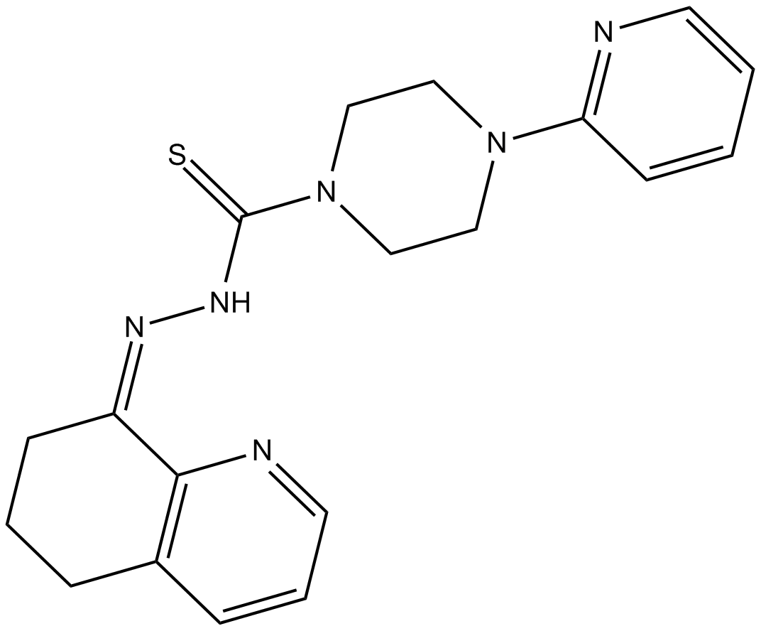
COTI-2
低毒性の抗がん剤である COTI-2 は、p53 変異型の経口投与可能な第 3 世代活性化剤です。 COTI-2 は、変異 p53 の再活性化と PI3K/AKT/mTOR 経路の阻害の両方によって作用します。 COTI-2 は、複数のヒト腫瘍細胞株でアポトーシスを誘導します。 COTI-2 は、p53 依存性および非依存性メカニズムを介して HNSCC で抗腫瘍活性を示します。 COTI-2 は変異型 p53 を野生型コンフォメーションに変換します。
-
GC15840
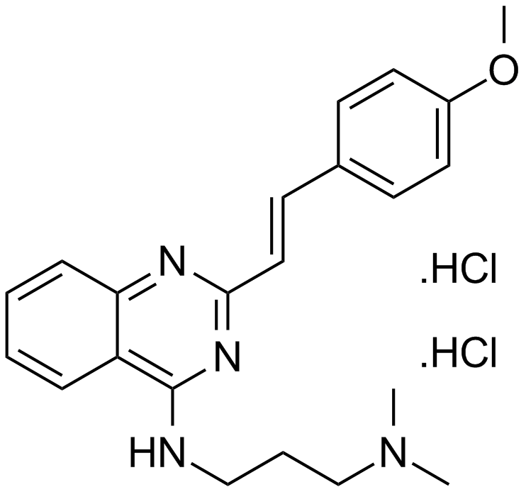
CP 31398 dihydrochloride
p53安定剤
-
GC13091
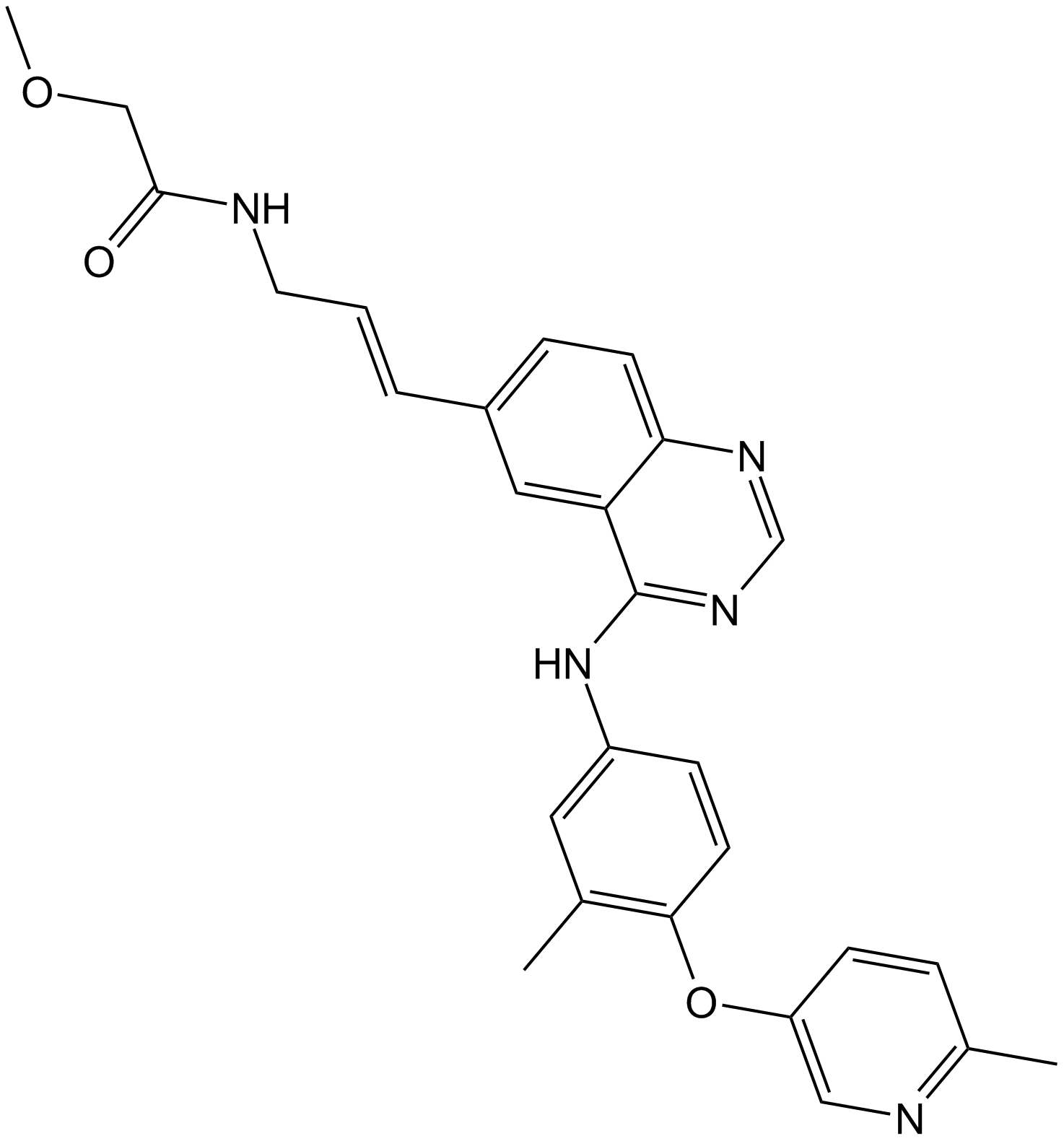
CP-724714
CP-724714 は、強力で選択的な経口活性 ErbB2 (HER2) チロシンキナーゼ阻害剤であり、IC50 は 10 nM です。 CP-724714 は、EGFR キナーゼに対して顕著な選択性を示します (IC50=6400 nM)。 CP-724714 は、無傷の細胞で ErbB2 受容体の自己リン酸化を強力に阻害します。抗腫瘍活動。
-
GC14500
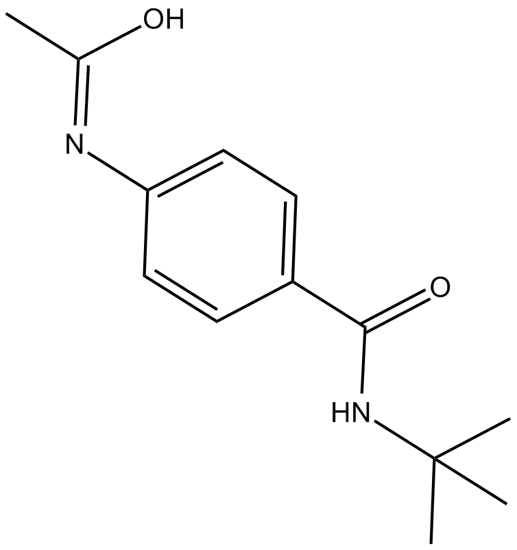
CPI-1189
CPI-1189 は、抗酸化作用と神経保護作用を持つ TNF-α 放出阻害剤です。
-
GC14699
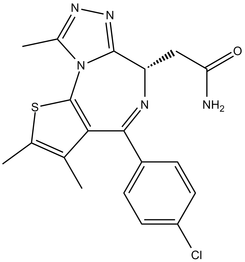
CPI-203
CPI-203 は、約 37 nM の IC50 値を持つ、BET ブロモドメインの強力で選択的な細胞透過性の新規阻害剤です (BRD4 α-スクリーンアッセイ)。
-
GC10021
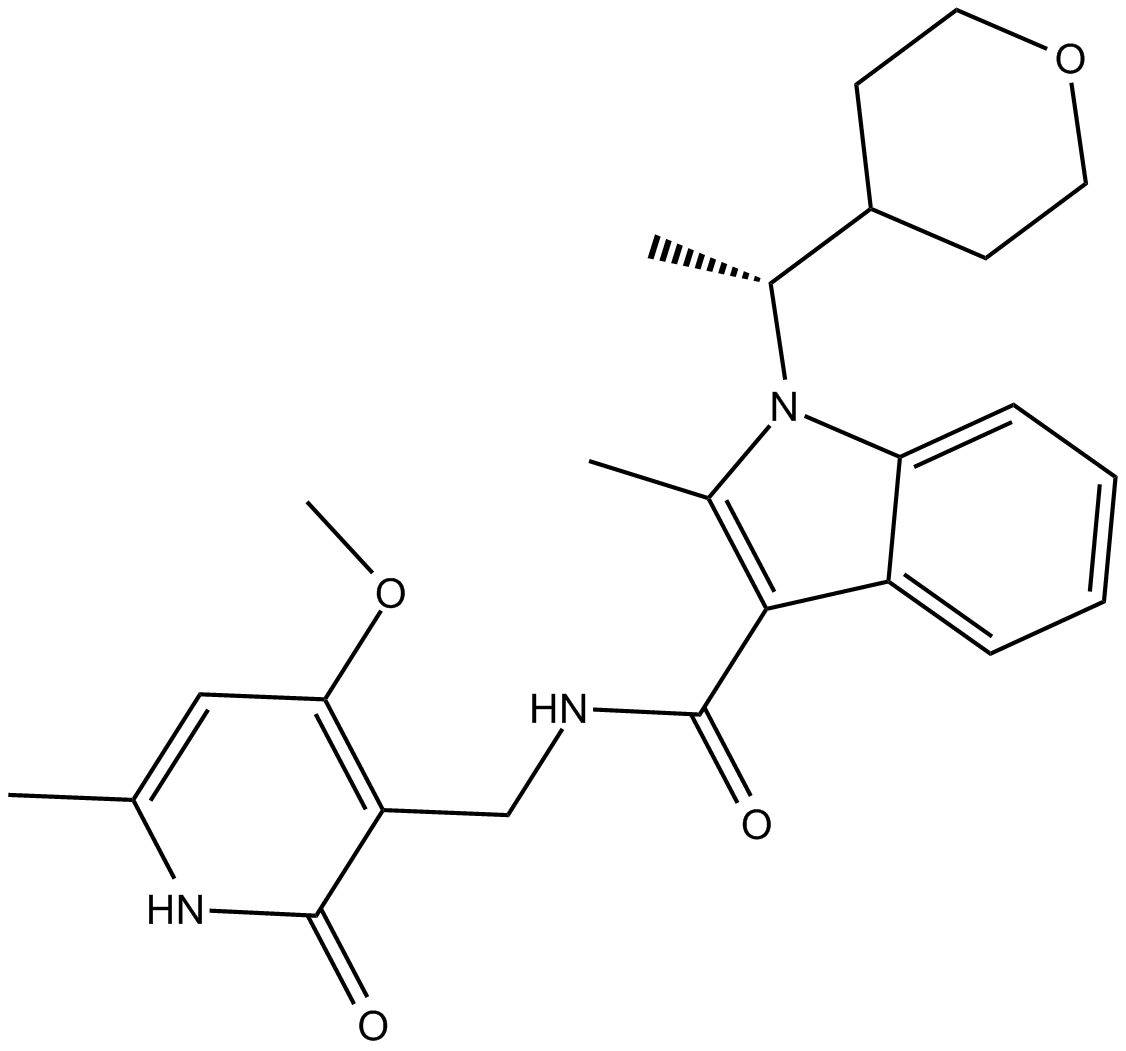
CPI-360
EZH2阻害剤
-
GC14921
CPI-613
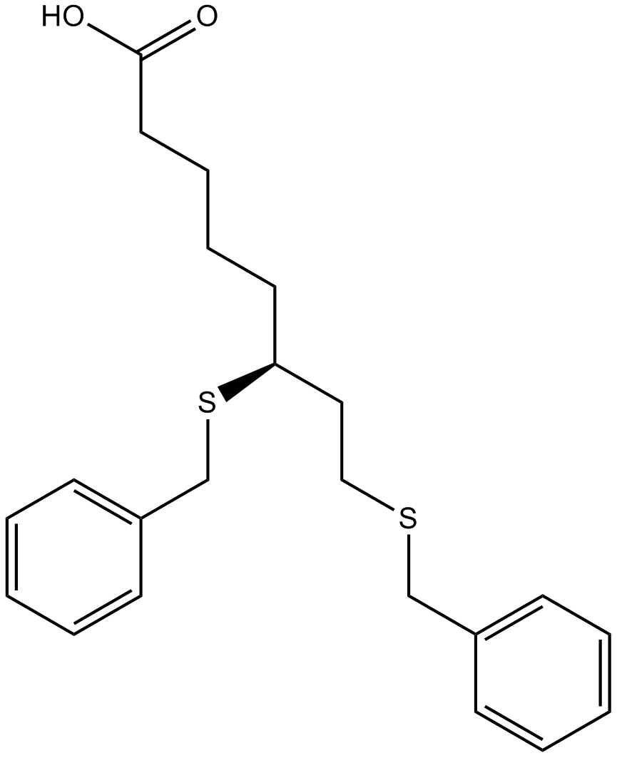
α-ケトグルタル酸脱水素酵素の阻害剤
-
GC39365
CPTH2
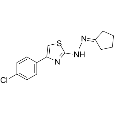
CPTH2 は、強力なヒストン アセチルトランスフェラーゼ (HAT) 阻害剤です。 CPTH2 は、Gcn5 によるヒストン H3 のアセチル化を選択的に阻害します。 CPTH2 はアポトーシスを誘導し、アセチルトランスフェラーゼ p300 (KAT3B) の阻害により明細胞腎癌 (ccRCC) 細胞株の浸潤性を低下させます。
-
GC35747
Crebanine
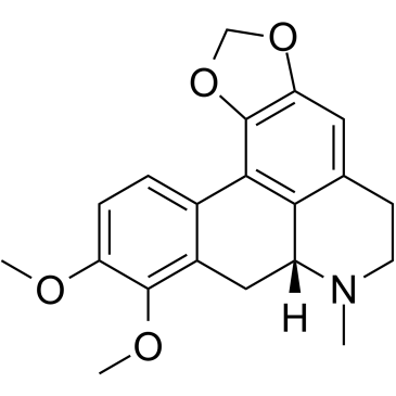
Stephania venosa 由来のアルカロイドであるクレバニンは、ヒト癌細胞の G1 停止とアポトーシスを誘導します。
-
GC34543
cRIPGBM
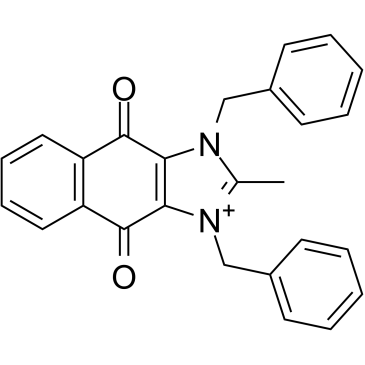
cRIPGBM は、受容体相互作用プロテインキナーゼ 2 (RIPK2) に結合することによる GBM 癌幹細胞 (CSC) のアポトーシスの細胞型選択的インデューサーである RIPGBM のプロアポトーシス誘導体であり、GBM-1 細胞で 68 nM の EC50 を示します。
-
GC13838
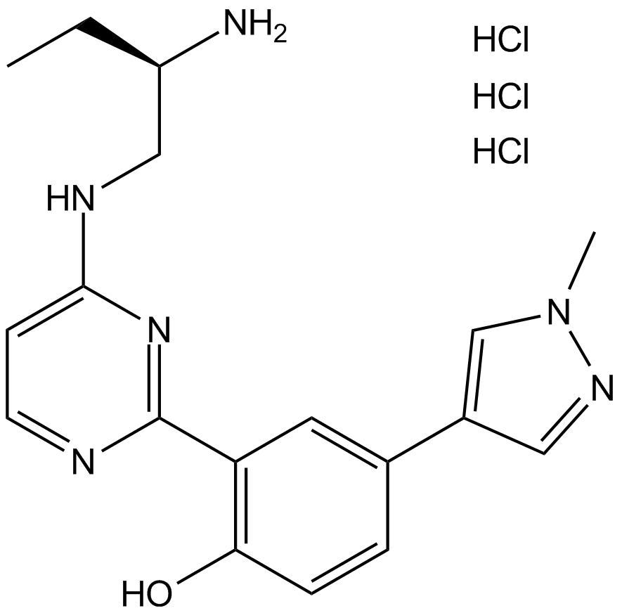
CRT 0066101
PKD阻害剤
-
GC35750
CRT0066101 dihydrochloride
CRT0066101 二塩酸塩は、PKD1、2、および 3 に対してそれぞれ 1、2.5、および 2 nM の IC50 値を持つ強力で特異的な PKD 阻害剤です。
-
GC45414
CRT0066854
CRT0066854 は、強力かつ選択的な非定型 PKC アイソザイム阻害剤です。

-
GC14355
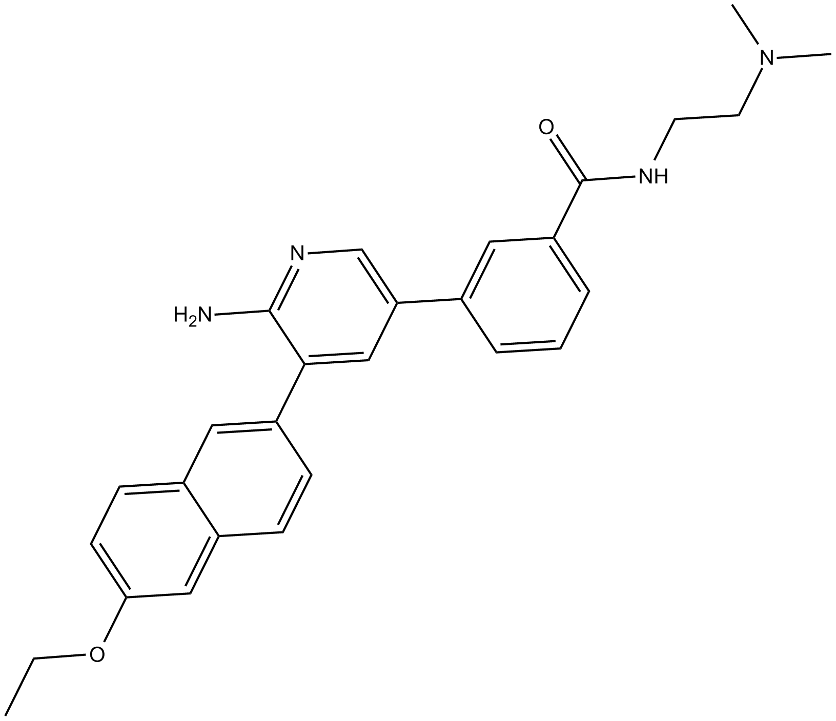
CRT5
ピラジンベンズアミドである CRT5 は、VEGF で処理された内皮細胞における PKD の 3 つのアイソフォームすべてに対する強力かつ選択的な阻害剤です (IC50 = PKD1、PKD2、および PKD3 に対してそれぞれ 1、2、および 1.5 nM)。
-
GC32911
CTX1
CTX1 は、HdmX を介した p53 抑制を克服する p53 アクティベーターです。 CTX1 は、マウスの急性骨髄性白血病 (AML) モデル系で強力な抗がん活性を示します。
-
GN10535
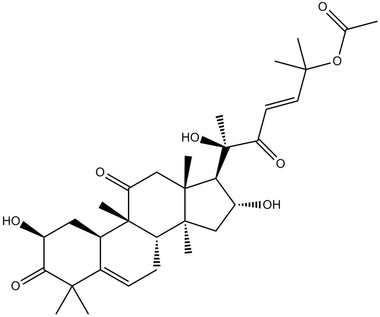
Cucurbitacin B
-
GC35758
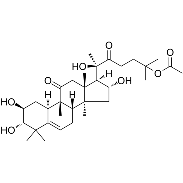
Cucurbitacin IIa
ククルビタシン IIa は、Hemsleya amalils Diels から分離されたトリテルペンであり、癌細胞のアポトーシスを誘導し、サバイビンの発現を減少させ、ホスホ ヒストン H3 を減少させ、癌細胞の切断された PARP を増加させます。
-
GN10788
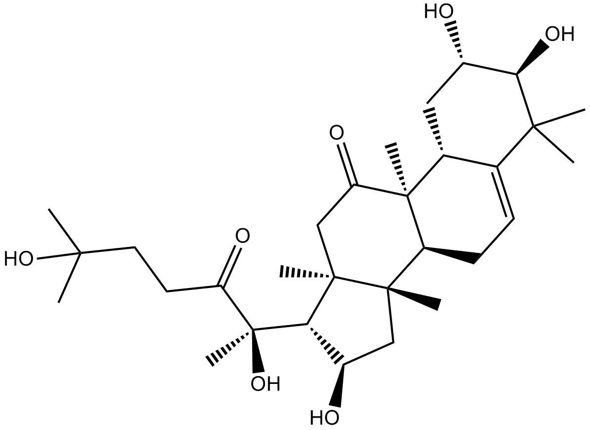
Cucurbitacin IIb
-
GC32781
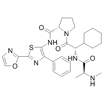
CUDC-427 (GDC-0917)
CUDC-427 (GDC-0917) は、さまざまながんの治療に使用される強力な第 2 世代の汎選択的 IAP アンタゴニストです。
-
GC12115
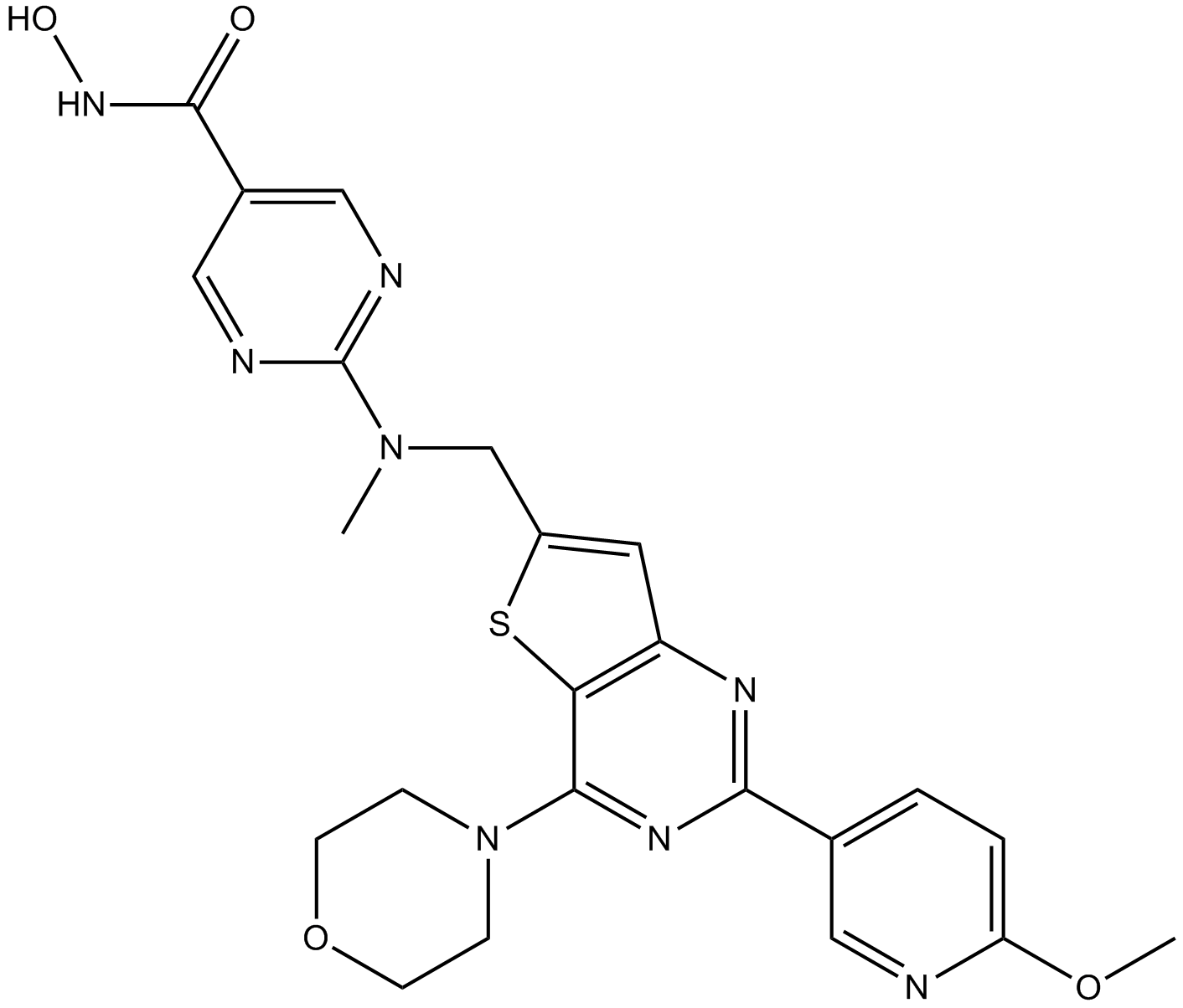
CUDC-907
HDACとPI3Kの二重阻害剤
-
GC11217
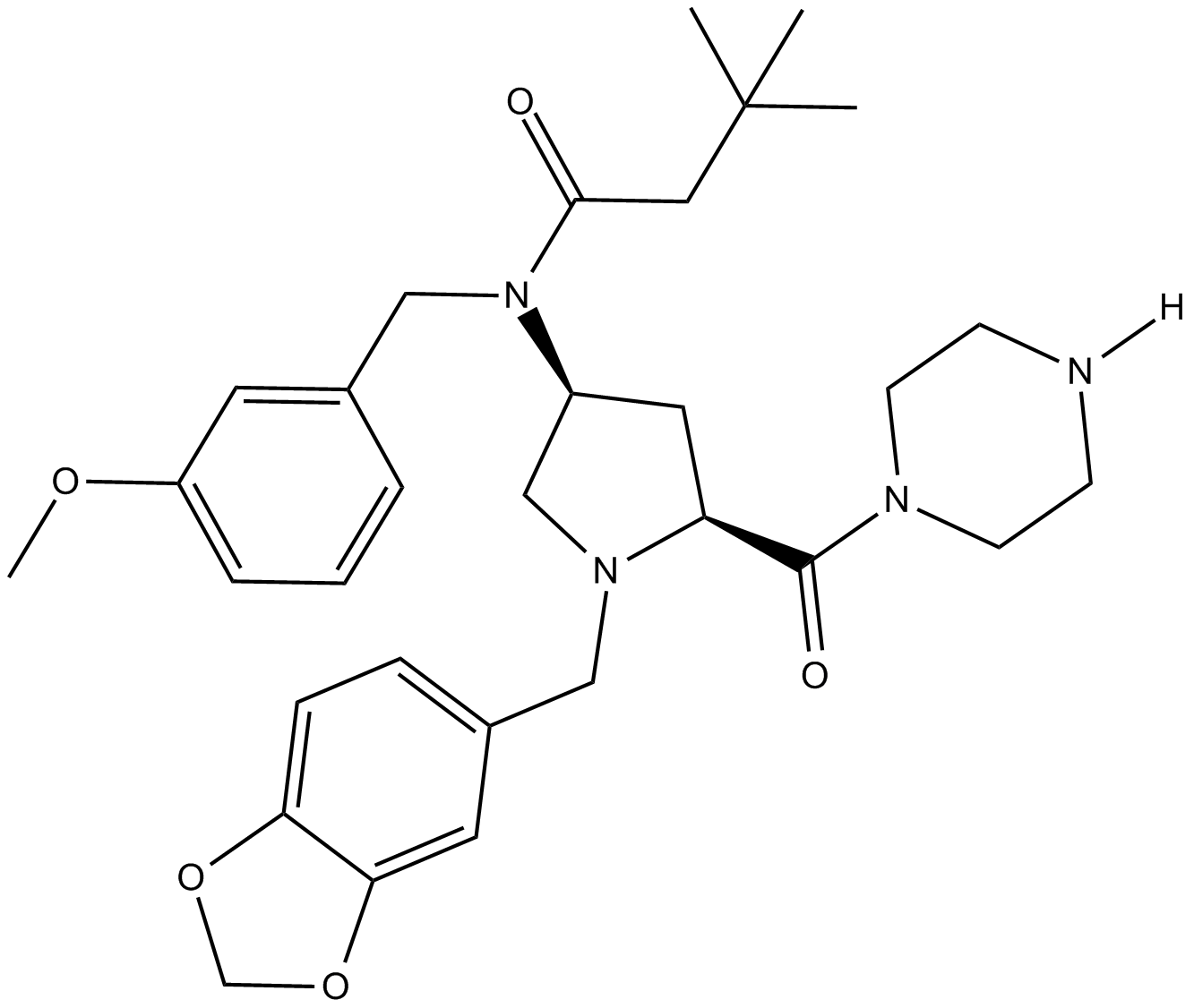
CUR 61414
CUR 61414 は、新規で強力な細胞透過性の Hedgehog シグナル伝達経路阻害剤です (IC50 = 100-200 nM)。
-
GC14787
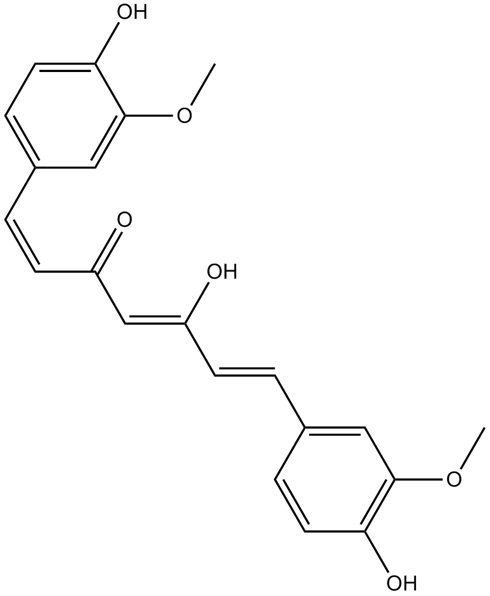
Curcumin
多様な生物学的活性を持つ黄色の色素
-
GC40226
Curcumin-d6
クルクミン D6 (ジフェルロイルメタン D6) は、クルクミン (ターメリック イエロー) とラベル付けされた重水素です。

-
GN10521
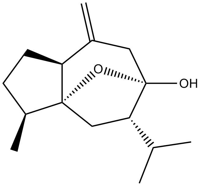
Curcumol
-
GC66356
Cusatuzumab
クサツズマブは、ヒト αCD70 モノクローナル抗体です。クサツズマブは、増強された抗体依存性細胞による細胞傷害活性を示します。クサツズマブは、白血病幹細胞 (LSC) を減少させ、骨髄分化とアポトーシスに関連する遺伝子シグネチャーを引き起こします。クサツズマブは、急性骨髄性白血病 (AML) の研究の可能性を秘めています。

-
GC63967
Cycleanine
シクレニンは強力な血管選択的カルシウム拮抗薬です。
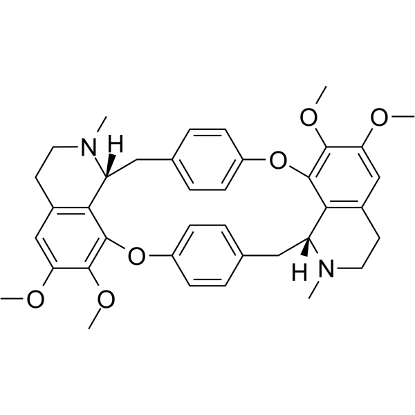
-
GC49716
Cyclo(RGDyK) (trifluoroacetate salt)
αVβ3インテグリンの環状ペプチドリガンド

-
GC17198
Cycloheximide
Cycloheximide is an antibiotic that inhibits protein synthesis at the translation level, acting exclusively on cytoplasmic (80s) ribosomes of eukaryotes.
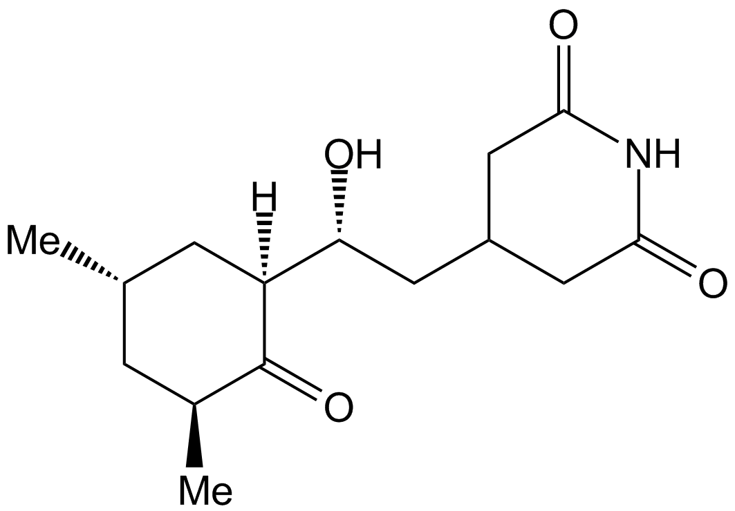
-
GC43346
Cyclopamine-KAAD
ヘッジホッグ シグナル伝達阻害剤であるシクロパミン KAAD は、平滑化されたアンタゴニストです。

-
GC47148
Cyclophosphamide-d4
シクロホスファミド-d4 は、重水素標識シクロホスファミドです。シクロホスファミドは、抗腫瘍活性、免疫抑制剤である窒素マスタードに化学的に関連する合成アルキル化剤です。

-
GC38419
Cyclovirobuxine D
シクロビロブキシン D (CVB-D) は、漢方薬のツゲの主要な有効成分です。
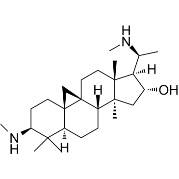
-
GC33330
Cynaropicrin
シナロピクリンは、腫瘍壊死因子 (TNF-α) の放出を阻害できるセスキテルペン ラクトンであり、マウスおよびヒトのマクロファージ細胞に対してそれぞれ 8.24 μM および 3.18 μM の IC50 を示します。シナロピクリンはまた、軟骨分解因子 (MMP13) の増加を阻害し、NF-κB シグナル伝達を抑制します。
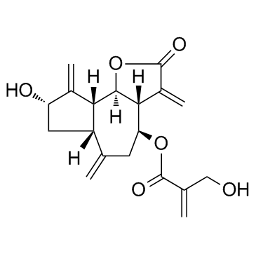
-
GC65565
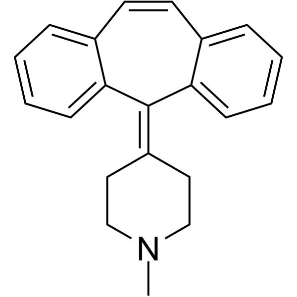
Cyproheptadine
シプロヘプタジンは、強力な経口活性 5-HT2A 受容体拮抗薬であり、抗うつ効果と抗セロトニン作動効果があります。
-
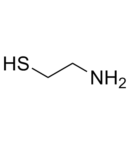
GC33779
Cysteamine (β-Mercaptoethylamine)
-
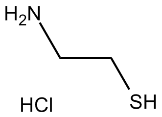
GC13502
Cysteamine HCl
システアミン HCl (2-アミノエタンチオール塩酸塩) は、腎症性シスチン症の治療用の経口活性剤であり、抗酸化剤です。
-
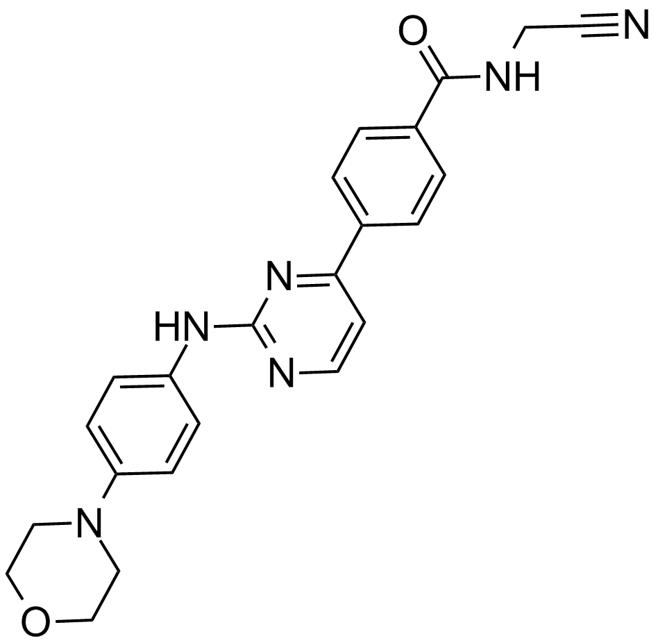
GC17050
CYT387
JAK1およびJAK2の強力な阻害剤
-
GC11383
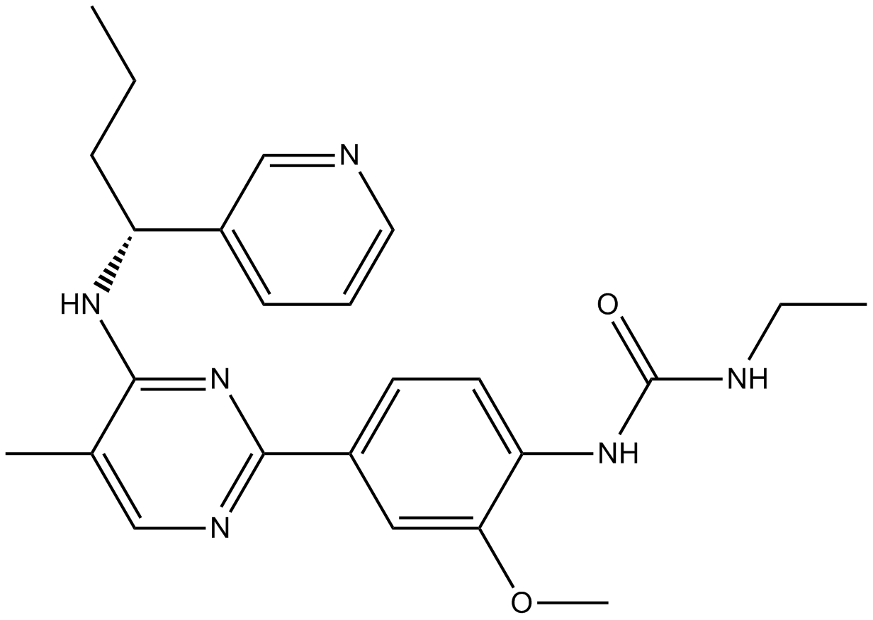
CYT997 (Lexibulin)
CYT997 (レキシブリン) (CYT-997) は、がん細胞株において 10 ~ 100 nM の IC50 を持つ強力な経口活性チューブリン重合阻害剤です。 in vitro および in vivo で強力な細胞毒性および血管破壊活性を示します。 CYT997 (レキシブリン) は細胞アポトーシスを誘導し、GC 細胞のミトコンドリア ROS 生成を誘導します。
-
GC13070
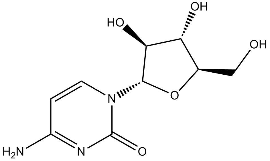
Cytarabine
細胞毒性剤、DNA合成を阻害する
-
GC43356
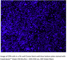
CytoCalcein™ Violet 450
CytoCalcein? Violet 450は、細胞の生存性を評価するために使用される蛍光色素です。
-
GC43357
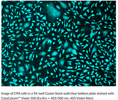
CytoCalcein™ Violet 500
CytoCalcein? Violet 500は、細胞の生存性を評価するために使用される蛍光色素です。
-
GC43361
Cytostatin (sodium salt)
サイトスタチンは、細胞外マトリックスへの細胞接着を阻害する自然な抗腫瘍剤であり、B16メラノーマ細胞のラミニンおよびコラーゲンIVに対する接着をin vitroでブロックします(IC50値はそれぞれ1.3および1.4μg/ml)、そしてマウスにおけるB16細胞の転移活性も抑制します。

-
GC43368
D,L-1′-Acetoxychavicol Acetate
D,L-1'-アセトキシチャビコール酢酸エステルは、ジンジャーのような植物の根茎から最初に単離された天然化合物です。

-
GC66824
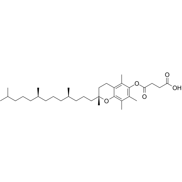
D-α-Tocopherol Succinate
D-α-コハク酸トコフェロール (ビタミン E コハク酸) は抗酸化剤のトコフェロールであり、ビタミン E の塩の形です。 D-α-コハク酸トコフェロールは癌の研究に使用できます。
-
GC12256
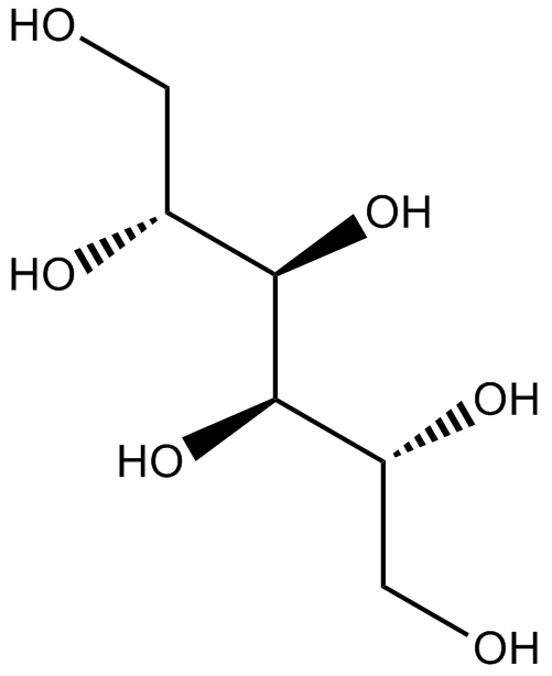
D-Mannitol
D-マンニトールは、浸透圧利尿剤であり、弱い腎血管拡張剤です。
-
GC60796
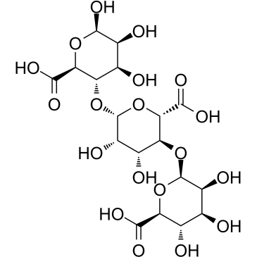
D-Trimannuronic acid
アルギン酸オリゴマーであるD-トリマンヌロン酸は、海藻から抽出されます。
-
GC13202
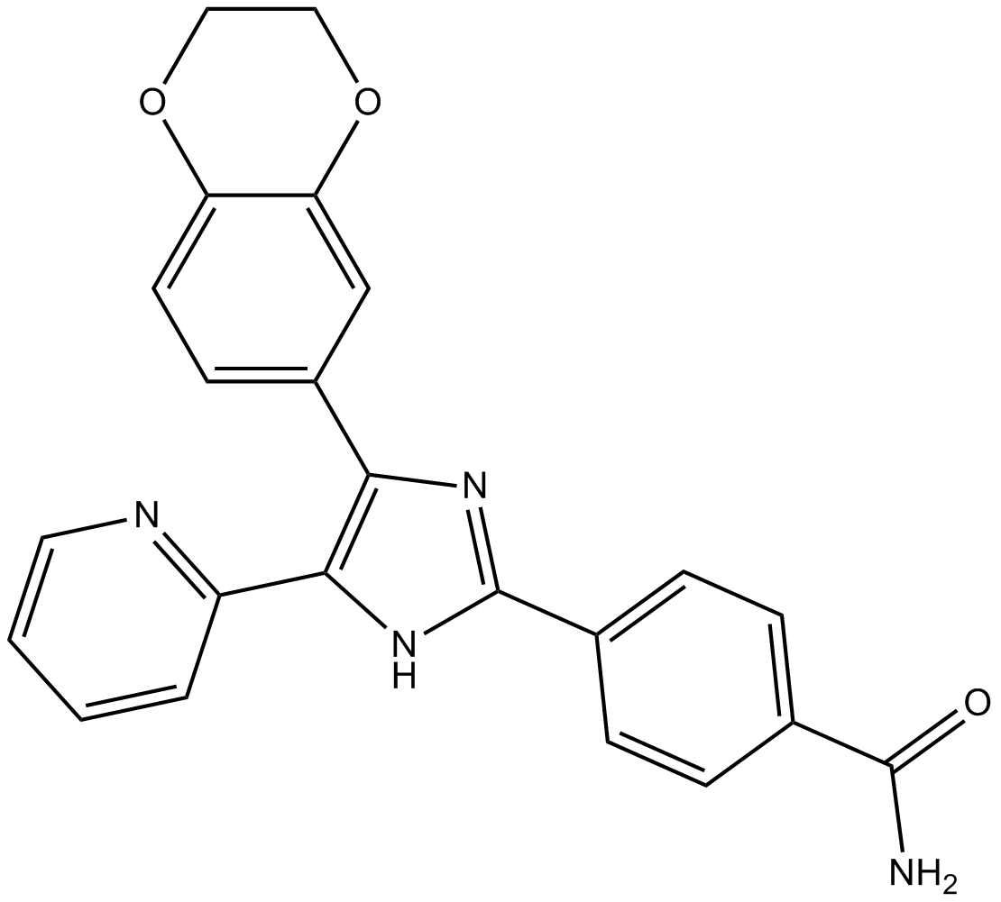
D4476
CK1とALK5の阻害剤
-
GC17851
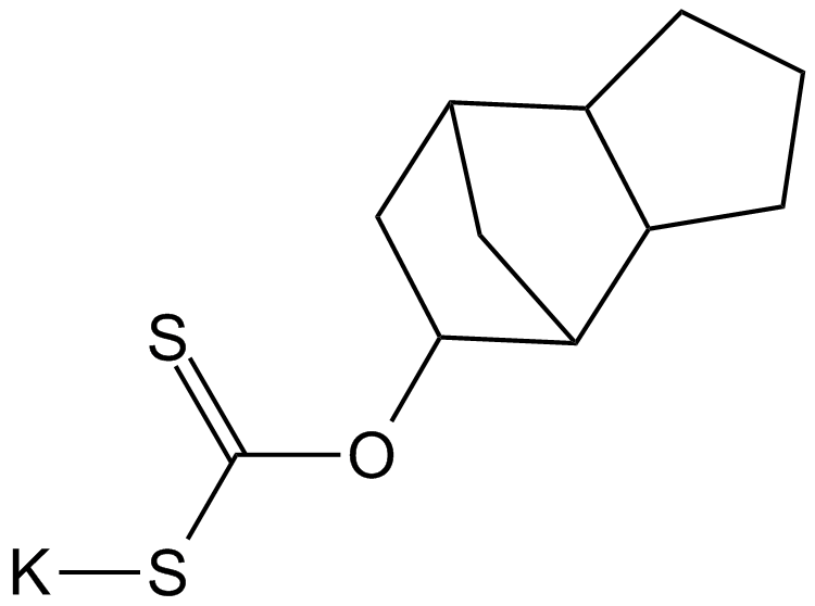
D609
PC特異的なPLCの競合阻害剤
-
GC50296
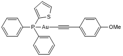
D9
D9 は、チオレドキシン還元酵素 (TrxR) の強力かつ選択的な阻害剤であり、EC50 は 2.8 nM です。 D9 は、in vitro と in vivo の両方で腫瘍増殖を阻害する能力を持っています。
-
GC18421
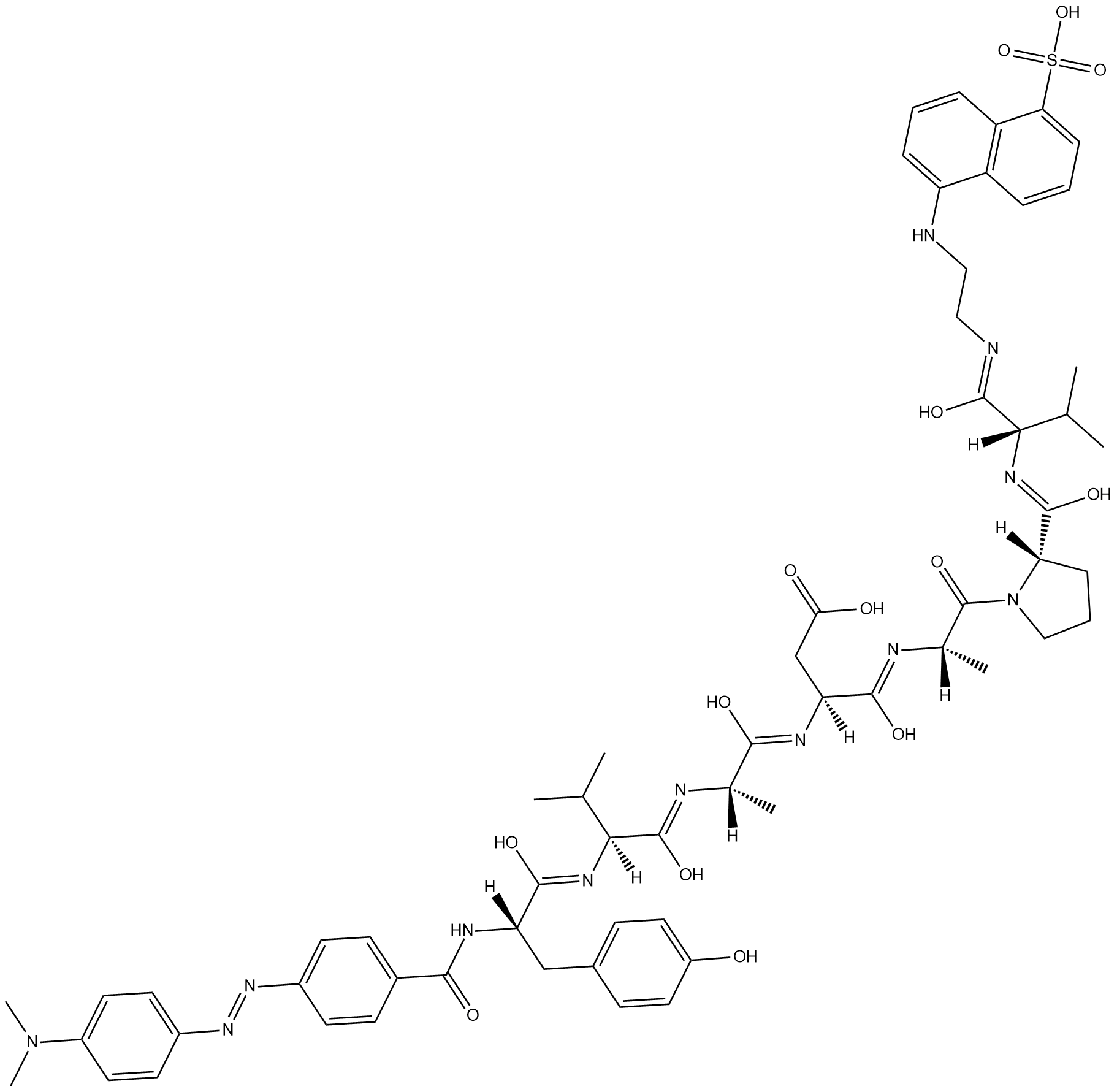
Dabcyl-YVADAPV-EDANS
Dabcyl-YVADAPV-EDANSは、カスパーゼ-1の蛍光基質です。
-
GC47166
Dabrafenib-d9
Dabrafenib-d9 (GSK2118436A-d9) は、Dabrafenib と標識された重水素です。ダブラフェニブ (GSK2118436A) は、C-Raf および B-RafV600E の IC50 がそれぞれ 5 nM および 0.6 nM の Raf の ATP 競合阻害剤です。

-
GC14485
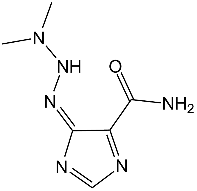
Dacarbazine
ダカルバジンは、細胞周期非特異的な抗腫瘍性アルキル化剤です。ダカルバジンは、T および B リンパ芽球応答を阻害し、IC50 値はそれぞれ 50 および 10 μg/ml です。ダカルバジンは、転移性悪性黒色腫の研究に使用できます。
-
GC47167
Dacarbazine-d6
ダカルバジン-d6 (イミダゾール カルボキサミド-d6) は、ダカルバジンと標識された重水素です。ダカルバジン(DTIC-Dome; DTIC)は抗腫瘍薬です。それはメラノーマに対して重要な活性を持っています。

-
GC68305
Dacetuzumab

-
GC10225
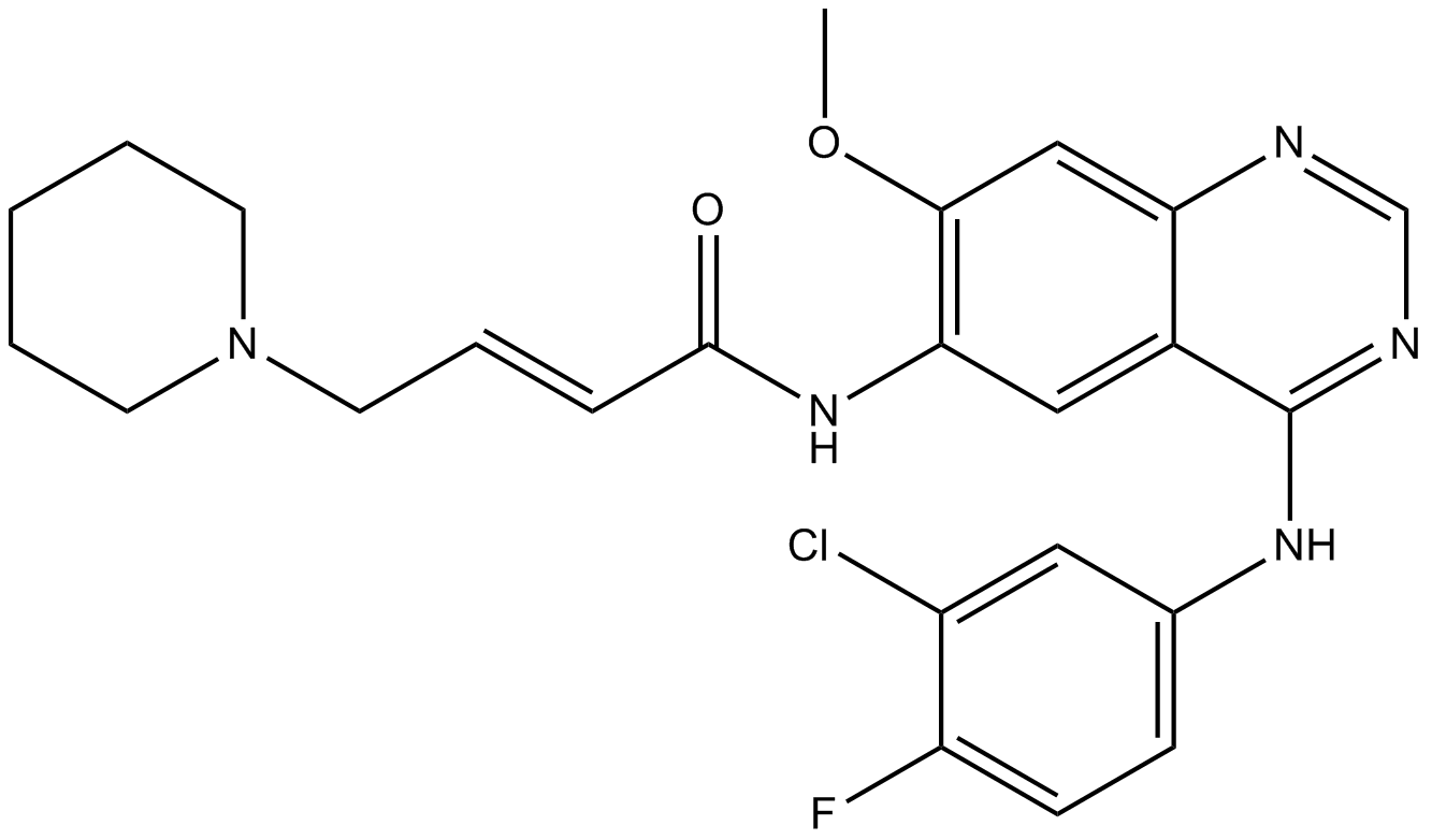
Dacomitinib (PF299804, PF299)
ダコミチニブ (PF299804、PF299) (PF-00299804) は、キナーゼの ERBB ファミリーの特異的かつ不可逆的な阻害剤であり、EGFR、ERBB2、および ERBB4 に対してそれぞれ 6 nM、45.7 nM、および 73.7 nM の IC50 を示します。
-
GC15211
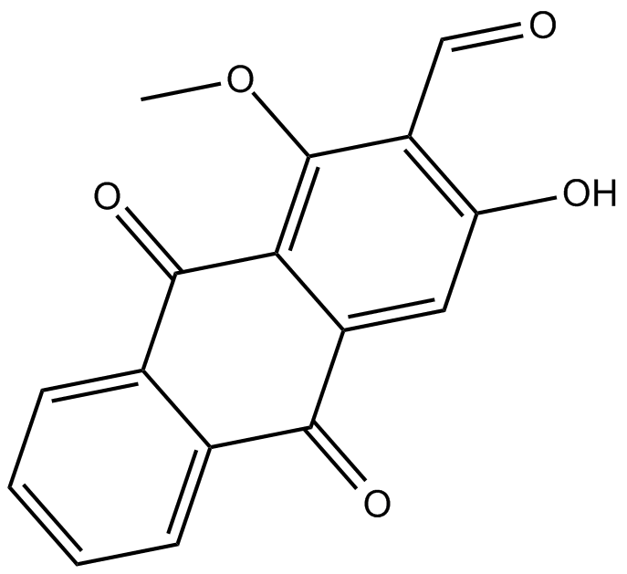
Damnacanthal
ダムナカンタールはモリンダ・シトリフォリアの根から分離されたアントラキノンです。
-
GN10318
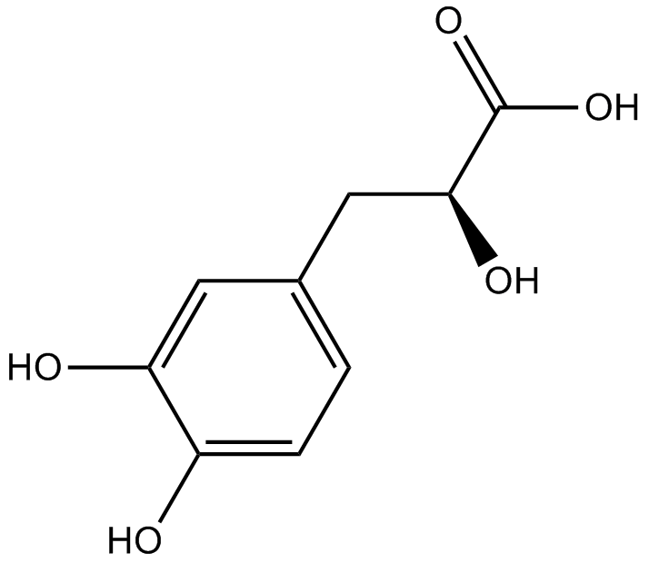
Danshensu
-
GC34010
Danshensu (Dan shen suan A)
Danshensu, an active ingredient of Salvia miltiorrhiza, shows wide cardiovascular benefit by activating Nrf2 signaling pathway.
-
GC15217
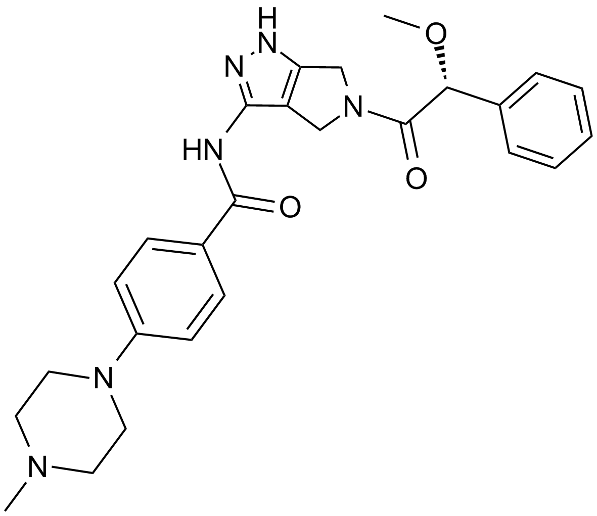
Danusertib (PHA-739358)
パン・オーロラキナーゼとAbl阻害剤
-
GC17650
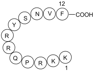
DAPK Substrate Peptide
DAPK1ペプチド基質
-
GC49883
DAPK Substrate Peptide (trifluoroacetate salt)
DAPK1ペプチド基質

-
GC12942
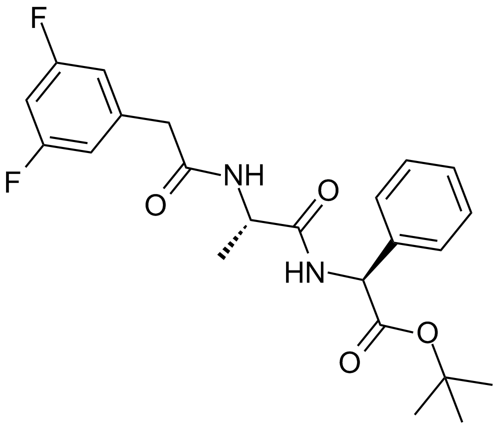
DAPT (GSI-IX)
γ-セクレターゼの阻害剤
-
GC43379
Darinaparsin
有機ヒ素であるダリナパルシン (ZIO-101) は、ミトコンドリアを標的とする薬剤です。ダリナパルシンは、がん細胞のアポトーシスを誘導し、抗がん効果があります。

-
GC15568
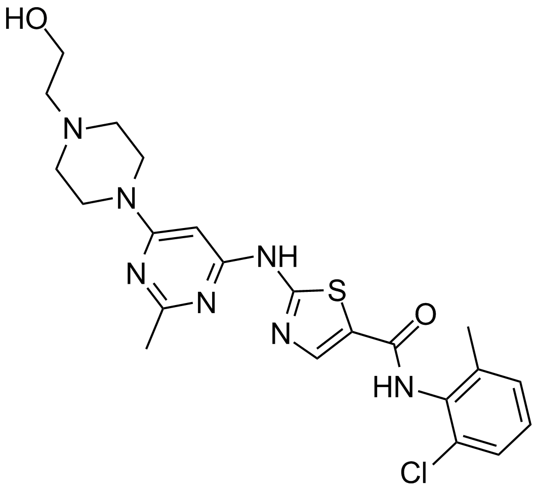
Dasatinib (BMS-354825)
AblとSrcの阻害剤
-
GC35812
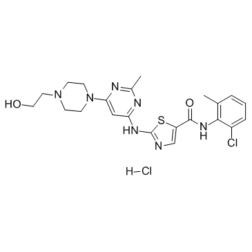
Dasatinib hydrochloride
ダサチニブ (BMS-354825) 塩酸塩は、強力な抗腫瘍活性を有する、非常に強力な ATP 競合性経口活性デュアル Src/Bcr-Abl 阻害剤です。 Kis は、Src と Bcr-Abl でそれぞれ 16 pM と 30 pM です。ダサチニブ塩酸塩は、Bcr-Abl および Src をそれぞれ <1.0 nM および 0.5 nM の IC50 で阻害します。ダサチニブ塩酸塩は、アポトーシスとオートファジーも誘導します。
-
GC10354
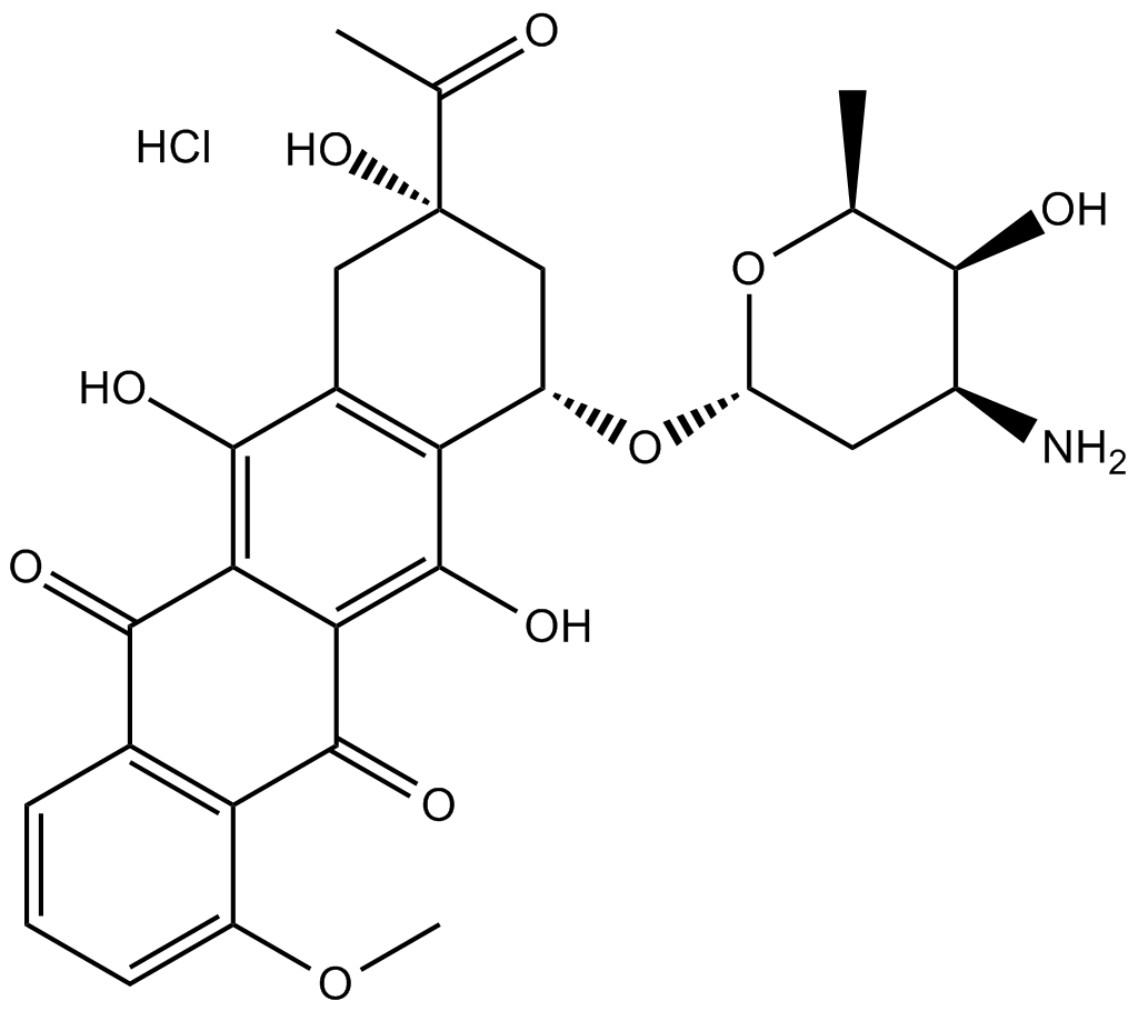
Daunorubicin HCl
ダウノルビシン (ダウノマイシン) 塩酸塩は、強力な抗腫瘍活性を持つトポイソメラーゼ II 阻害剤です。
