Apoptosis
As one of the cellular death mechanisms, apoptosis, also known as programmed cell death, can be defined as the process of a proper death of any cell under certain or necessary conditions. Apoptosis is controlled by the interactions between several molecules and responsible for the elimination of unwanted cells from the body.
Many biochemical events and a series of morphological changes occur at the early stage and increasingly continue till the end of apoptosis process. Morphological event cascade including cytoplasmic filament aggregation, nuclear condensation, cellular fragmentation, and plasma membrane blebbing finally results in the formation of apoptotic bodies. Several biochemical changes such as protein modifications/degradations, DNA and chromatin deteriorations, and synthesis of cell surface markers form morphological process during apoptosis.
Apoptosis can be stimulated by two different pathways: (1) intrinsic pathway (or mitochondria pathway) that mainly occurs via release of cytochrome c from the mitochondria and (2) extrinsic pathway when Fas death receptor is activated by a signal coming from the outside of the cell.
Different gene families such as caspases, inhibitor of apoptosis proteins, B cell lymphoma (Bcl)-2 family, tumor necrosis factor (TNF) receptor gene superfamily, or p53 gene are involved and/or collaborate in the process of apoptosis.
Caspase family comprises conserved cysteine aspartic-specific proteases, and members of caspase family are considerably crucial in the regulation of apoptosis. There are 14 different caspases in mammals, and they are basically classified as the initiators including caspase-2, -8, -9, and -10; and the effectors including caspase-3, -6, -7, and -14; and also the cytokine activators including caspase-1, -4, -5, -11, -12, and -13. In vertebrates, caspase-dependent apoptosis occurs through two main interconnected pathways which are intrinsic and extrinsic pathways. The intrinsic or mitochondrial apoptosis pathway can be activated through various cellular stresses that lead to cytochrome c release from the mitochondria and the formation of the apoptosome, comprised of APAF1, cytochrome c, ATP, and caspase-9, resulting in the activation of caspase-9. Active caspase-9 then initiates apoptosis by cleaving and thereby activating executioner caspases. The extrinsic apoptosis pathway is activated through the binding of a ligand to a death receptor, which in turn leads, with the help of the adapter proteins (FADD/TRADD), to recruitment, dimerization, and activation of caspase-8 (or 10). Active caspase-8 (or 10) then either initiates apoptosis directly by cleaving and thereby activating executioner caspase (-3, -6, -7), or activates the intrinsic apoptotic pathway through cleavage of BID to induce efficient cell death. In a heat shock-induced death, caspase-2 induces apoptosis via cleavage of Bid.
Bcl-2 family members are divided into three subfamilies including (i) pro-survival subfamily members (Bcl-2, Bcl-xl, Bcl-W, MCL1, and BFL1/A1), (ii) BH3-only subfamily members (Bad, Bim, Noxa, and Puma9), and (iii) pro-apoptotic mediator subfamily members (Bax and Bak). Following activation of the intrinsic pathway by cellular stress, pro‑apoptotic BCL‑2 homology 3 (BH3)‑only proteins inhibit the anti‑apoptotic proteins Bcl‑2, Bcl-xl, Bcl‑W and MCL1. The subsequent activation and oligomerization of the Bak and Bax result in mitochondrial outer membrane permeabilization (MOMP). This results in the release of cytochrome c and SMAC from the mitochondria. Cytochrome c forms a complex with caspase-9 and APAF1, which leads to the activation of caspase-9. Caspase-9 then activates caspase-3 and caspase-7, resulting in cell death. Inhibition of this process by anti‑apoptotic Bcl‑2 proteins occurs via sequestration of pro‑apoptotic proteins through binding to their BH3 motifs.
One of the most important ways of triggering apoptosis is mediated through death receptors (DRs), which are classified in TNF superfamily. There exist six DRs: DR1 (also called TNFR1); DR2 (also called Fas); DR3, to which VEGI binds; DR4 and DR5, to which TRAIL binds; and DR6, no ligand has yet been identified that binds to DR6. The induction of apoptosis by TNF ligands is initiated by binding to their specific DRs, such as TNFα/TNFR1, FasL /Fas (CD95, DR2), TRAIL (Apo2L)/DR4 (TRAIL-R1) or DR5 (TRAIL-R2). When TNF-α binds to TNFR1, it recruits a protein called TNFR-associated death domain (TRADD) through its death domain (DD). TRADD then recruits a protein called Fas-associated protein with death domain (FADD), which then sequentially activates caspase-8 and caspase-3, and thus apoptosis. Alternatively, TNF-α can activate mitochondria to sequentially release ROS, cytochrome c, and Bax, leading to activation of caspase-9 and caspase-3 and thus apoptosis. Some of the miRNAs can inhibit apoptosis by targeting the death-receptor pathway including miR-21, miR-24, and miR-200c.
p53 has the ability to activate intrinsic and extrinsic pathways of apoptosis by inducing transcription of several proteins like Puma, Bid, Bax, TRAIL-R2, and CD95.
Some inhibitors of apoptosis proteins (IAPs) can inhibit apoptosis indirectly (such as cIAP1/BIRC2, cIAP2/BIRC3) or inhibit caspase directly, such as XIAP/BIRC4 (inhibits caspase-3, -7, -9), and Bruce/BIRC6 (inhibits caspase-3, -6, -7, -8, -9).
Any alterations or abnormalities occurring in apoptotic processes contribute to development of human diseases and malignancies especially cancer.
References:
1.Yağmur Kiraz, Aysun Adan, Melis Kartal Yandim, et al. Major apoptotic mechanisms and genes involved in apoptosis[J]. Tumor Biology, 2016, 37(7):8471.
2.Aggarwal B B, Gupta S C, Kim J H. Historical perspectives on tumor necrosis factor and its superfamily: 25 years later, a golden journey.[J]. Blood, 2012, 119(3):651.
3.Ashkenazi A, Fairbrother W J, Leverson J D, et al. From basic apoptosis discoveries to advanced selective BCL-2 family inhibitors[J]. Nature Reviews Drug Discovery, 2017.
4.McIlwain D R, Berger T, Mak T W. Caspase functions in cell death and disease[J]. Cold Spring Harbor perspectives in biology, 2013, 5(4): a008656.
5.Ola M S, Nawaz M, Ahsan H. Role of Bcl-2 family proteins and caspases in the regulation of apoptosis[J]. Molecular and cellular biochemistry, 2011, 351(1-2): 41-58.
対象は Apoptosis
- Pyroptosis(15)
- Caspase(77)
- 14.3.3 Proteins(3)
- Apoptosis Inducers(71)
- Bax(15)
- Bcl-2 Family(136)
- Bcl-xL(13)
- c-RET(15)
- IAP(32)
- KEAP1-Nrf2(73)
- MDM2(21)
- p53(137)
- PC-PLC(6)
- PKD(8)
- RasGAP (Ras- P21)(2)
- Survivin(8)
- Thymidylate Synthase(12)
- TNF-α(141)
- Other Apoptosis(1144)
- Apoptosis Detection(0)
- Caspase Substrate(0)
- APC(6)
- PD-1/PD-L1 interaction(60)
- ASK1(4)
- PAR4(2)
- RIP kinase(47)
- FKBP(22)
製品は Apoptosis
- Cat.No. 商品名 インフォメーション
-
GC38182
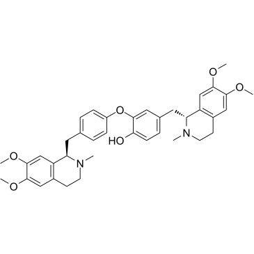
Dauricine
-
GC49913
Davunetide (acetate)
神経保護作用を持つADNP由来のペプチド

-
GC63364
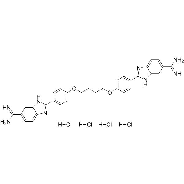
DB2115 tertahydrochloride
L-フコース類似体である 2-デオキシ-2-フルオロ-L-フコースは、フコシル化阻害剤です。 2-デオキシ-2-フルオロ-L-フコースは、哺乳動物細胞における GDP-フコースの de novo 合成を阻害します。フコシル化は、多くのヒトがんの進行を示す比較的明確なバイオマーカーです。たとえば、膵臓がんや肝細胞がんなどです。
-
GC62222
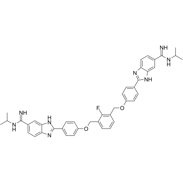
DB2313
DB2313 は、アポトーシスが 14 nM の強力な転写因子 PU.1 阻害剤です。 DB2313 は、PU.1 と標的遺伝子プロモーターとの相互作用を妨害します。 DB2313 は、急性骨髄性白血病 (AML) 細胞のアポトーシスを誘導し、抗がん効果があります。
-
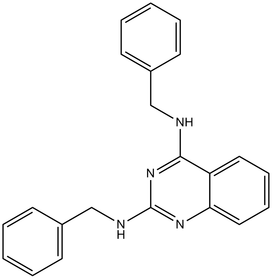
GC10824
DBeQ
DBeQ は、p97(wt) および p97(C522A) の IC50 値がそれぞれ 1.5 μM および 1.6 μM である、選択的、強力、可逆的、および ATP 競合性の p97 阻害剤です。 DBeQ は Vps4 も 11.5 μM の IC50 で阻害します。
-
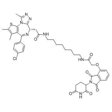
GC32719
dBET6
dBET6 は、Cereblon と BET のリガンドによって結合された非常に強力で選択的な細胞透過性の PROTAC であり、IC50 は 14 nM であり、抗腫瘍活性があります。
-
GC19485
DC661
DC661 は、強力なパルミトイルタンパク質チオエステラーゼ 1 (PPT1) 阻害剤であり、オートファジーを阻害し、抗リソソーム剤として作用します。抗がん作用。

-
GC16795
DCA
DCAは、がん細胞のミトコンドリアにおける代謝調節因子であり、抗がん活性を持ちます。DCAはPDHKを阻害し、腫瘍微小環境中の乳酸濃度を低下させます。DCAは反応性酸素種(ROS)の生成を増加させ、がん細胞アポトーシスを促進します。また、DCAはNKCC阻害剤としても機能します。

-
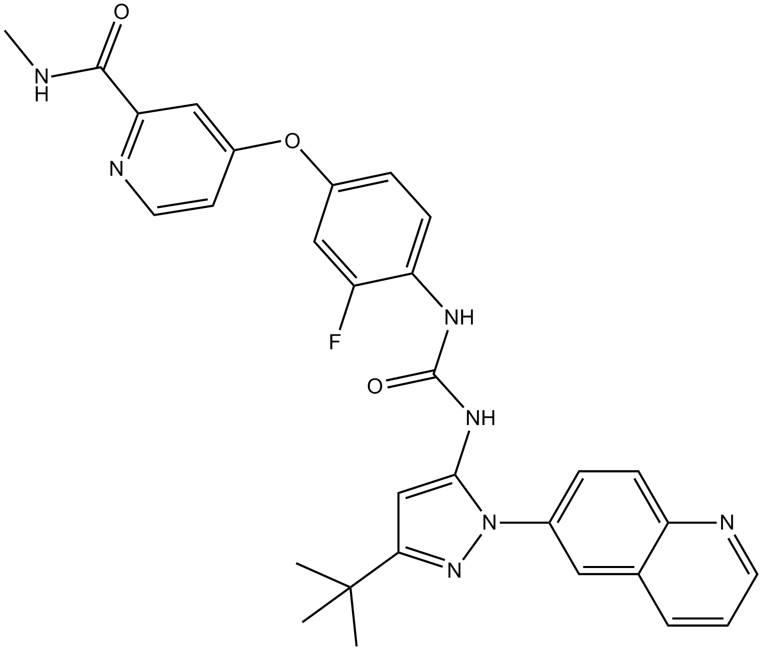
GC14007
DCC-2036 (Rebastinib)
DCC-2036 (レバスチニブ) (DCC-2036) は、Abl1WT および Abl1T315I に対する経口活性の非 ATP 競合的 Bcr-Abl 阻害剤であり、IC50 はそれぞれ 0.8 nM および 4 nM です。 DCC-2036 (レバスチニブ) は、SRC、KDR、FLT3、および Tie-2 も阻害し、c-Kit に対しては低い活性を示します。
-
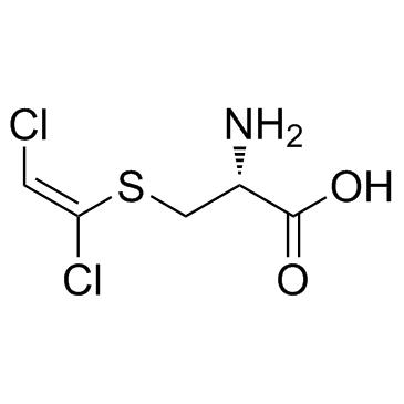
GC33934
DCVC
DCVC (S-[(1E)-1,2-ジクロロエテニル]--L-システイン) は、トリクロロエチレン (TCE) の生理活性代謝物です。
-
GC64971
DDO-7263
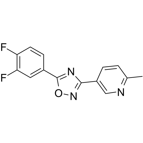
1,2,4-オキサジアゾール誘導体である DDO-7263 は、強力な Nrf2-ARE 活性化剤です。
-
GC52191
Deacetylanisomycin
デアセチルアニソマイシンは、植物の強力な成長調節因子であり、アニソマイシンの不活性な誘導体です。

-
GC15255
Decitabine(NSC127716, 5AZA-CdR)
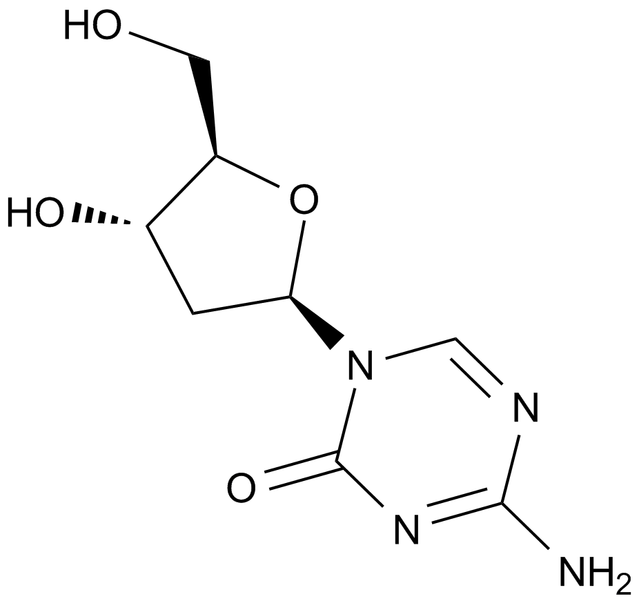
5-アザシチジンの2'デオキシアナログ
-
GC31892
Decursin ((+)-Decursin)
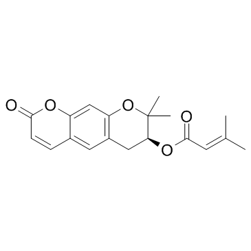
デカーシン ((+)-デカーシン) ((+)-デカーシン ((+)-デカーシン)) は強力な抗腫瘍剤です。
-
GC18556
Degarelix (acetate)
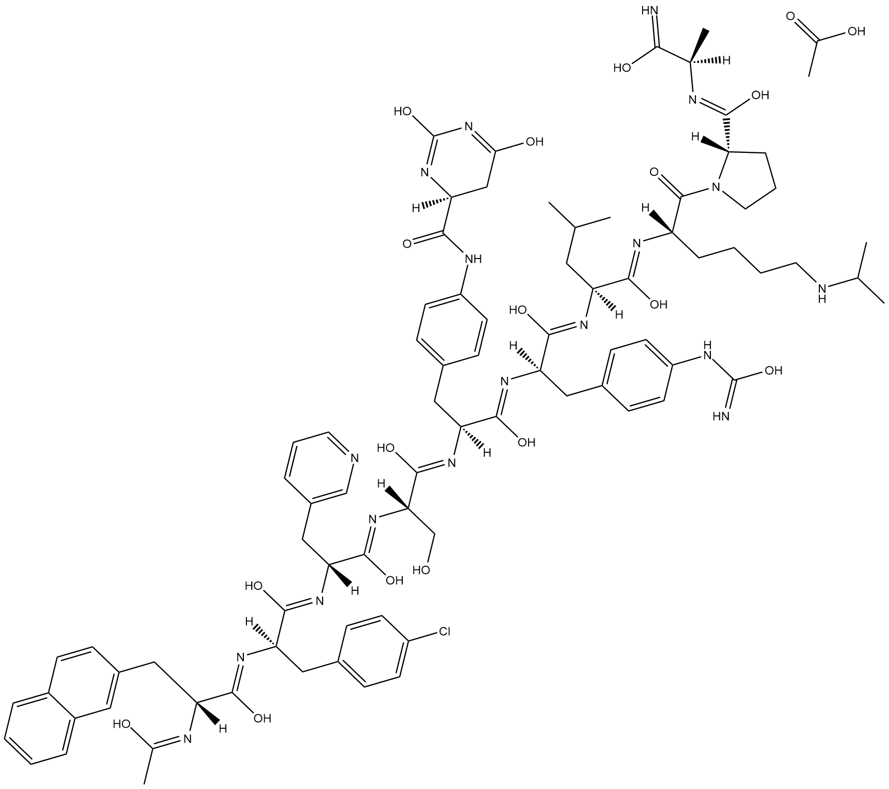
デガレリックスは、合成ゴナドトロピン放出ホルモン受容体(GNRHR)拮抗剤です(人間の受容体を発現するHEK293細胞におけるIC50 = 3 nM)。
-
GC15484
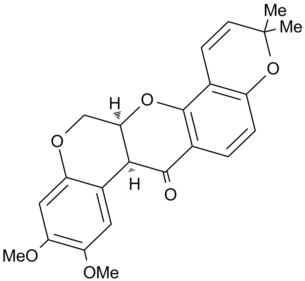
Deguelin
強力な抗増殖作用を持つロテノイド化合物
-
GC35829
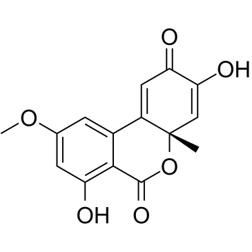
Dehydroaltenusin
デヒドロアルテヌシンは、真核生物の DNA ポリメラーゼ α の低分子選択的阻害剤であり、IC50 値が 0.68 μM の真菌によって産生される抗生物質の一種です。哺乳動物の pol α 活性に対するデヒドロアルテヌシンの阻害作用様式は、DNA テンプレート プライマーに関しては競合的であり (Ki=0.23 μM)、2'-デオキシリボヌクレオシド 5'-三リン酸基質に関しては非競合的です (Ki=0.18 μM)。 .デヒドロアルテヌシンは、癌細胞周期を S 期で停止させ、アポトーシスを引き起こします。デヒドロアルテヌシンは、in vivo でヒト腺癌腫瘍に対する抗腫瘍活性を持っています。
-
GN10040
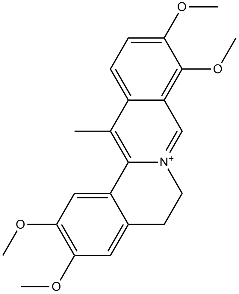
Dehydrocorydaline
-
GC35834
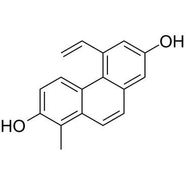
Dehydroeffusol
Dehydroeffusol は、薬草 Juncus effuses からのフェナントレンです。 Dehydroeffusol は、腫瘍抑制性小胞体ストレスと中等度のアポトーシスを選択的に誘導することにより、胃癌細胞の増殖と腫瘍原性を阻害します。非常に低い毒性を示します。
-
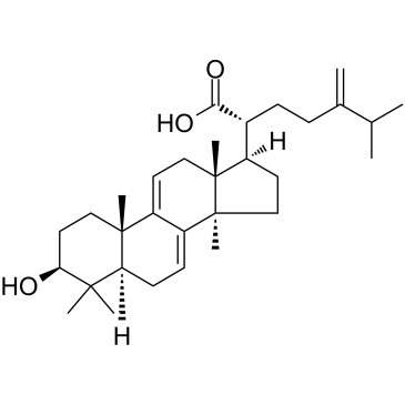
GC38452
Dehydrotrametenolic acid
デヒドロトラメテノール酸は、Poria cocos の菌核から分離されたステロールです。
-
GC16214
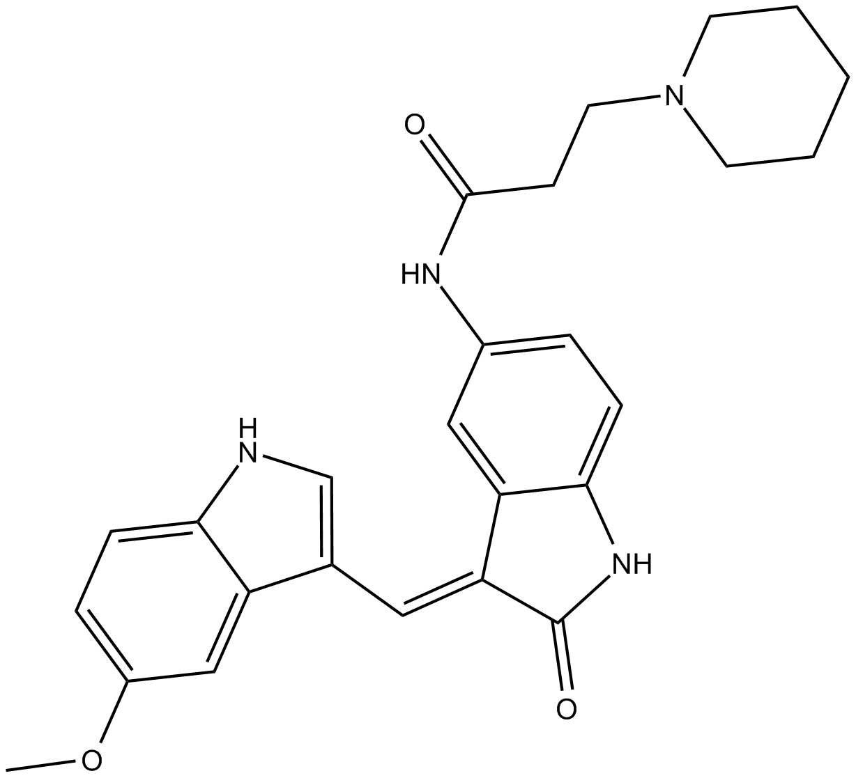
DEL-22379
DEL-22379 は ERK 二量体化阻害剤です。 DEL-22379は、低いナノモル濃度でも結合が検出可能ですが、低マイクロモル範囲で推定されるKdでERK2に容易に結合します。 ERK2 の二量体化は、約 0.5 μM の IC50 で徐々に阻害されます。
-
GC43406
Delphinidin (chloride)
アントシアニジンの 1 つであるデルフィニジン (塩化物) は、ベリーや赤ワインから分離されます。

-
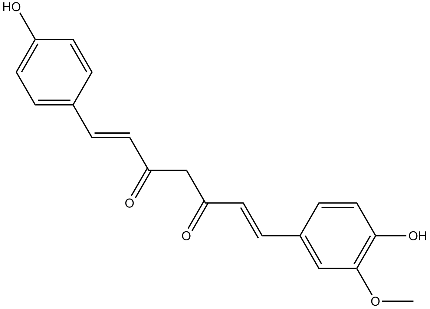
GN10529
Demethoxycurcumin
-
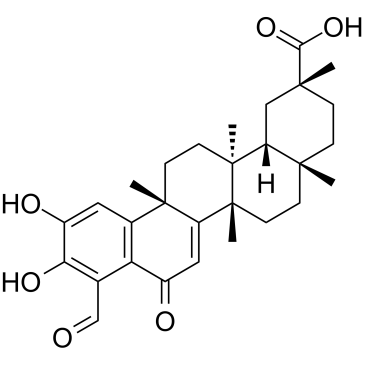
GC38160
Demethylzeylasteral
多様な生物学的活性を持つノルトリテルペノイド
-
GC43408
Deoxycholic Acid (sodium salt hydrate)
胆汁酸の一種であるデオキシコール酸(コラン酸)ナトリウム水和物は、腸内代謝の副産物であり、Gタンパク質共役胆汁酸受容体TGR5を活性化します。

-
GC47187
Deoxycholic Acid-d4
デオキシコール酸-d4は、重水素標識デオキシコール酸です。

-
GC68306
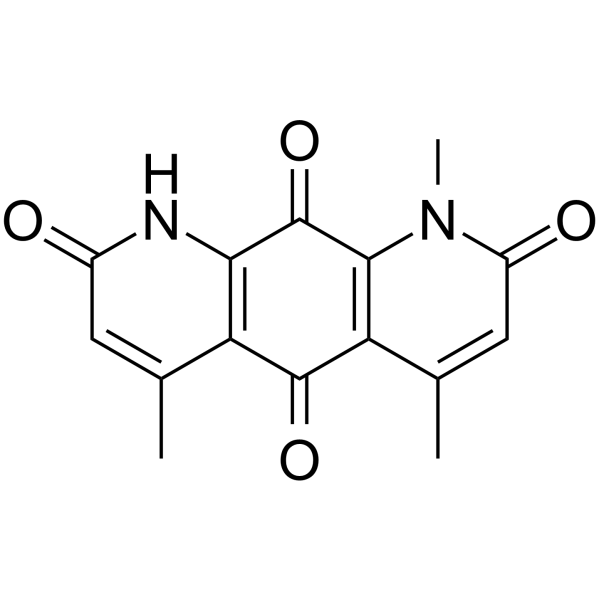
Deoxynyboquinone
-
GC38564
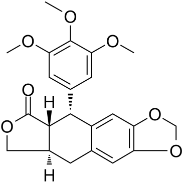
Deoxypodophyllotoxin
ポドフィロトキシンの誘導体であるデオキシポドフィロトキシン (DPT) は、Sinopodophullumhexandrum (Berberidaceae) の根茎から分離された強力な抗有糸分裂、抗炎症、および抗ウイルス特性を持つリグナンです。
-
GC16077
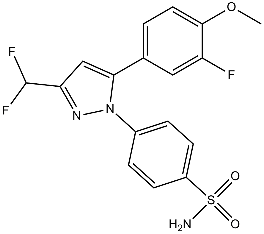
Deracoxib
選択的シクロオキシゲナーゼ 2 阻害剤であるデラコキシブは、非麻薬性、非ステロイド性抗炎症薬 (NSAID) です。
-
GC34157
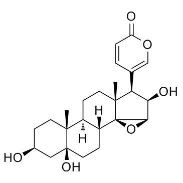
Desacetylcinobufotalin (Deacetylcinobufotalin)
デスアセチルシノブフォタリン(デアセチルシノブフォタリン)は天然化合物です。アポトーシス誘導剤であり、HepG2 細胞に対して顕著な阻害効果を示し、IC50 値は 0.0279μmol/ml です。
-
GC62927
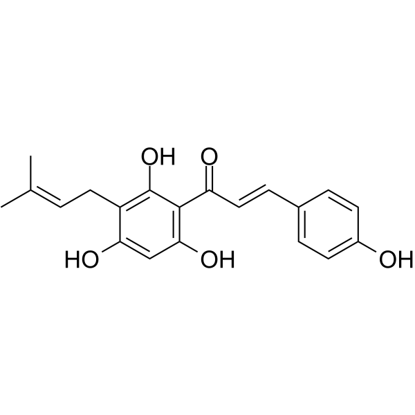
Desmethylxanthohumol
デスメチルキサントフモールは、ホップ毬花 (Humulus lupulus L.) から分離されたプレニル化ヒドロキシカルコンです。デスメチルキサントフモールは、強力なアポトーシス誘導剤です。デスメチルキサントフモールには、抗原形質、抗増殖、および抗酸化生物活性があります。
-
GC38221
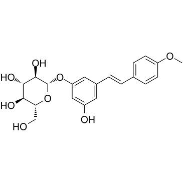
Desoxyrhaponticin
Desoxyrhaponticin は、チベットの栄養食品 Rheum tanguticum Maxim からのスチルベン配糖体です。デオキシラポンティシンは、脂肪酸シンターゼ (FASN) 阻害剤であり、ヒト癌細胞に対してアポトーシス効果があります。
-
GC10661
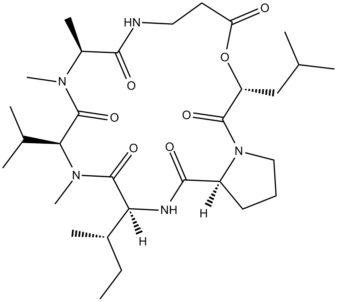
Destruxin B
昆虫病原菌Metarhizium anisopliaeから分離されたデストルキシンBは、殺虫作用と抗がん作用を持つシクロデプシペプチドの1つです。
-
GC10520
Dextran sulfate sodium salt (M.W 200000)
デキストラン硫酸ナトリウム塩は、分子量範囲が 35000 ~ 45000 の無水グルコースのポリマーです。

-
GC38250
Dextran sulfate sodium salt (MW 16000-24000)
硫酸化ポリ糖類
-
GC38252
Dextran sulfate sodium salt (MW 35000-45000)
硫酸化多糖類
-
GC38253
Dextran sulfate sodium salt (MW 4500-5500)
硫酸化多糖類
-
GC38251
Dextran sulfate sodium salt (MW 450000-550000)
硫酸化多糖類
-
GC43436
Diacetylcercosporin
ジアセチルセルコスポリンは、CercosporaとSeptoriaによって生産されるペリレンキノンであり、多様な生物学的活性を持っています。

-
GC45992
Diallyl Tetrasulfide
多様な生物学的活性を持つ有機硫黄化合物

-
GC43439
Diallyl Trisulfide
ジアリルトリスルフィドはニンニクから分離されます。

-
GC35859
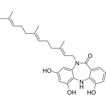
Diazepinomicin
ジアゼピノマイシン (TLN-4601) は、Micromonospora sp. によって産生される二次代謝産物です。ジアゼピノマイシン (TLN-4601) は、EGF 誘導 Ras-ERK MAPK シグナル伝達経路を阻害し、アポトーシスを誘導します。 K-Ras 変異モデルの抗腫瘍剤です。
-
GC61419
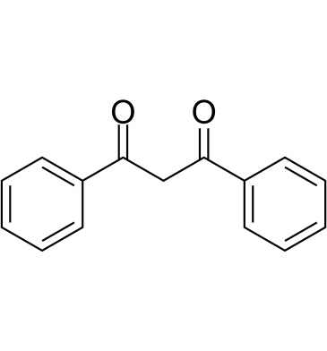
Dibenzoylmethane
甘草の微量成分であるジベンゾイルメタンは、Nrf2を活性化し、さまざまな癌や酸化的損傷を防ぎます.
-
GC64139
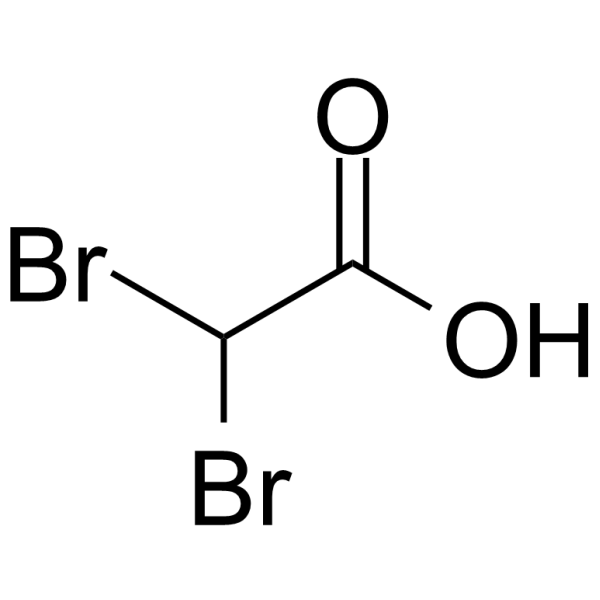
Dibromoacetic acid
-
GC14622
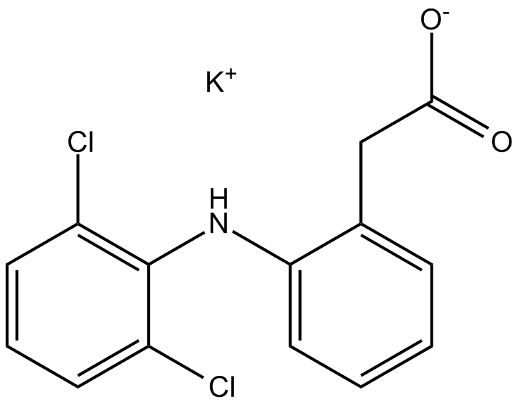
Diclofenac Potassium
ジクロフェナク カリウムは、強力で非選択的な抗炎症剤であり、COX 阻害剤として作用し、CHO 細胞におけるヒト COX-1 および COX-2 の IC50 は 4 および 1.3 nM、ヒツジ COX- の IC50 は 5.1 および 0.84 μM です。 1 と COX-2、それぞれ 。
-
GN10751
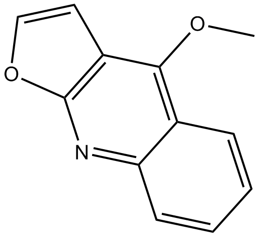
Dictamnine
-
GC49153
Didemnin B
Didemnin Bは、海洋チュニケートが生産する環状デプシペプチドであり、特異的にGTP結合型のEEF1Aに結合する。

-
GC39808
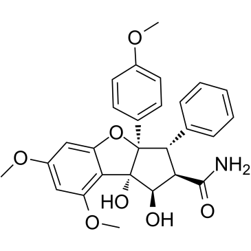
Didesmethylrocaglamide
ロカグラミドの誘導体であるジデスメチルロカグラミドは、強力な真核生物開始因子 4A (eIF4A) 阻害剤です。ジデスメチルロカグラミドは、IC50 が 5 nM の強力な増殖阻害活性を持っています。ジデスメチルロカグラミドは、複数の成長促進シグナル伝達経路を抑制し、腫瘍細胞のアポトーシスを誘導します。抗腫瘍活性。
-
GC38179
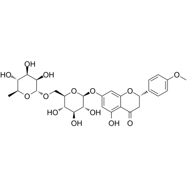
Didymin
柑橘類に含まれるフラボノイド配糖体であるジジミンには、抗酸化作用があります。ディジミンは、神経芽細胞腫において N-Myc を阻害し、RKIP をアップレギュレートすることによってアポトーシスを誘導します。
-
GC15578
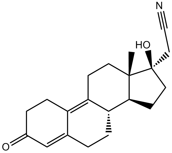
Dienogest
Dienogest (STS-557) は、COX-2、mPGES-1、およびアロマターゼの遺伝子発現を効果的に減少させる、経口的に活性で選択的なプロゲステロン受容体アゴニストです。
-
GC43452
Diffractaic Acid
Uの主成分である回折酸。

-
GC64375
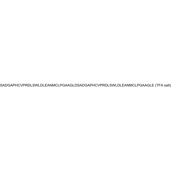
Difopein TFA
14-3-3 タンパク質 (高度に保存された真核生物調節分子) の特異的かつ競合的な阻害剤であるジフォペイン (TFA) は、14-3-3 が標的タンパク質に結合する能力をブロックし、14-3-3/リガンド相互作用を阻害します。 . Difopein (TFA) はアポトーシスの誘導を引き起こし、シスプラチンの細胞を殺す能力を高めます。
-
GC49842
Digoxigenin Bisdigitoxoside
Na+/K+-ATPase阻害剤

-
GN10056
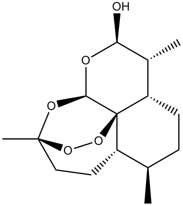
Dihydroartemisinin
-
GC38617
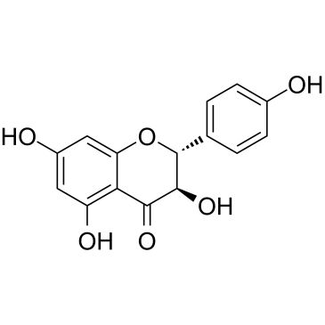
Dihydrokaempferol
ジヒドロケンフェロールは、バウヒニア チャンピオンニ (Benth) から分離されます。
-
GC43462
Dihydrolipoic Acid
ジヒドロリポ酸 (DHLA) は、ほぼすべての酸素中心のラジカルを消去できる優れた抗酸化剤です 。

-
GC38620
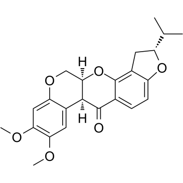
Dihydrorotenone
天然の農薬であるジヒドロロテノンは、強力なミトコンドリア阻害剤です。
-
GC41112
Dihydroxy Melphalan
ジヒドロキシメルファランは、メルファランの不活性な分解生成物です。

-
GC43463
DiIC1(5)
DiIC1(5)は、ミトコンドリア膜電位の破壊を検出するためのシグナルオフ型蛍光プローブです。

-
GC38772
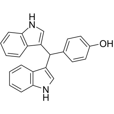
DIM-C-pPhOH
DIM-C-pPhOH は、核内受容体 4A1 (NR4A1) 拮抗薬です。 DIM-C-pPhOH は、がん細胞の増殖と mTOR シグナル伝達を阻害し、アポトーシスと細胞ストレスを誘導します。 DIM-C-pPhOH は、ACHN 細胞と 786-O 細胞でそれぞれ 13.6 μM と 13.0 μM の IC50 値で細胞増殖を抑制します。
-
GC16590
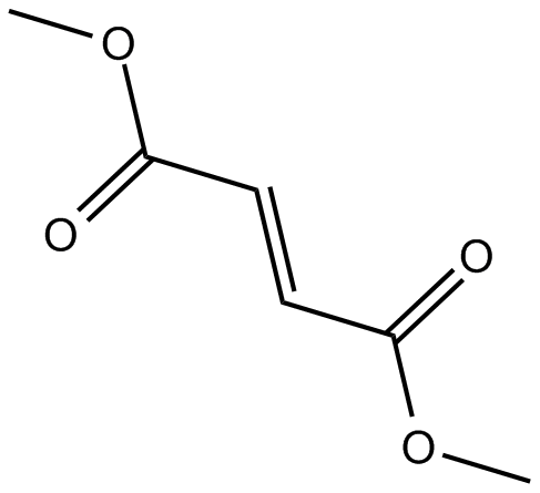
Dimethyl Fumarate
フマル酸ジメチル (DMF) は、経口活性で脳浸透性の Nrf2 活性化剤であり、抗酸化遺伝子発現のアップレギュレーションを誘導します。
-
GC62936
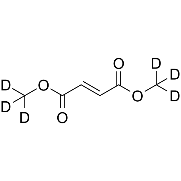
Dimethyl fumarate D6
フマル酸ジメチル D6 は、フマル酸ジメチルと標識された重水素です。
-
GC25351
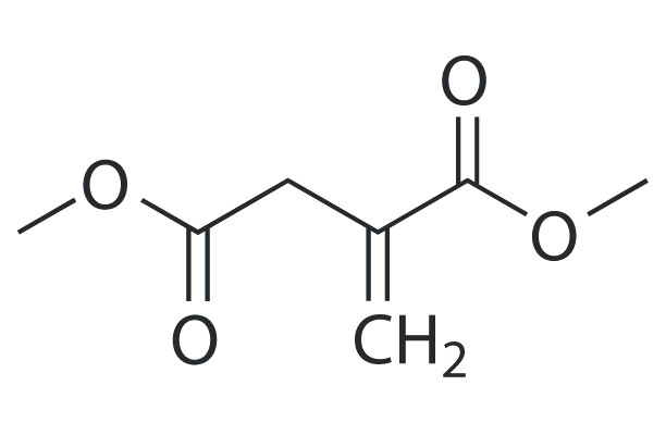
Dimethyl itaconate
ジメチルイタコネートは、LPS誘導性Nod様受容体蛋白質3(NLRP3)インフラマソームおよび核因子2 / ヘムオキシゲナーゼ-1(NRF2 / HO-1)経路の調節を介して、神経毒性から神経保護的な主要な星状細胞を再プログラムすることができます。
-
GN10531
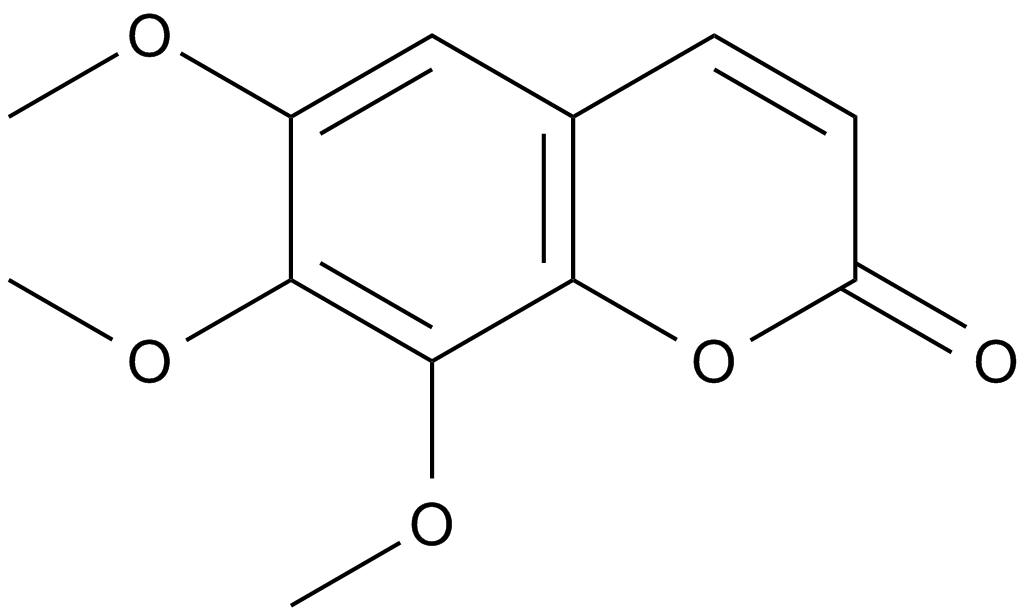
Dimethylfraxetin
-
GC17648
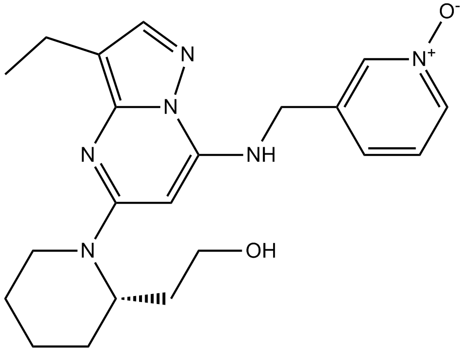
Dinaciclib(SCH727965)
Dinaciclib(SCH727965) (SCH 727965) は CDK の強力な阻害剤であり、CDK2、CDK5、CDK1、CDK9 の IC50 はそれぞれ 1 nM、1 nM、3 nM、4 nM です。
-
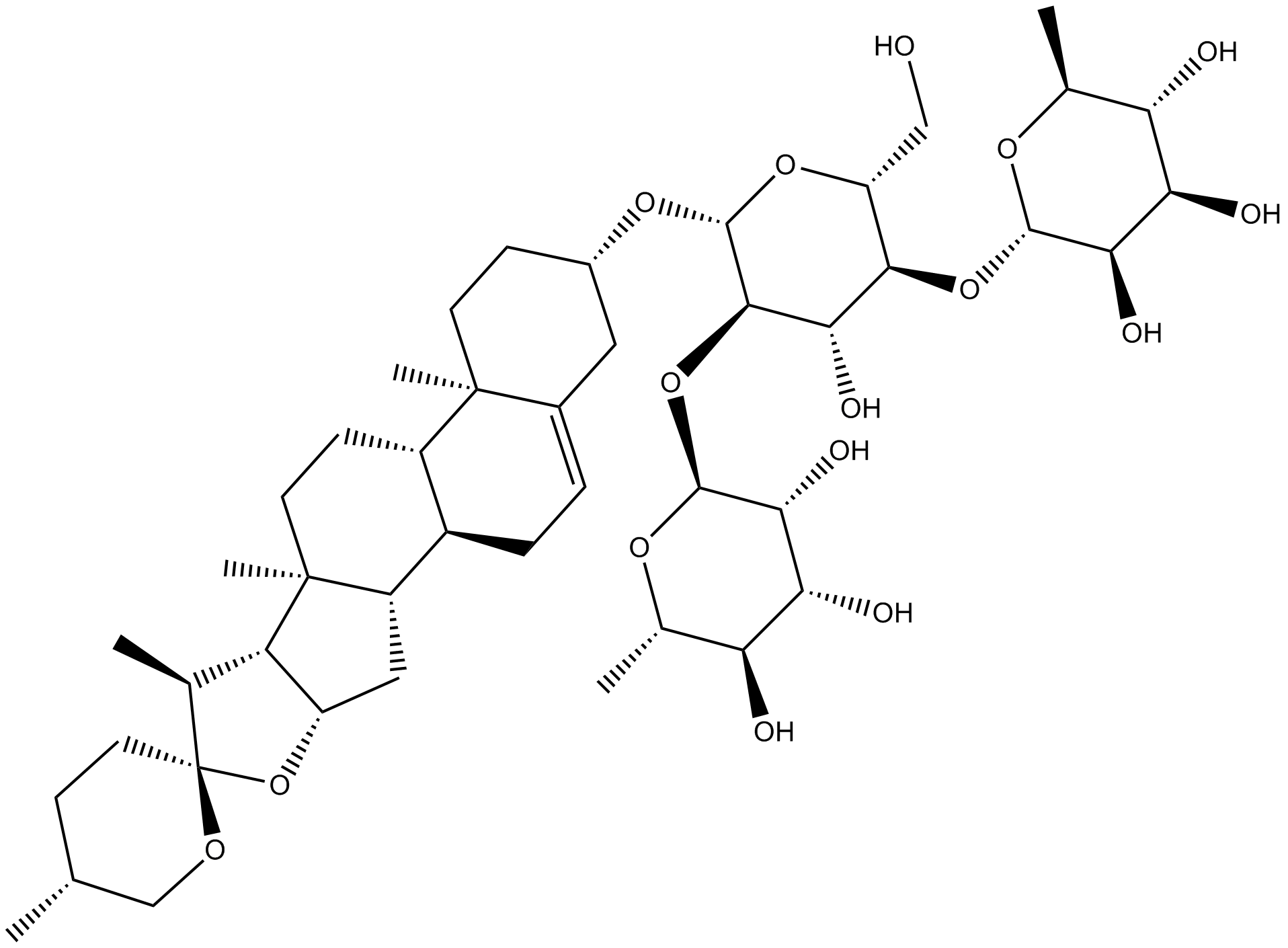
GN10284
Dioscin
-
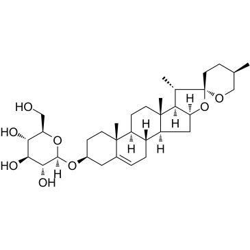
GC38408
Diosgenin glucoside
-
GC60782
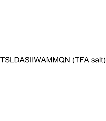
Disitertide TFA
ジシテルチド (P144) TFA は、その受容体との相互作用をブロックするように特別に設計された、ペプチド性トランスフォーミング増殖因子-ベータ 1 (TGF-β1) 阻害剤です。ジシテルチド (P144) TFA は、PI3K 阻害剤であり、アポトーシス誘導剤でもあります。
-
GC10801
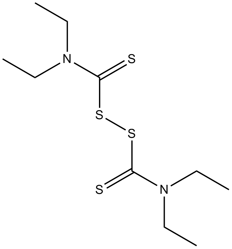
Disulfiram
アルデヒド脱水素酵素の不可逆的阻害剤
-
GC39295
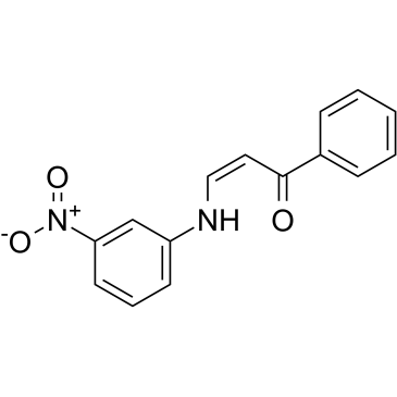
DJ001
DJ001 は、IC50 が 1.43 μM の非常に特異的、選択的かつ非競合的なプロテイン チロシン ホスファターゼ-σ (PTPσ) 阻害剤です。
-
GC49164
DM4 (hydrate)
メイタンシンの誘導体

-
GC35882
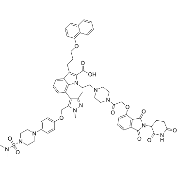
dMCL1-2
dMCL1-2 は、30 nM の KD で MCL1 に結合するセレブロンに基づく、骨髄細胞性白血病 1 (MCL1) (Bcl-2 ファミリーのメンバー) の強力かつ選択的な PROTAC です。 dMCL1-2 は、MCL1 の分解によって細胞アポトーシス機構を活性化します。
-
GC40192
DMPAC-Chol
DMPAC-Cholは陽イオン性のコレステロール誘導体であり、遺伝子トランスフェクションのためにリポソーム形成に使用されています。

-
GC61466
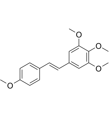
DMU-212
DMU-212 はレスベラ トロールのメチル化誘導体であり、抗有糸分裂、抗増殖、抗酸化、アポトーシス促進活性を備えています。 DMU-212 は、アポトーシスの誘導と ERK1/2 タンパク質の活性化を介して有糸分裂停止を誘導します。 DMU-212 は経口活性があります。
-
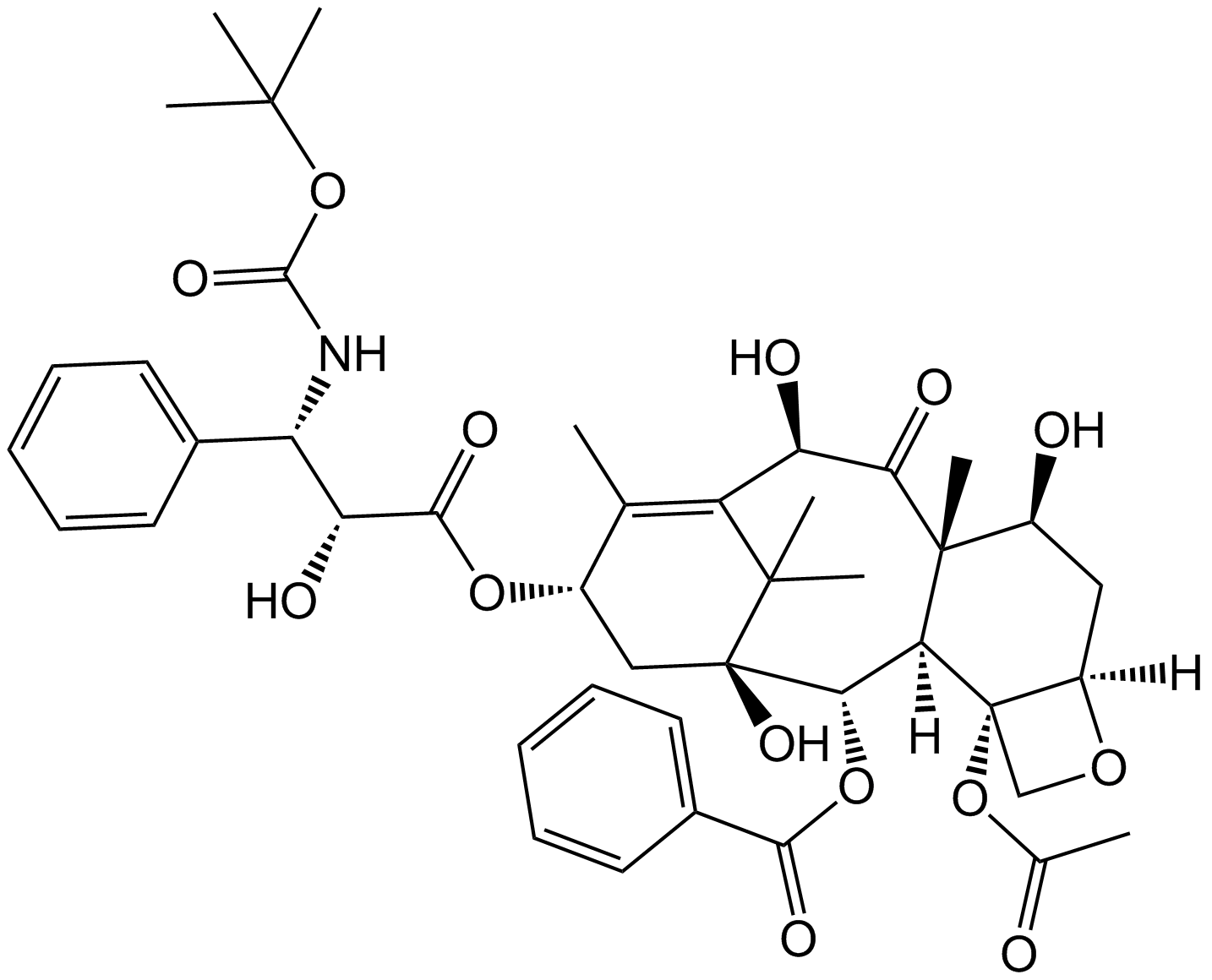
GC16684
Docetaxel
ドセタキセルは、マイクロチューブの解重合を阻害し、bcl-2およびbcl-xL遺伝子発現の影響を抑制することにより、抗腫瘍剤です。
-
GA21397
Dolastatin 15
Dolabella auricularia 由来のデプシペプチドである Dolastatin 15 (DLS 15) は、抗チューブリン剤 Dolastatin 10 に構造的に関連する強力な抗有糸分裂剤です。 Dolastatin 15 は、多発性骨髄腫細胞の細胞周期停止とアポトーシスを誘導します。ドラスタチン 15 は、ADC 細胞毒素として使用できます。
-
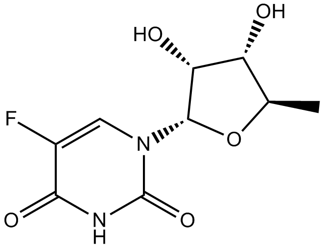
GC11099
Doxifluridine
ドキシフルリジン (Ro 21-9738; 5'-DFUR) は、PC9-DPE2 細胞のチミジンホスホリラーゼ活性化剤であり、IC50 は 0.62 μM です。
-
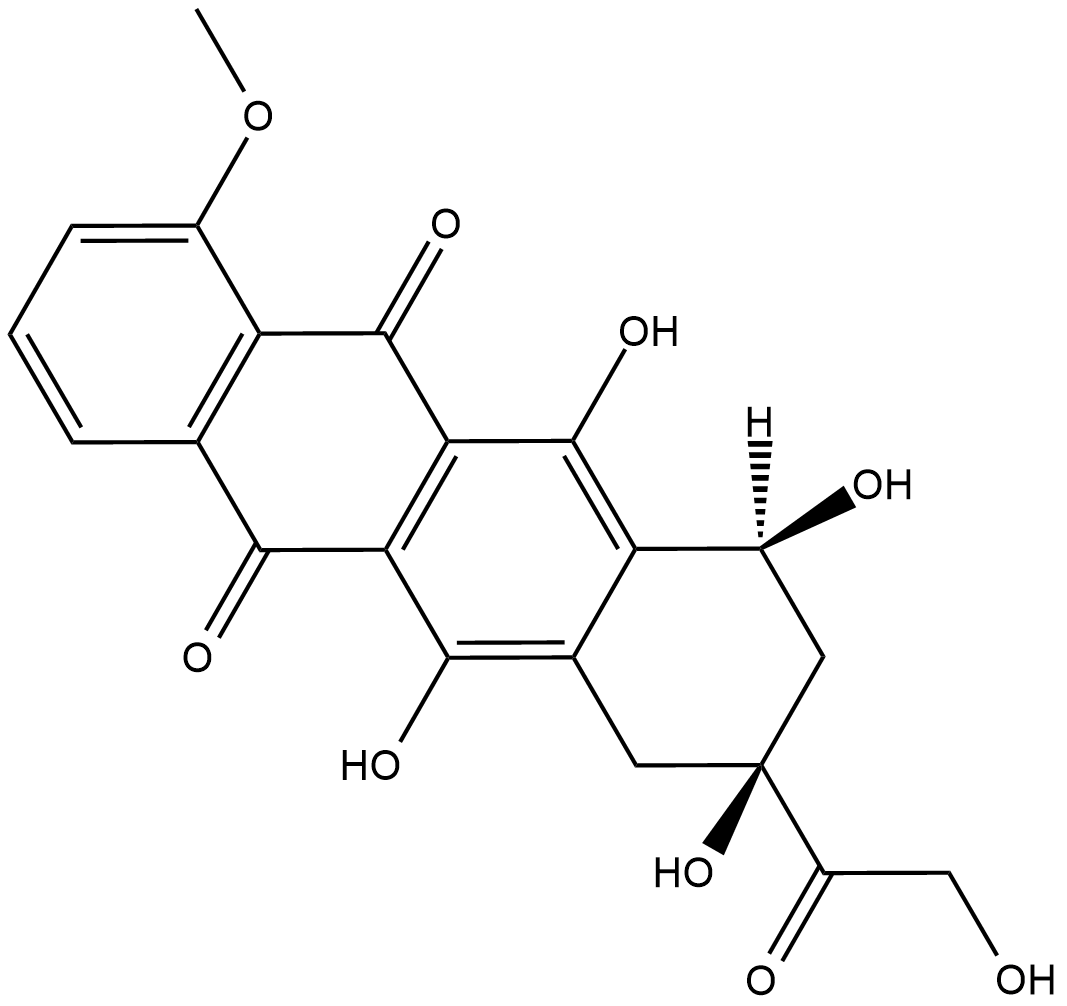
GC18439
Doxorubicinone
ドキソルビシノンは、抗癌化学療法剤ドキソルビシンの代謝産物です。ドキソルビシンは、強力なヒト DNA トポイソメラーゼ I およびトポイソメラーゼ II 阻害剤であり、IC50 はそれぞれ 0.8 μM および 2.67 μM です。
-
GC15399
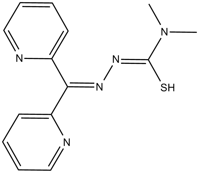
Dp44mT
Dp44mT は、選択的な抗がん作用を持つ鉄キレート剤です。
-
GC33384
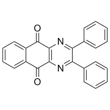
DPBQ
DPBQ は p53 を活性化し、倍数体特異的な方法でアポトーシスを引き起こしますが、トポイソメラーゼを阻害したり DNA に結合したりしません。 DPBQ は p53 の発現とリン酸化を誘発し、この効果は 4 倍体細胞に特異的です。
-
GC35898
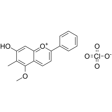
Dracorhodin perchlorate
ドラコロジン過塩素酸塩(Dracohodin perchlorate)は、生薬龍の血から抽出された天然物です。ドラコロジン過塩素酸塩は、細胞増殖を阻害し、細胞周期の停止とアポトーシスを誘導します。
-
GC39707
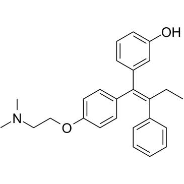
Droloxifene
タモキシフェン誘導体であるドロロキシフェンは、経口的に活性で選択的なエストロゲン受容体モジュレーターです。ドロロキシフェンは、抗エストロゲン作用および抗着床作用を示します。ドロロキシフェンは、MCF-7 細胞で p53 の発現とアポトーシスを誘導します。ドロロキシフェンは、卵巣摘出ラットの骨量減少を防ぎます。
-
GC13706
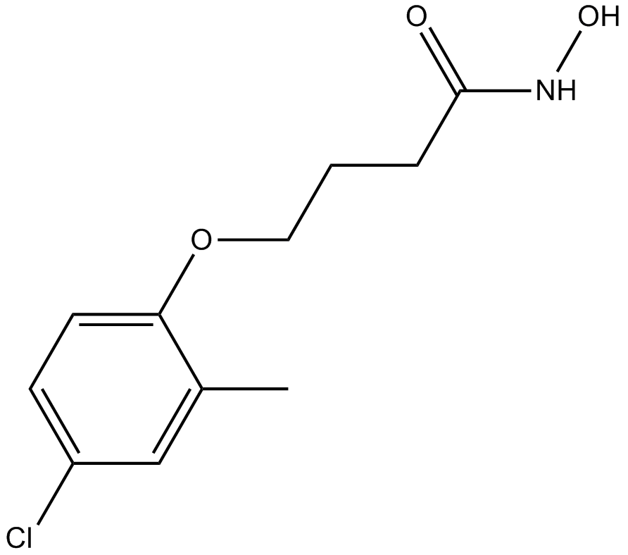
Droxinostat
HDAC3、HDAC6、およびHDAC8の阻害剤
-
GC35908
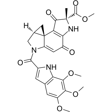
Duocarmycin A
有名な抗腫瘍抗生物質の 1 つであるデュオカルマイシン A は、DNA アルキル化剤であり、DNA の AT リッチ配列の 3' 末端にあるアデニン N3 を効率的にアルキル化します。化学療法剤としてのデュオカルマイシン A は、クロマチン凝縮、DNA ヒストグラム パターンでのサブ G1 蓄積、プロカスパーゼ 3 および 9 レベルの低下など、HLC-2 細胞に典型的なアポトーシス変化をもたらします。
-
GC35913
Durvalumab
デュルバルマブ (MEDI 4736) は、ヒト化抗 PD-L1 モノクローナル抗体です。デュルバルマブ (MEDI4736) は、PD-L1 と PD-1 および CD80 の両方への結合を完全にブロックし、IC50 はそれぞれ 0.1 および 0.04 nM です。

-
GC10893
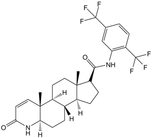
Dutasteride
デュタステリド (GG745) は、両方の 5α-レダクターゼ アイソザイムの強力な阻害剤です。デュタステリドは、DHT と構造的に類似しているため、アンドロゲン受容体 (AR) にオフターゲット効果を及ぼす可能性があります。
-
GC62590
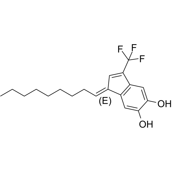
E64FC26
E64FC26 は、PDIA1、PDIA3、PDIA4、TXNDC5、および PDIA6 に対してそれぞれ 1.9、20.9、25.9、16.3、および 25.4 μM の IC50 を持つ、タンパク質ジスルフィドイソメラーゼ (PDI) ファミリーの強力な汎阻害剤です。 E64FC26 は抗骨髄腫活性を示します。
-
GC39377
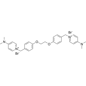
EB-3D
EB-3D は強力かつ選択的なコリンキナーゼ α (ChoKα) 阻害剤であり、ChoKα1 に対する IC50 は 1 μM です。 EB-3D は、ChoKα 発現、AMPK 活性化、アポトーシス、小胞体ストレス、および脂質代謝に影響を及ぼします。 EB-3D は、T 白血病細胞株のパネルで強力な抗増殖活性を示します。抗がん作用。
-
GC50282
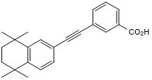
EC 19
合成レチノイド;幹細胞の分化を誘導する
-
GC39174
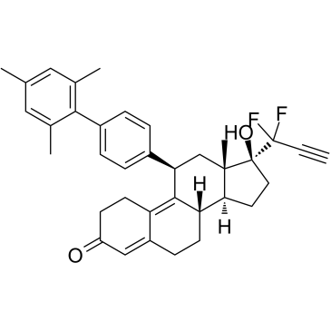
EC359
EC359 は 10.2 nM の Kd を持つ、強力で選択的、高親和性で経口的に活性な白血病抑制因子受容体 (LIFR) 阻害剤であり、LIFR と直接相互作用して LIF/LIFR 相互作用を効果的にブロックします。
-
GC38149
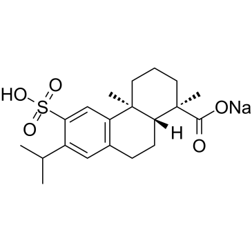
Ecabet sodium
Ecabet ナトリウム (TA-2711) は現在、ROS 産生を阻害し、ヘリコバクター ピロリの除菌を改善することにより、一部の胃腸疾患に適用されています 。
-
GN10504
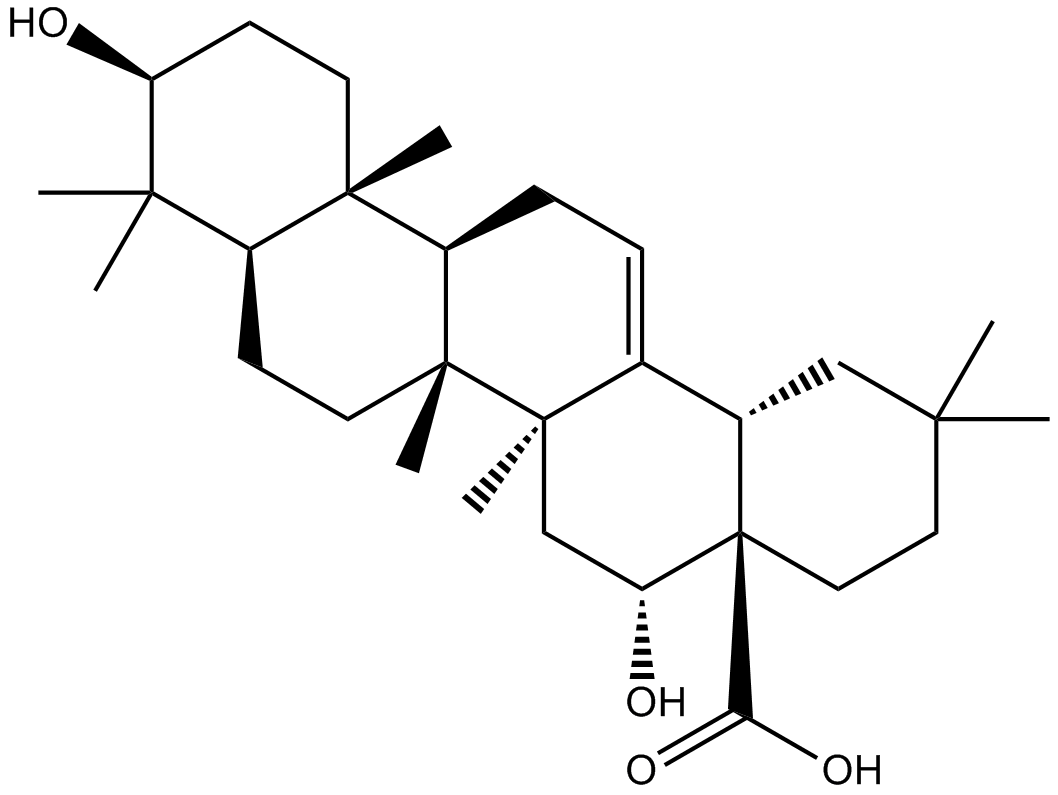
Echinocystic acid
-
GC47279
Echinosporin
抗菌および抗がん活性を持つ細菌代謝産物

-
GC63871
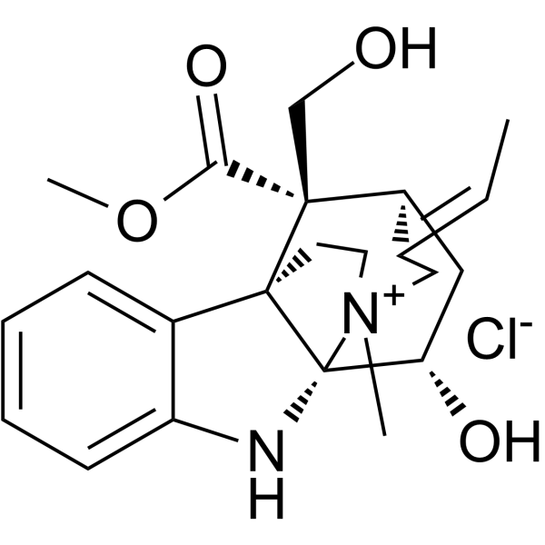
Echitamine chloride
塩化エキタミンは、強力な抗腫瘍活性を持つアルストニアに存在する主要なモノテルペン インドール アルカロイドです。塩化エキタミンは、DNA の断片化と細胞のアポトーシスを誘導します。塩化エキタミンは、10.92 μ の IC50 で膵臓リパーゼを阻害します;M.
-
GC63470
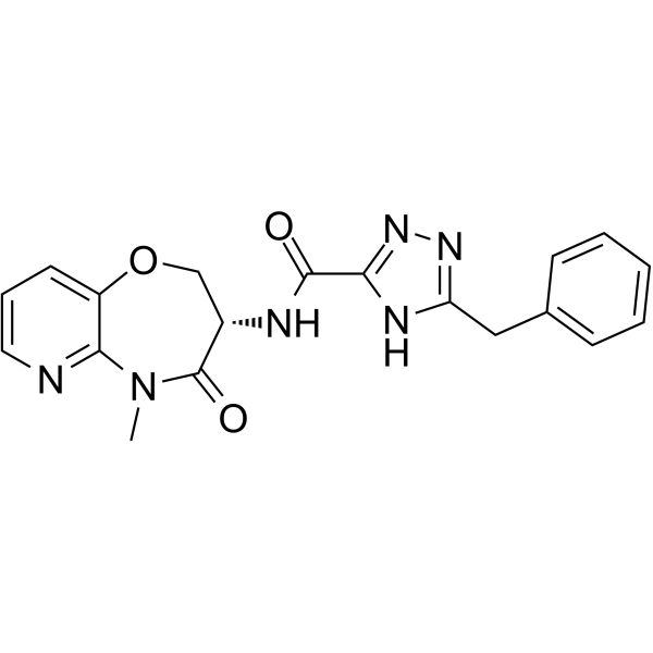
Eclitasertib
Eclitasertib (DNL-758) は、IC50 が 1 μM 未満の強力な受容体相互作用プロテインキナーゼ 1 (RIPK1) 阻害剤です (特許 WO2017136727A2 より、実施例 42)。
-
GC33156
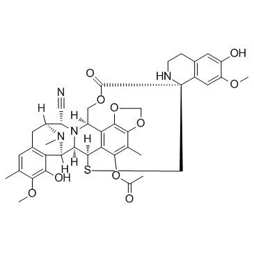
Ecteinascidin 770 (Ecteinascidine 770)
エクテナサイジン 770 (Ecteinascidine 770) (ET-770) は、強力な抗がん作用を持つ 1,2,3,4-テトラヒドロイソキノリン アルカロイドです。 U373MG 細胞を 4.83 nM の IC50 で阻害します。
-
GC12838
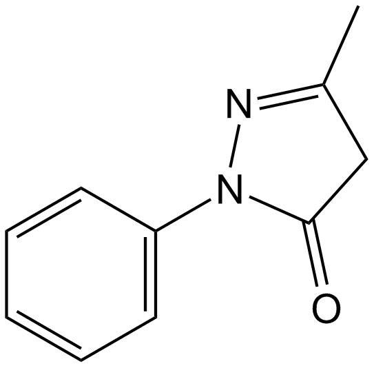
Edaravone
エダラボンは強力な新規フリーラジカルスカベンジャーであり、組織プラスミノーゲン活性化因子で処理されたラットの MMP-9 関連の脳出血を抑制します。
-
GC62562
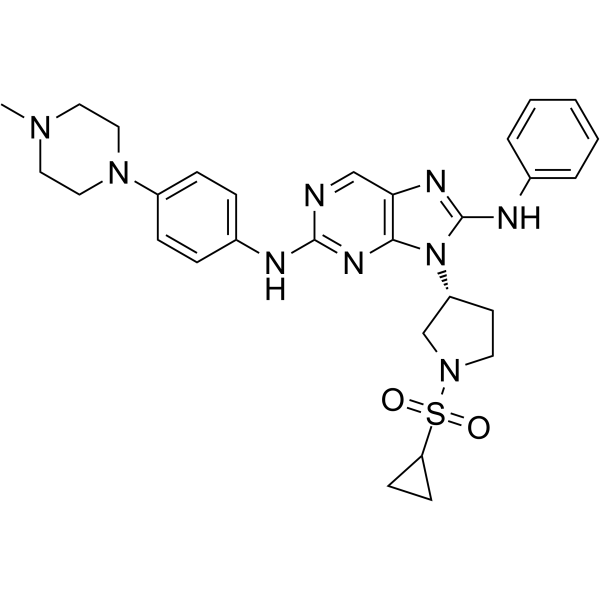
EGFR-IN-11
EGFR-IN-11 は、三重変異体 EGFRL858R/T790M/C797S に対する IC50 が 18 nM の第 4 世代 EGFR-チロシンキナーゼ阻害剤 (EGFR-TKI) です。 EGFR-IN-11 は、EGFR リン酸化を有意に抑制し、アポトーシスを誘導し、G0/G1 で細胞周期を停止させます。
-
GC14756
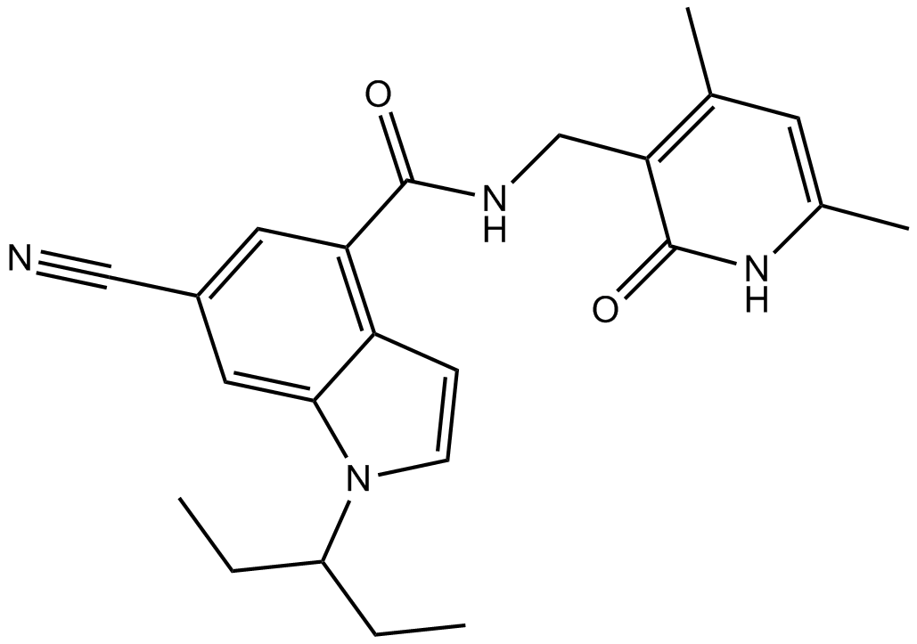
EI1
EI1 (KB-145943) は、EZH2 (WT) と EZH2 (Y641F) の IC50 がそれぞれ 15 nM と 13 nM の強力で選択的な EZH2 阻害剤です。
-
GC64864
EJMC-1
EJMC-1 は、42 μM の IC50 値を持つ中程度に強力な TNF-α 阻害剤です 。
