Apoptosis
As one of the cellular death mechanisms, apoptosis, also known as programmed cell death, can be defined as the process of a proper death of any cell under certain or necessary conditions. Apoptosis is controlled by the interactions between several molecules and responsible for the elimination of unwanted cells from the body.
Many biochemical events and a series of morphological changes occur at the early stage and increasingly continue till the end of apoptosis process. Morphological event cascade including cytoplasmic filament aggregation, nuclear condensation, cellular fragmentation, and plasma membrane blebbing finally results in the formation of apoptotic bodies. Several biochemical changes such as protein modifications/degradations, DNA and chromatin deteriorations, and synthesis of cell surface markers form morphological process during apoptosis.
Apoptosis can be stimulated by two different pathways: (1) intrinsic pathway (or mitochondria pathway) that mainly occurs via release of cytochrome c from the mitochondria and (2) extrinsic pathway when Fas death receptor is activated by a signal coming from the outside of the cell.
Different gene families such as caspases, inhibitor of apoptosis proteins, B cell lymphoma (Bcl)-2 family, tumor necrosis factor (TNF) receptor gene superfamily, or p53 gene are involved and/or collaborate in the process of apoptosis.
Caspase family comprises conserved cysteine aspartic-specific proteases, and members of caspase family are considerably crucial in the regulation of apoptosis. There are 14 different caspases in mammals, and they are basically classified as the initiators including caspase-2, -8, -9, and -10; and the effectors including caspase-3, -6, -7, and -14; and also the cytokine activators including caspase-1, -4, -5, -11, -12, and -13. In vertebrates, caspase-dependent apoptosis occurs through two main interconnected pathways which are intrinsic and extrinsic pathways. The intrinsic or mitochondrial apoptosis pathway can be activated through various cellular stresses that lead to cytochrome c release from the mitochondria and the formation of the apoptosome, comprised of APAF1, cytochrome c, ATP, and caspase-9, resulting in the activation of caspase-9. Active caspase-9 then initiates apoptosis by cleaving and thereby activating executioner caspases. The extrinsic apoptosis pathway is activated through the binding of a ligand to a death receptor, which in turn leads, with the help of the adapter proteins (FADD/TRADD), to recruitment, dimerization, and activation of caspase-8 (or 10). Active caspase-8 (or 10) then either initiates apoptosis directly by cleaving and thereby activating executioner caspase (-3, -6, -7), or activates the intrinsic apoptotic pathway through cleavage of BID to induce efficient cell death. In a heat shock-induced death, caspase-2 induces apoptosis via cleavage of Bid.
Bcl-2 family members are divided into three subfamilies including (i) pro-survival subfamily members (Bcl-2, Bcl-xl, Bcl-W, MCL1, and BFL1/A1), (ii) BH3-only subfamily members (Bad, Bim, Noxa, and Puma9), and (iii) pro-apoptotic mediator subfamily members (Bax and Bak). Following activation of the intrinsic pathway by cellular stress, pro‑apoptotic BCL‑2 homology 3 (BH3)‑only proteins inhibit the anti‑apoptotic proteins Bcl‑2, Bcl-xl, Bcl‑W and MCL1. The subsequent activation and oligomerization of the Bak and Bax result in mitochondrial outer membrane permeabilization (MOMP). This results in the release of cytochrome c and SMAC from the mitochondria. Cytochrome c forms a complex with caspase-9 and APAF1, which leads to the activation of caspase-9. Caspase-9 then activates caspase-3 and caspase-7, resulting in cell death. Inhibition of this process by anti‑apoptotic Bcl‑2 proteins occurs via sequestration of pro‑apoptotic proteins through binding to their BH3 motifs.
One of the most important ways of triggering apoptosis is mediated through death receptors (DRs), which are classified in TNF superfamily. There exist six DRs: DR1 (also called TNFR1); DR2 (also called Fas); DR3, to which VEGI binds; DR4 and DR5, to which TRAIL binds; and DR6, no ligand has yet been identified that binds to DR6. The induction of apoptosis by TNF ligands is initiated by binding to their specific DRs, such as TNFα/TNFR1, FasL /Fas (CD95, DR2), TRAIL (Apo2L)/DR4 (TRAIL-R1) or DR5 (TRAIL-R2). When TNF-α binds to TNFR1, it recruits a protein called TNFR-associated death domain (TRADD) through its death domain (DD). TRADD then recruits a protein called Fas-associated protein with death domain (FADD), which then sequentially activates caspase-8 and caspase-3, and thus apoptosis. Alternatively, TNF-α can activate mitochondria to sequentially release ROS, cytochrome c, and Bax, leading to activation of caspase-9 and caspase-3 and thus apoptosis. Some of the miRNAs can inhibit apoptosis by targeting the death-receptor pathway including miR-21, miR-24, and miR-200c.
p53 has the ability to activate intrinsic and extrinsic pathways of apoptosis by inducing transcription of several proteins like Puma, Bid, Bax, TRAIL-R2, and CD95.
Some inhibitors of apoptosis proteins (IAPs) can inhibit apoptosis indirectly (such as cIAP1/BIRC2, cIAP2/BIRC3) or inhibit caspase directly, such as XIAP/BIRC4 (inhibits caspase-3, -7, -9), and Bruce/BIRC6 (inhibits caspase-3, -6, -7, -8, -9).
Any alterations or abnormalities occurring in apoptotic processes contribute to development of human diseases and malignancies especially cancer.
References:
1.Yağmur Kiraz, Aysun Adan, Melis Kartal Yandim, et al. Major apoptotic mechanisms and genes involved in apoptosis[J]. Tumor Biology, 2016, 37(7):8471.
2.Aggarwal B B, Gupta S C, Kim J H. Historical perspectives on tumor necrosis factor and its superfamily: 25 years later, a golden journey.[J]. Blood, 2012, 119(3):651.
3.Ashkenazi A, Fairbrother W J, Leverson J D, et al. From basic apoptosis discoveries to advanced selective BCL-2 family inhibitors[J]. Nature Reviews Drug Discovery, 2017.
4.McIlwain D R, Berger T, Mak T W. Caspase functions in cell death and disease[J]. Cold Spring Harbor perspectives in biology, 2013, 5(4): a008656.
5.Ola M S, Nawaz M, Ahsan H. Role of Bcl-2 family proteins and caspases in the regulation of apoptosis[J]. Molecular and cellular biochemistry, 2011, 351(1-2): 41-58.
対象は Apoptosis
- Pyroptosis(15)
- Caspase(77)
- 14.3.3 Proteins(3)
- Apoptosis Inducers(71)
- Bax(15)
- Bcl-2 Family(136)
- Bcl-xL(13)
- c-RET(15)
- IAP(32)
- KEAP1-Nrf2(73)
- MDM2(21)
- p53(137)
- PC-PLC(6)
- PKD(8)
- RasGAP (Ras- P21)(2)
- Survivin(8)
- Thymidylate Synthase(12)
- TNF-α(141)
- Other Apoptosis(1144)
- Apoptosis Detection(0)
- Caspase Substrate(0)
- APC(6)
- PD-1/PD-L1 interaction(60)
- ASK1(4)
- PAR4(2)
- RIP kinase(47)
- FKBP(22)
製品は Apoptosis
- Cat.No. 商品名 インフォメーション
-
GC33043
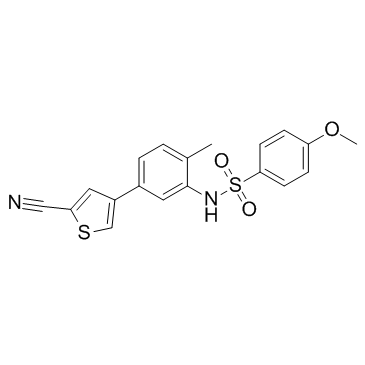
EL-102
EL-102 は、低酸素誘導因子 1 (Hif1α) 阻害剤です。 EL-102 はアポトーシスを誘導し、チューブリンの重合を阻害し、前立腺癌に対する活性を示します。 EL-102はがんの研究に使用できます。
-
GC13885
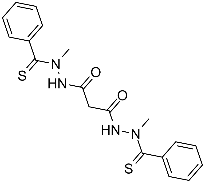
Elesclomol (STA-4783)
アポトーシス誘導剤
-
GC16827
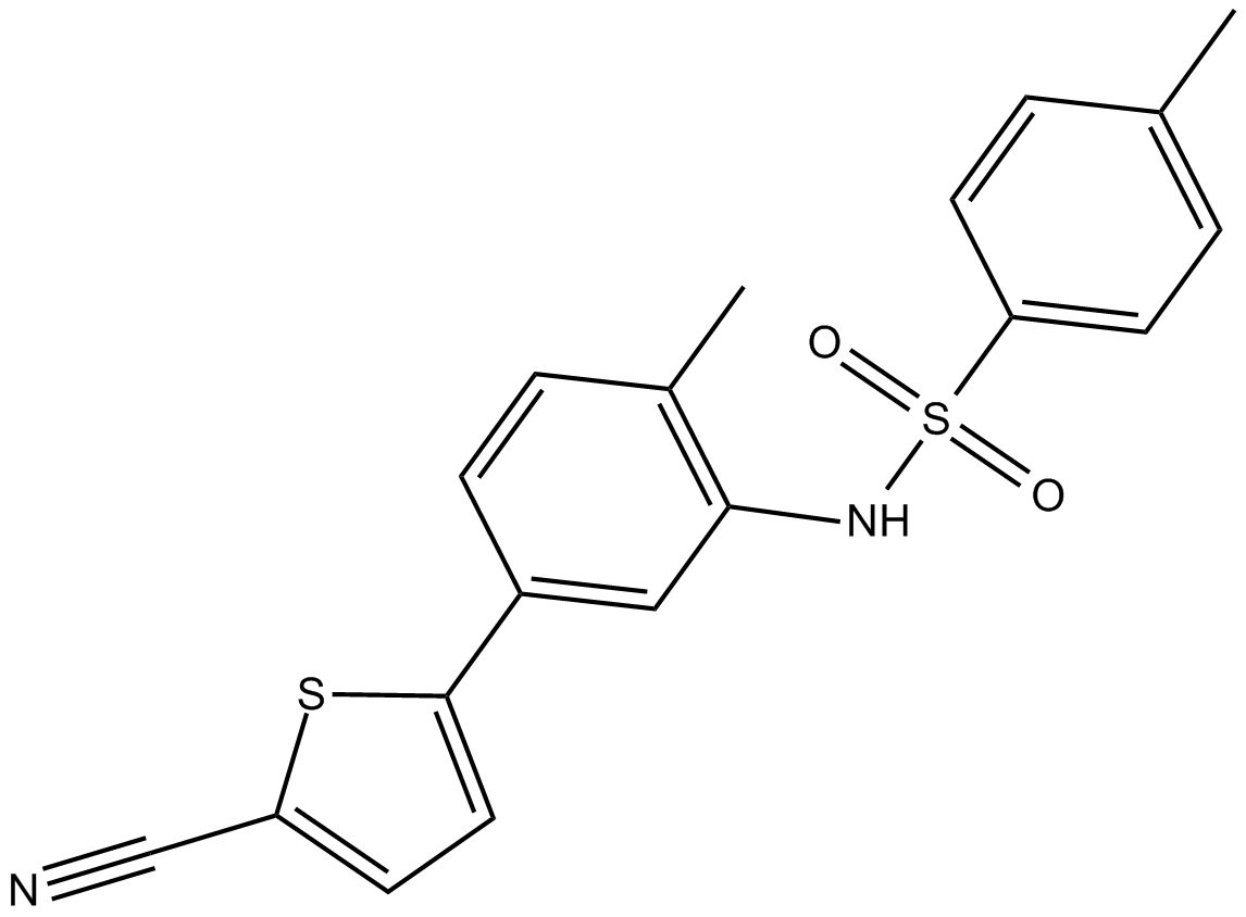
ELR510444
ELR510444 は新規の微小管破壊剤です。 MDA-MB-231 細胞増殖を 30.9 nM の IC50 で阻害します。 P-糖タンパク質薬物輸送体の基質ではなく、βIII-チューブリン過剰発現細胞株で活性を保持します。
-
GC48434
Elsinochrome A
真菌代謝産物

-
GC13163
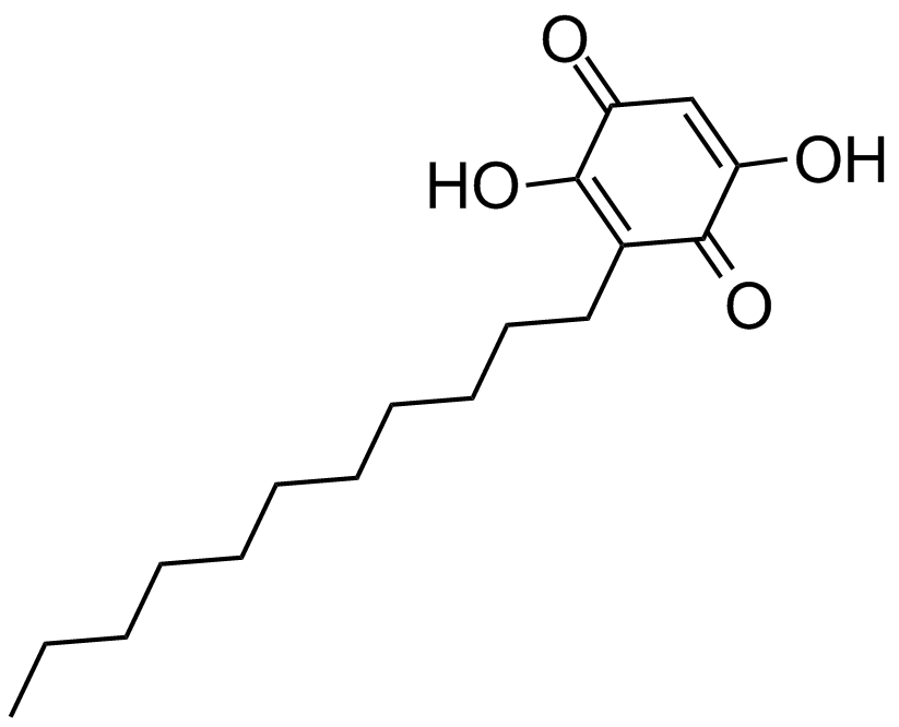
Embelin
A benzoquinone with diverse biological activities
-
GC35980
Emricasan
エムリカサン (PF 03491390) は、経口活性で不可逆的な汎カスパーゼ阻害剤です。

-
GC16519
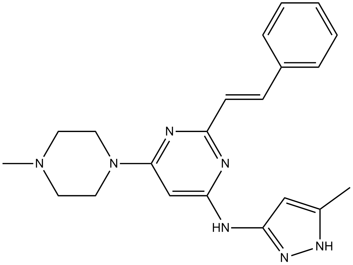
ENMD-2076
多機能キナーゼ阻害剤
-
GC43610
Enniatin A1
フザリウム マイコトキシンから分離されたエニアチン A1 は、D-α-ヒドロキシイソ吉草酸と N-メチル-L-アミノ酸が交互に結合した環状ヘキサデプシペプチドです。エニアチン A1 は、アポトーシスの誘導と ERK シグナル伝達経路の破壊による抗発がん特性を持っています。 Enniatin A1 は、ラット肝ミクロソームで 49 μM の IC50 で ACAT を阻害します。

-
GC16823
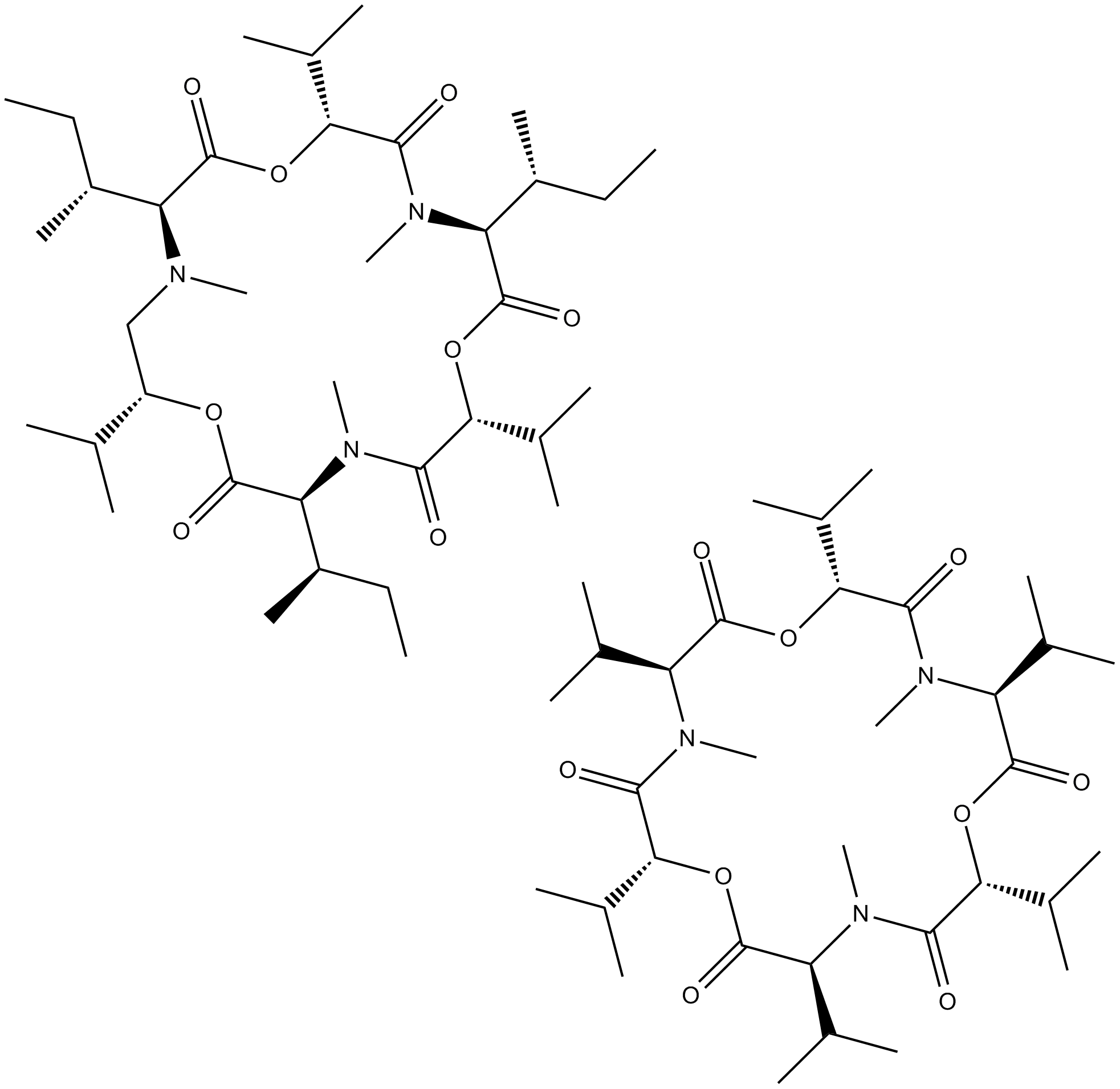
Enniatin Complex
Enniatin 複合体は、真菌の Fusarium 種から大部分が分離されたシクロヘキサデプシペプチドの混合物であり、イオノフォア、抗生物質、および in vitro 脂質低下特性を持っています。
-
GC11625
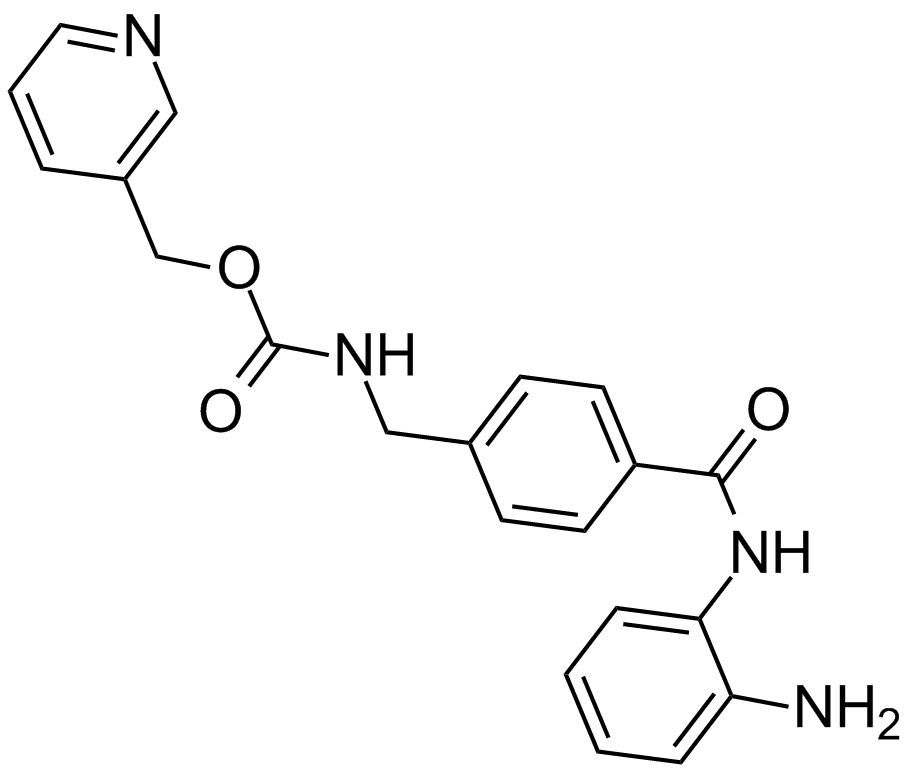
Entinostat (MS-275,SNDX-275)
ヒストン脱アセチル化酵素阻害剤
-
GC66344
Envafolimab
Envafolimab (ASC 22; KN 035) は、ヒト化単一ドメイン抗 PD-L1 抗体の組換えタンパク質です。 Envafolimab は、抗 PD-L1 ドメインとヒト IgG1 抗体の Fc フラグメントとの融合によって作成されます。 Envafolimab は、PD-L1 と PD-1 の間の相互作用を 5.25nm の IC50 値でブロックします。エンバフォリマブは抗腫瘍活性を示します。 Envafolimab は、固形腫瘍の研究の可能性を秘めています。

-
GC11499
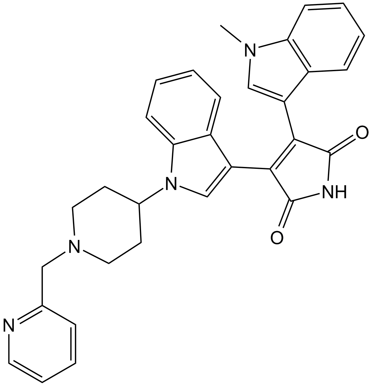
Enzastaurin (LY317615)
エンザスタウリン (LY317615) (LY317615) は、強力で選択的な PKCβ です。 PKCα、PKCγ よりも 6 ~ 20 倍の選択性を示す、6 nM の IC50 を持つ阻害剤。およびPKCε。
-
GC31390
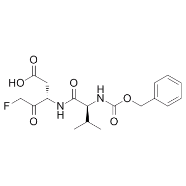
EP1013 (F1013)
EP1013 (F1013) (F1013) は、1 型糖尿病の研究に使用される広域スペクトルのカスパーゼ選択的阻害剤です。
-
GC11834
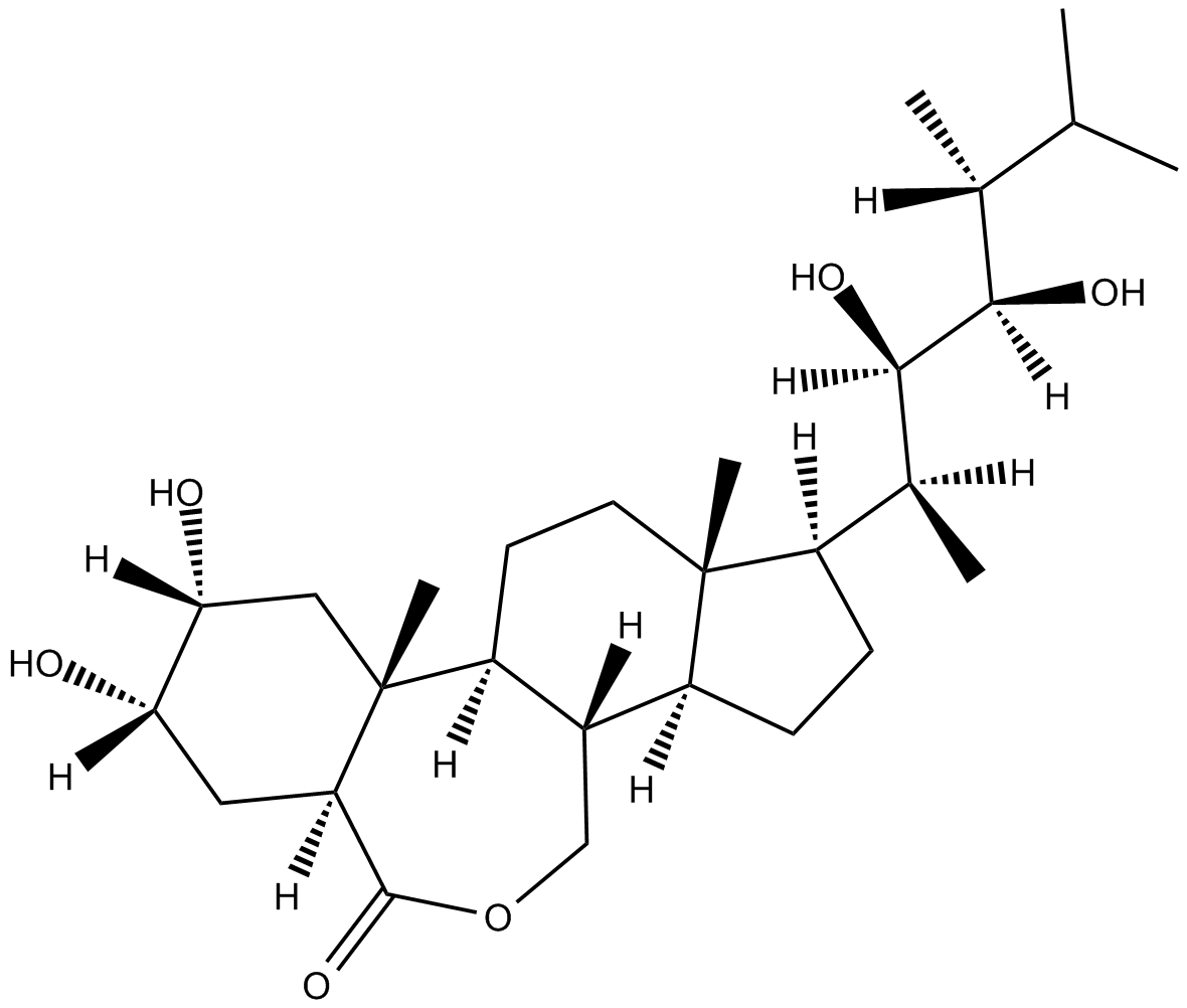
Epibrassinolide
Epibrassinolide (24-Epibrassinolide) は、どこにでも存在する植物成長ホルモンであり、植物の重金属や農薬ストレスを軽減する大きな可能性を示しています。エピブラシノリドは、非腫瘍細胞の増殖に影響を与えることなく、さまざまな癌細胞の潜在的なアポトーシス誘導因子です。
-
GC35997
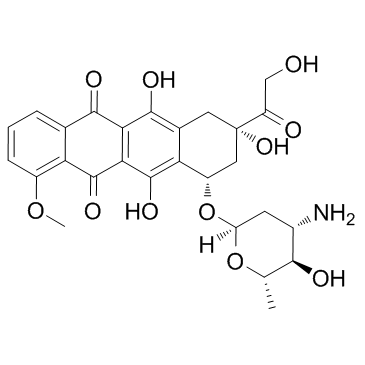
Epirubicin
ドキソルビシンの半合成 L-アラビノ誘導体であるエピルビシン (4'-エピドキソルビシン) は、トポイソメラーゼを阻害することによって抗腫瘍剤を持っています。エピルビシンは DNA と RNA の合成を阻害します。エピルビシンは、フォークヘッド ボックス タンパク質 p3 (Foxp3) 阻害剤であり、制御性 T 細胞の活性を阻害します。
-
GC11202
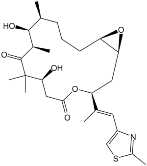
Epothilone A
エポチロン A は [3H] パクリタキセルのチューブリン ポリマーへの結合の競合的阻害剤であり、Ki は 0.6 ~ 1.4 μM です。
-
GC17240
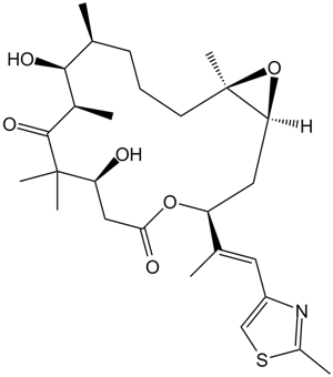
Epothilone B (EPO906, Patupilone)
エポチロン B (EPO906、Patupilone) は、Ki が 0.71μ の微小管安定剤です。
-
GC13383
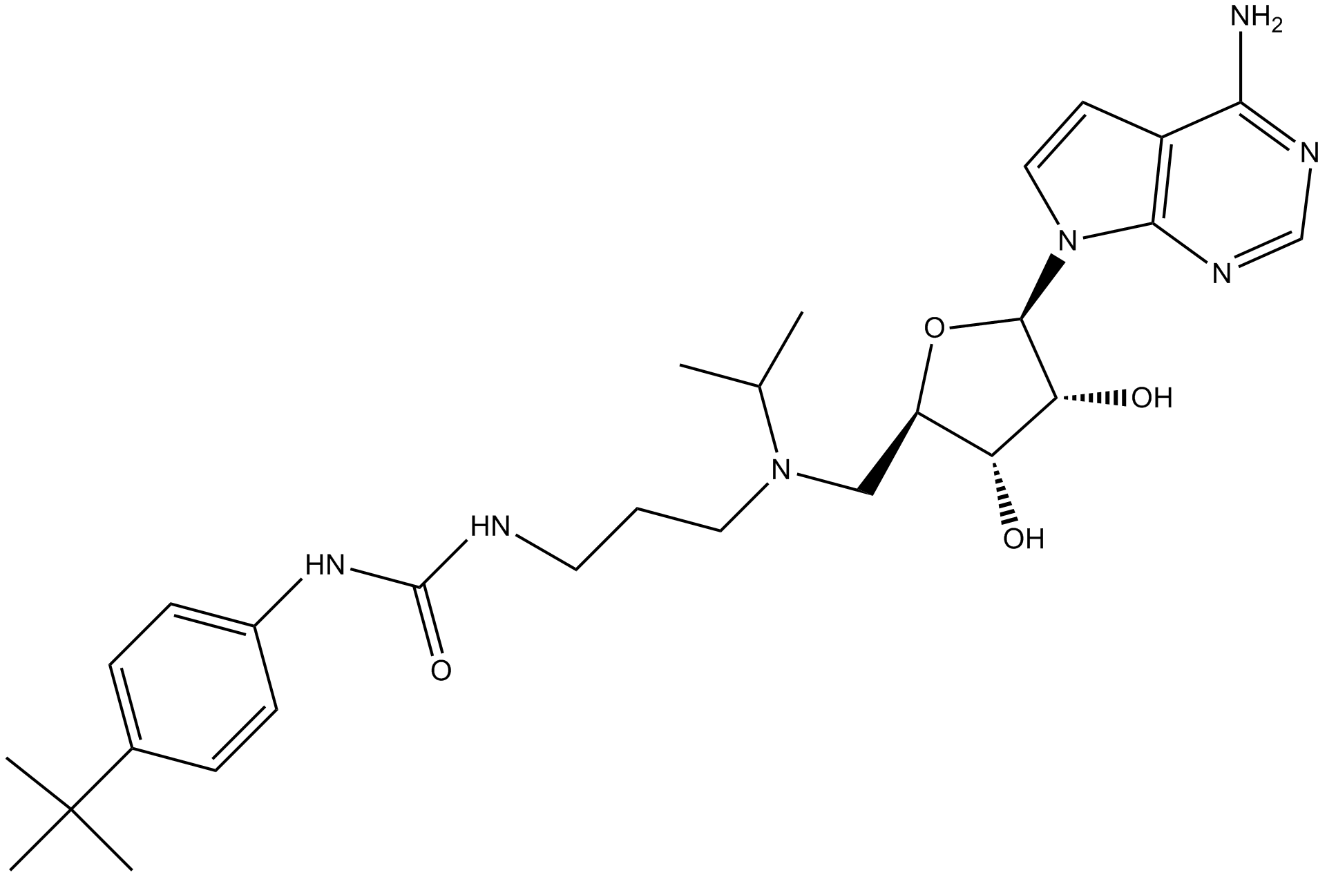
EPZ004777
DOT1Lの強力な阻害剤
-
GC10389
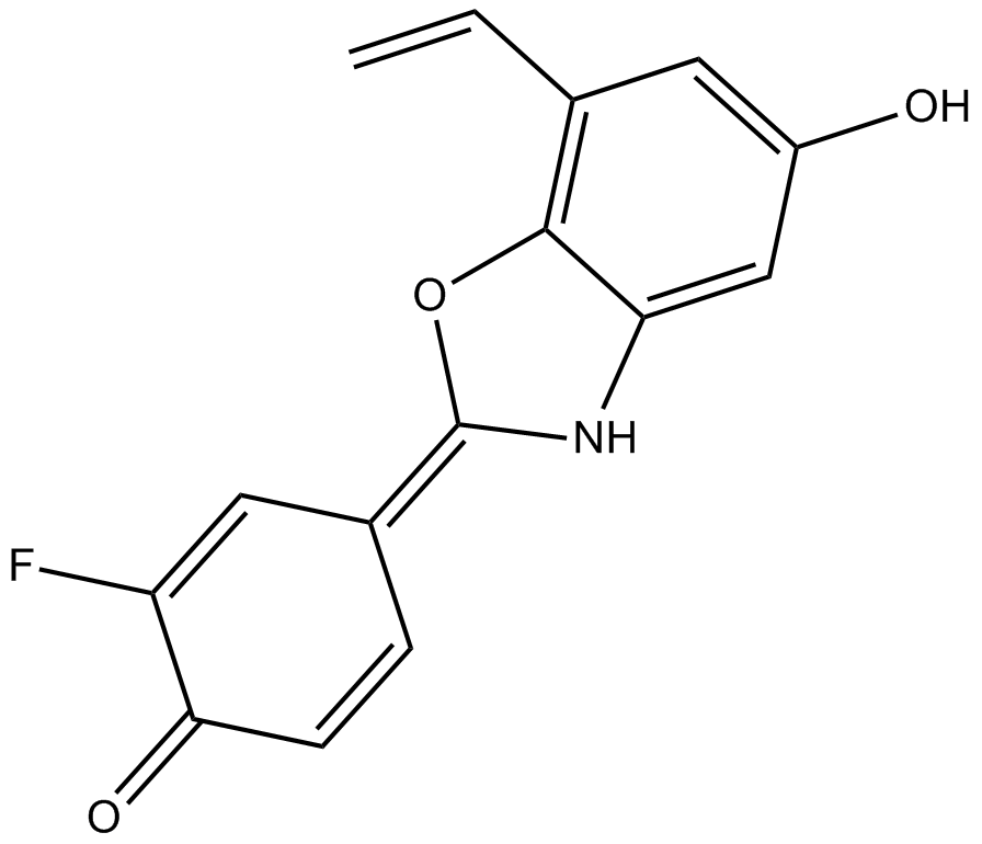
ERB 041
ERB 041 (ERB-041) は、強力で選択的なエストロゲン受容体 (ER) β です。ヒト、ラット、マウス ERβ に対してそれぞれ 5.4、3.1、3.7 nM の IC50 を持つアゴニスト。 ERB 041 は、ERβ に対して 200 倍を超える選択性を示します。以上 ERα。 ERB 041 は、WNT/β-カテニン シグナル伝達経路を弱めることによって作用する強力な皮膚がんの化学予防剤です。 ERB 041 は卵巣癌のアポトーシスを誘導します。
-
GC52516
Erbstatin
チロシンキナーゼ阻害剤

-
GC19142
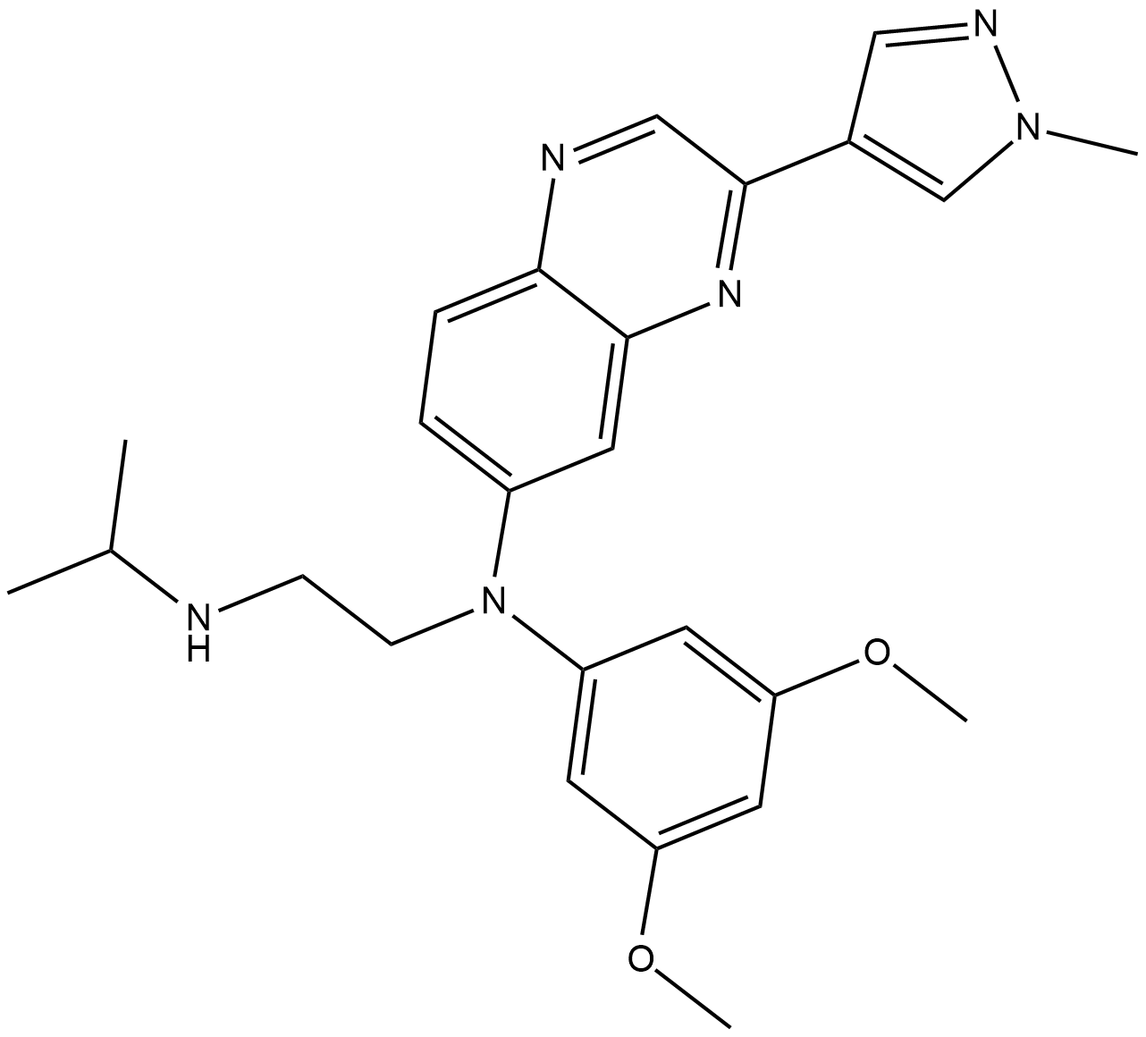
Erdafitinib
Erdafitinib (JNJ-42756493) は強力な経口投与可能な FGFR ファミリー阻害剤です。それぞれ 1.2、2.5、3.0、5.7 nM の IC50 で FGFR1/2/3/4 を阻害します。
-
GC63845
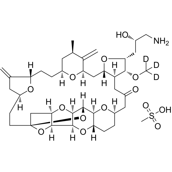
Eribulin-d3 mesylate
エリブリン-d3メシル酸塩は、重水素で標識されたエリブリンメシル酸塩です。エリブリン メシル酸塩は、癌の研究に使用される微小管ターゲッティング エージェントです。
-
GC60153
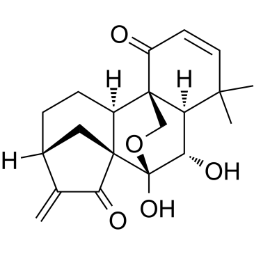
Eriocalyxin B
Eriocalyxin B は、漢方薬 Isodon eriocalyx から分離された ent-カウレン ジテルペノイドです。
-
GC38181
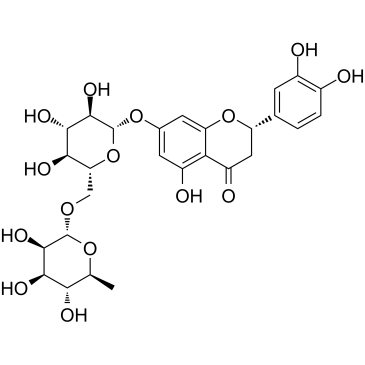
Eriocitrin
エリオシトリンは、強力な抗酸化剤であるレモンから分離されたフラボノイドです。エリオシトリンは、p53、サイクリン A、サイクリン D3、および CDK6 のアップレギュレーションを介して S 期の細胞周期を停止させることにより、肝細胞癌細胞株の増殖を阻害する可能性があります。エリオシトリンは、ミトコンドリアが関与する内因性シグナル伝達経路を活性化することにより、アポトーシスを引き起こします。
-
GN10470
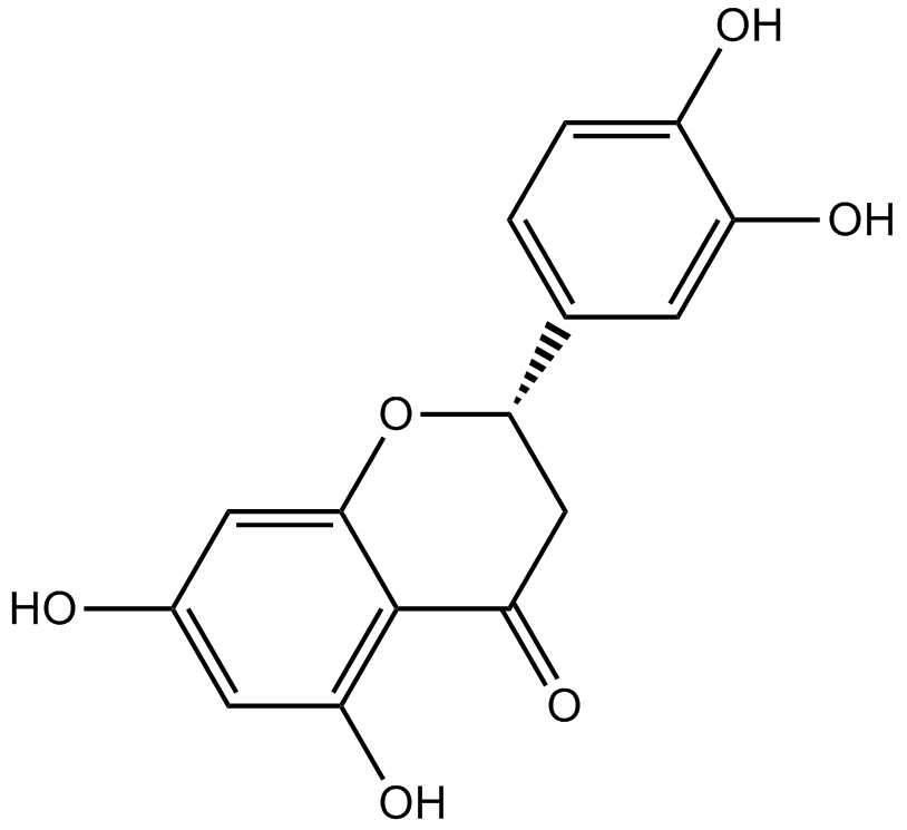
Eriodictyol
-
GC62957
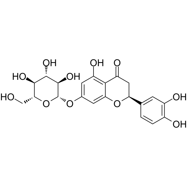
Eriodictyol-7-O-glucoside
エリオディクチオール-7-O-グルコシド (エリオディクチオール 7-O-β-D-グルコシド) は、フラボノイドであり、強力なフリーラジカルスカベンジャーです。
-
GC38447
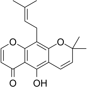
Eriosematin
Eriosematin は、Flemingia philippinensis の根に由来する化合物で、抗増殖活性とアポトーシス誘導特性を備えています 。
-
GC43625
Erucin
エルシン (ERU) は、特にルッコラに豊富に含まれるイソチオシアネートです。

-
GC11935
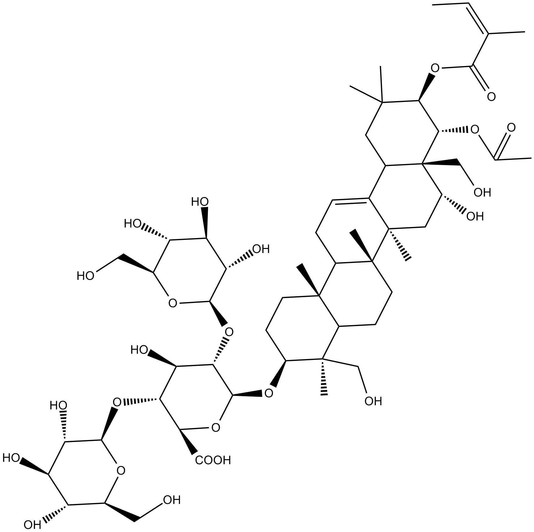
Escin
セイヨウトチノキ (Aesculus hippocastanum) の種子から分離されたトリテルペノイド サポニンの天然化合物であるエスシンは、血管保護、抗炎症、抗浮腫、および抗侵害受容剤として使用できます 。
-
GC34579
Etanercept
TNF に結合する二量体融合タンパク質であるエタネルセプトは、TNF 阻害剤として作用します。
-
GC38083
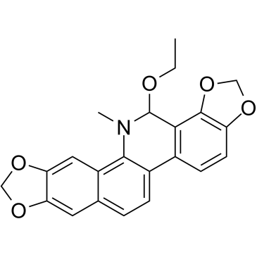
Ethoxysanguinarine
エトキシサンギナリンは、主に Macleaya cordata に含まれるベンゾフェナントリジン アルカロイドの天然物です。エトキシサンギナリンは、プロテインホスファターゼ 2A (CIP2A) の阻害剤です。エトキシサンギナリンは細胞アポトーシスを誘導し、結腸直腸癌細胞の増殖を阻害します。
-
GC61669
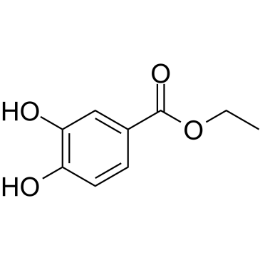
Ethyl 3,4-dihydroxybenzoate
抗酸化物質である 3,4-ジヒドロキシ安息香酸エチル (プロトカテク酸エチル) は、ピーナッツ種子の種皮に見られるプロリルヒドロキシラーゼ阻害剤です。
-
GC60824
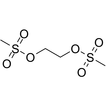
Ethylene dimethanesulfonate
エチレンジメタンスルホネートは、エチレングリコールの穏やかなアルキル化、非揮発性メタンスルホン酸ジエステルです。
-
GC12105
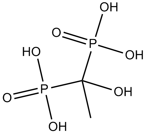
Etidronate
エチドロネート(エチドロネート)は、経口および静脈内で活性なビスフォスフォネートです。
-
GC15617
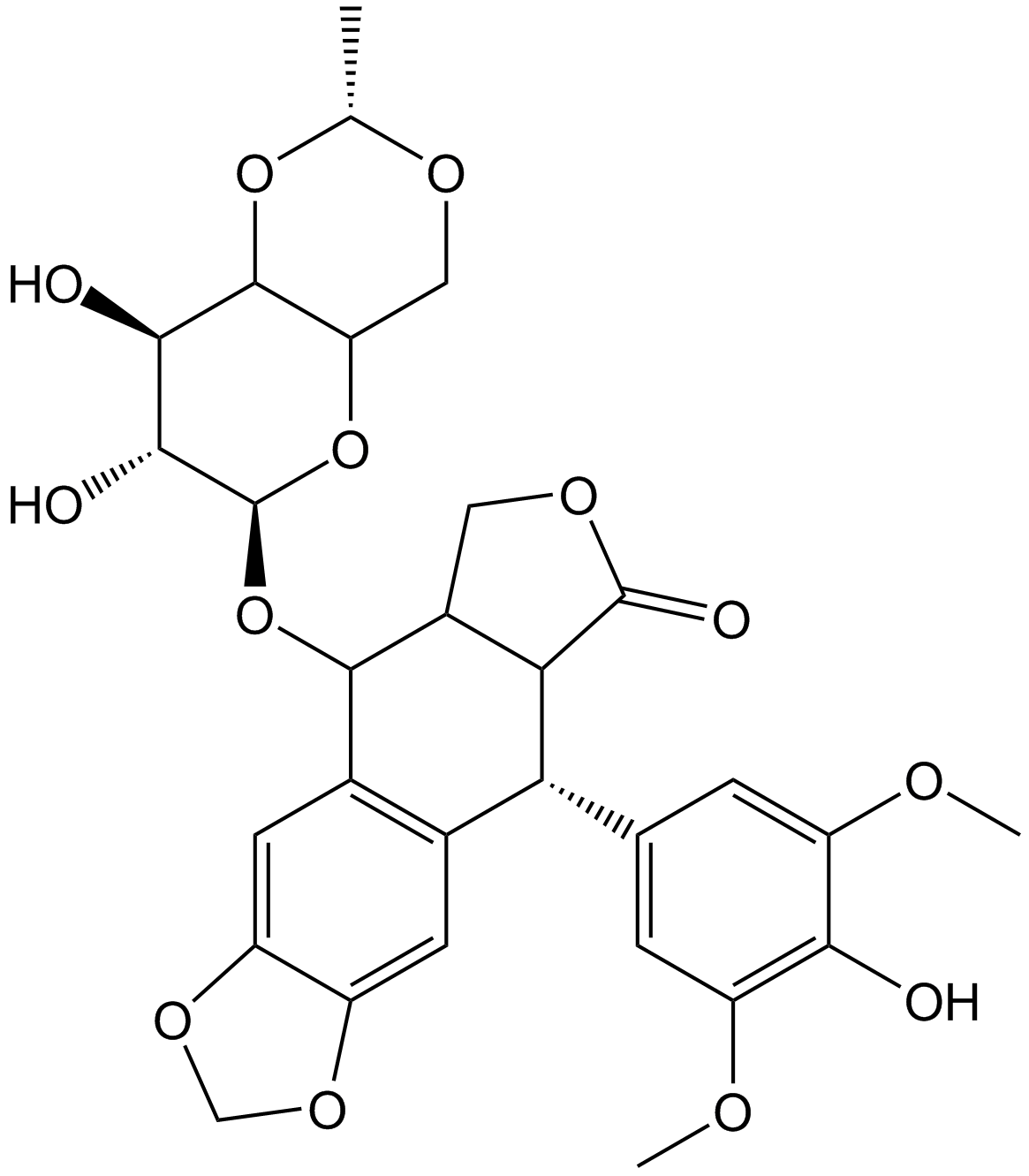
Etoposide
トポイソメラーゼII阻害剤
-
GC60826
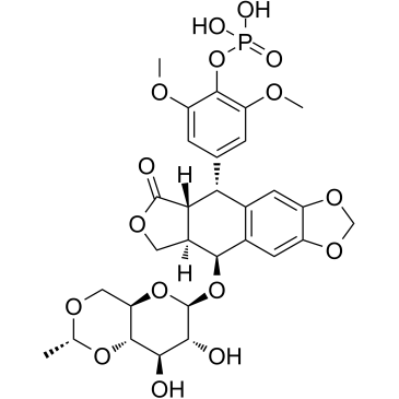
Etoposide phosphate
リン酸エトポシド(BMY-40481)は、強力な抗がん化学療法剤であり、DNA 鎖の再ライゲーションを防ぐ選択的トポイソメラーゼ II 阻害剤です。
-
GC10855
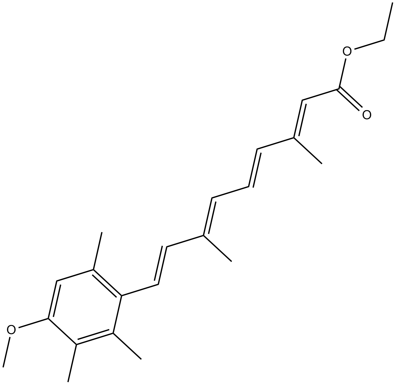
Etretinate
エトレチネート (Ro 10-9359) は、重度の乾癬治療の可能性がある第二世代のレチノイドです。
-
GC30910
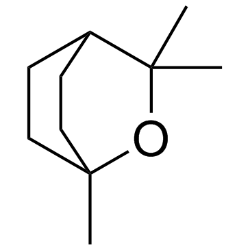
Eucalyptol (1,8-Cineole)
多様な生物学的活性を持つ二環式モノテルペン
-
GC16517
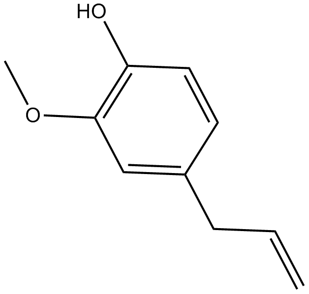
Eugenol
オイゲノールはクローブに含まれる精油で、抗菌、駆虫、抗酸化作用があります。
-
GC43643
Eupenifeldin
Eupenifeldin は、Eupenicillium brefeldianum ATCC 74184 の培養物から分離された五環性ビストロポロンです。Eupenifeldin は、HCT-116 細胞株に対して細胞毒性があります。 Eupenifeldin は、白血病の研究の可能性を秘めています。

-
GC38165
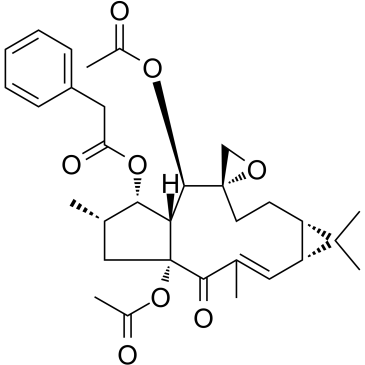
Euphorbia Factor L1
-
GC60157
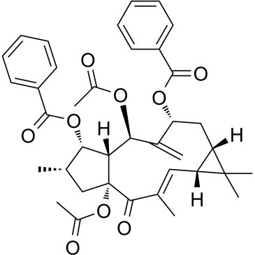
Euphorbia Factor L2
ユーフォルビア因子 L2 は、ケッパー ユーフォルビア種子 (ユーフォルビア ラチリス L. の種子) から分離されたラチラン ジテルペノイドであり、伝統的にがんの治療に適用されてきました。ユーフォルビア因子 L2 は強力な細胞毒性を示し、ミトコンドリア経路を介してアポトーシスを誘導します。
-
GC38392
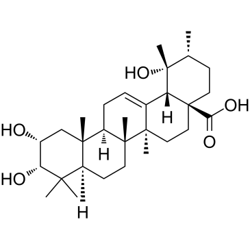
Euscaphic acid
DNA ポリメラーゼ阻害剤であるエウスカフィン酸は、R. alceaefolius Poir の根に由来するトリテルペンです。 Euscaphic は、ウシ DNA ポリメラーゼ α (pol α) およびラット DNA ポリメラーゼ β (pol β) を 61 および 108 μM の IC50 値で阻害します。ユースカフィン酸はアポトーシスを誘導します。
-
GC13601
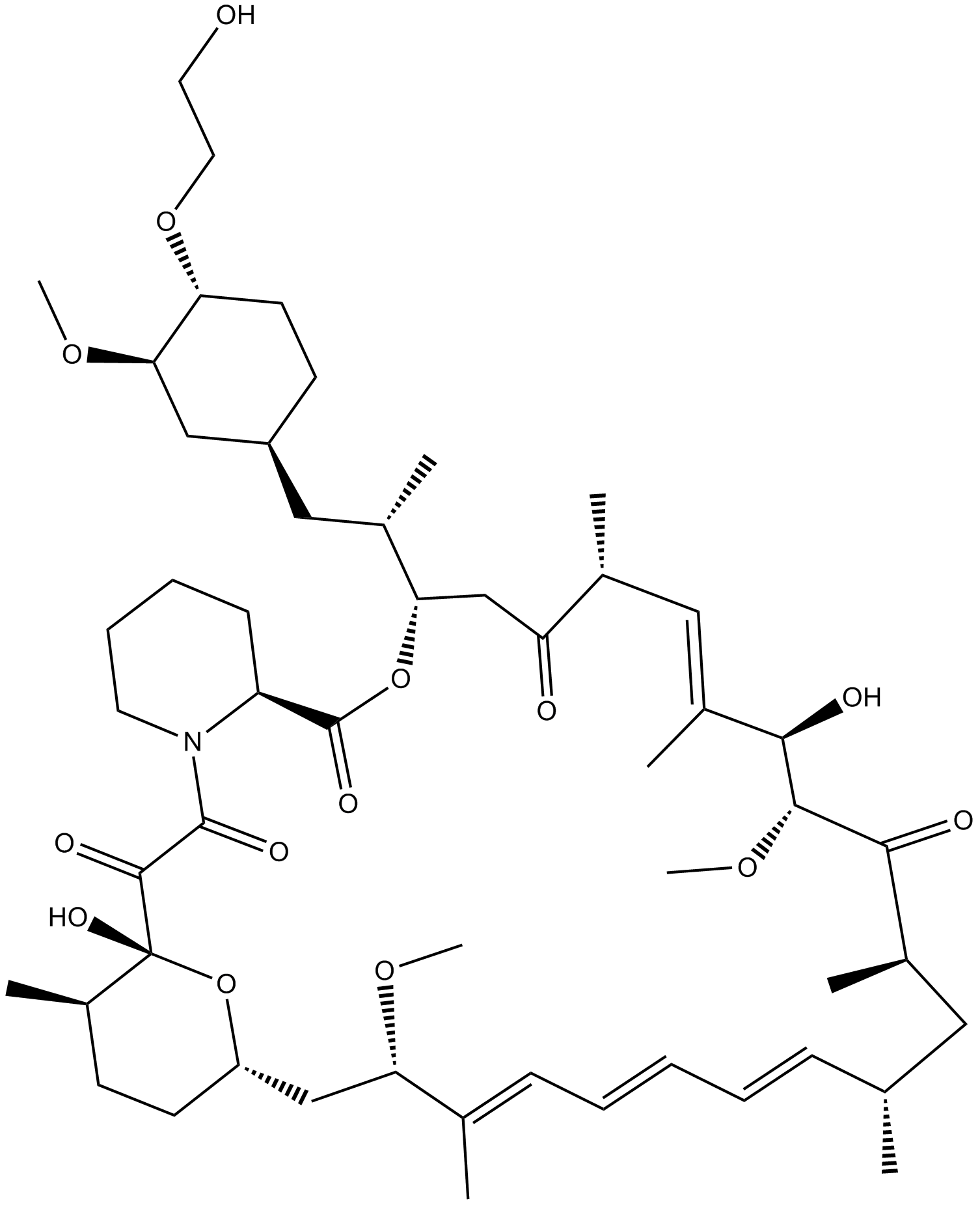
Everolimus (RAD001)
ラパマイシン誘導体
-
GC15605
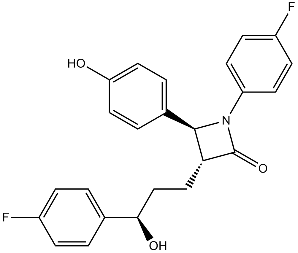
Ezetimibe
エゼチミベ (SCH 58235) は強力なコレステロール吸収阻害剤です。
-
GC60159
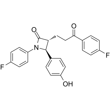
Ezetimibe ketone
エゼチミベ ケトン (EZM-K) は、エゼチミベの第 I 相代謝物です。
-
GC66354
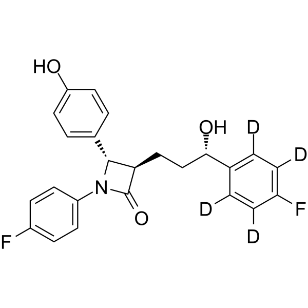
Ezetimibe-d4-1
Ezetimibe-d4 は、Ezetimibe と標識された重水素です。エゼチミベ (SCH 58235) は強力なコレステロール吸収阻害剤です。エゼチミベはニーマン・ピック C1-like1 (NPC1L1) 阻害剤であり、強力な Nrf2 活性化剤です。
-
GC14791
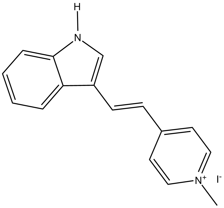
F16
F16 は neu 過剰発現細胞の強力な成長阻害剤であり、乳腺上皮の増殖だけでなく、さまざまなマウス乳癌細胞株やヒト乳癌細胞株の増殖も選択的に阻害します。
-
GC62203
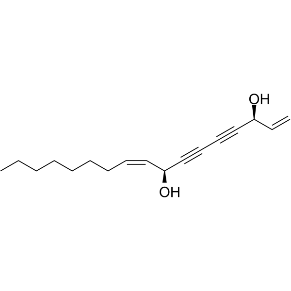
Falcarindiol
経口活性ポリアセチレンオキシリピンであるファルカリンジオールは、PPARγを活性化し、細胞内のコレステロール輸送体ABCA1の発現を増加させます。
-
GC38437
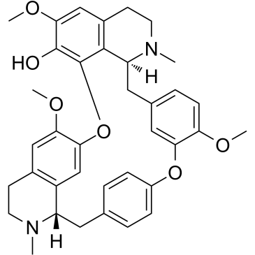
Fangchinoline
多様な生物学的活性を持つアルカロイド
-
GC60836
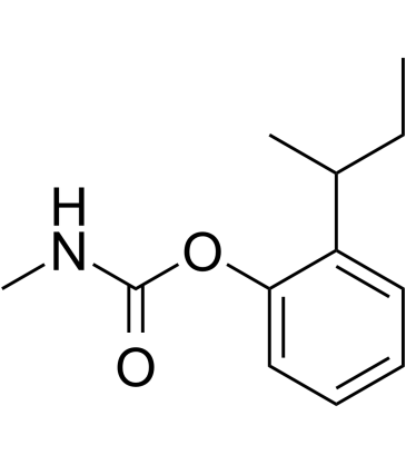
Fenobucarb
フェノブカルブはカーバメート殺虫剤です。
-
GC14499
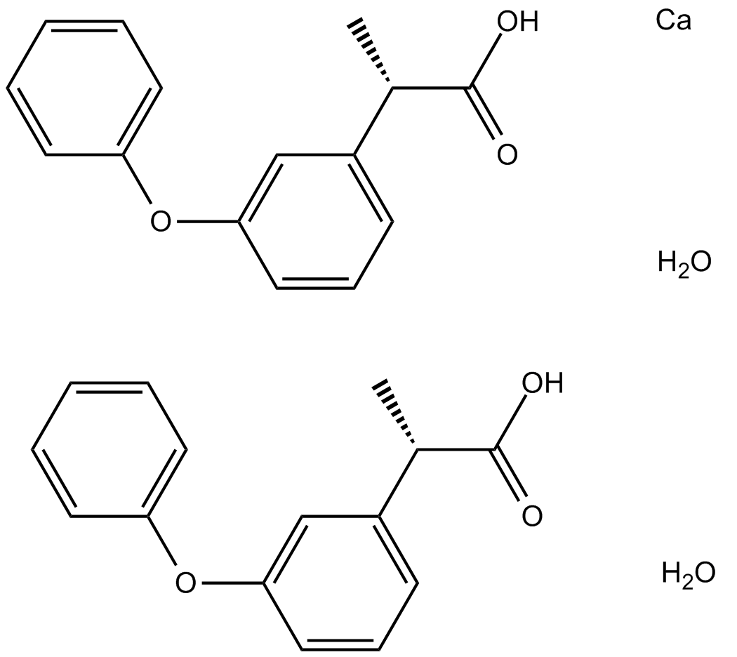
Fenoprofen calcium hydrate
フェノプロフェンカルシウム水和物は、非ステロイド性の抗炎症性抗関節炎薬です。
-
GC18641
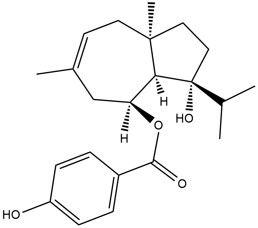
Ferutinin
天然のテルペノイド化合物であるフェルチニンは、それぞれ 33.1 nM および 180.5 nM の IC50 を持つエストロゲン受容体 ERα アゴニストおよびエストロゲン ERβ 受容体アゴニスト/アンタゴニストです。
-
GC12940
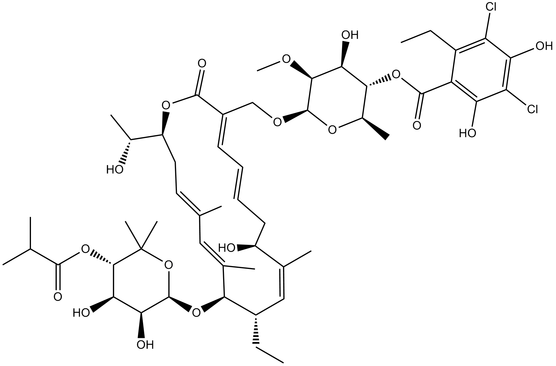
Fidaxomicin
大環状抗生物質であるフィダキソマイシン (OPT-80) は、経口活性で強力な RNA ポリメラーゼ阻害剤です。
-
GC36046
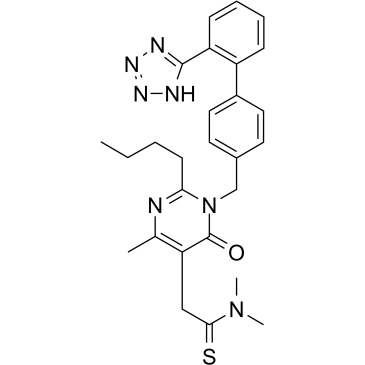
Fimasartan
フィマサルタン (BR-A-657) は、高血圧および心不全の治療に使用される非ペプチドのアンギオテンシン II 受容体拮抗薬です。
-
GN10030
Fisetin
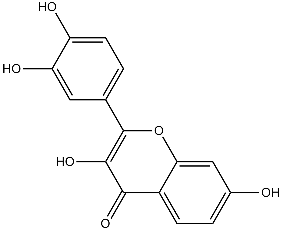
-
GC49344
Fisetin-d5
フィセチンの定量化のための内部標準

-
GC60845
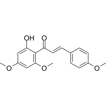
Flavokawain A
有望な抗発がん剤であるフラボカワイン A は、抗腫瘍活性を持つカバ抽出物からのカルコンです。 Flavokawain A は、Bax タンパク質依存性およびミトコンドリア依存性アポトーシス経路の関与により、細胞アポトーシスを誘導します。フラボカイン A は、膀胱癌の研究の可能性を秘めています。
-
GC41261
Flavokawain B
フラボカバイン B (フラボカバイン B) は、カバカバ植物の根抽出物から分離されたカルコンであり、さまざまな癌細胞株の増殖を阻害する強力なアポトーシス誘導剤です。フラボカバイン B (フラボカバイン B) は、強力な抗血管新生活性を示します。フラボカバイン B (フラボカバイン B) は、ヒト脳内皮細胞 (HUVEC) の遊走と管形成を非常に低濃度で非毒性の濃度で阻害します。

-
GC36050
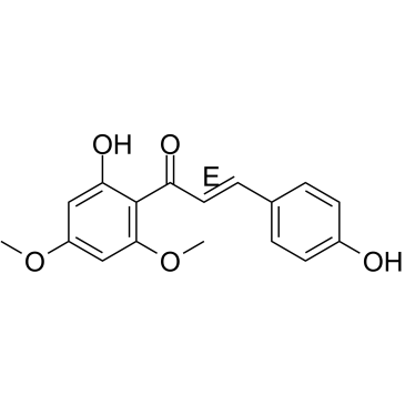
Flavokawain C
フラボカイン C は、カバ根に含まれる天然のカルコンです。フラボカイン C は、HCT 116 細胞に対して 12.75 μM の IC50 で、ヒト癌細胞株に対して細胞毒性を発揮します。
-
GC16875
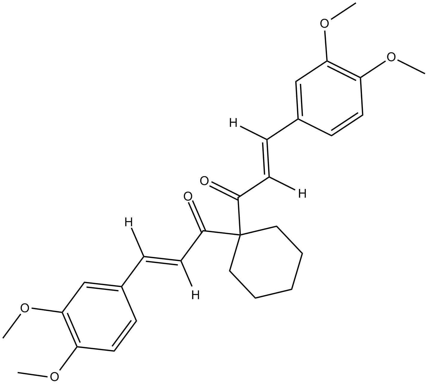
FLLL32
クルクミンの合成類似体である FLLL32 は、抗腫瘍活性を持つ JAK2/STAT3 二重阻害剤です。 FLLL32 は、乳癌細胞における IFNα および IL-6 による STAT3 リン酸化の誘導を阻害できます。
-
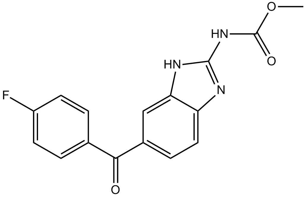
GC13210
Flubendazole
フルベンダゾールは安全で有効な駆虫薬であり、ヒト、げっ歯類、反芻動物の駆虫に広く使用されています。
-
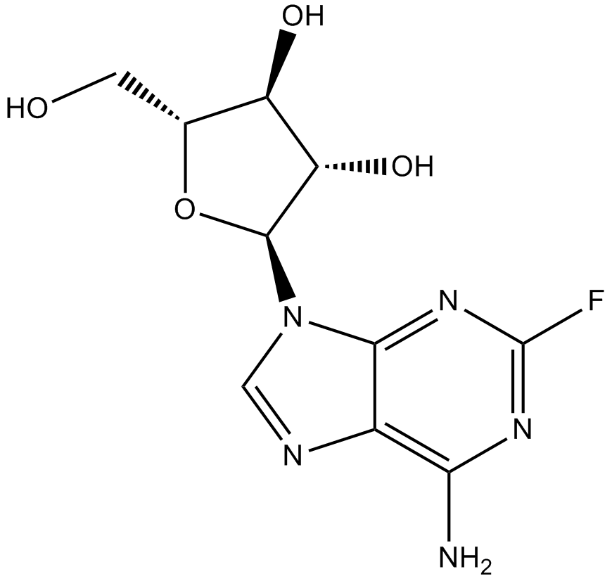
GC14144
Fludarabine
フルダラビン (NSC 118218) は DNA 合成阻害剤であり、リンパ増殖性悪性腫瘍における抗腫瘍活性を持つフッ素化プリン類似体です。フルダラビンは、正常な休止リンパ球または活性化リンパ球において、サイトカインによる STAT1 の活性化および STAT1 依存性遺伝子転写を阻害します。
-
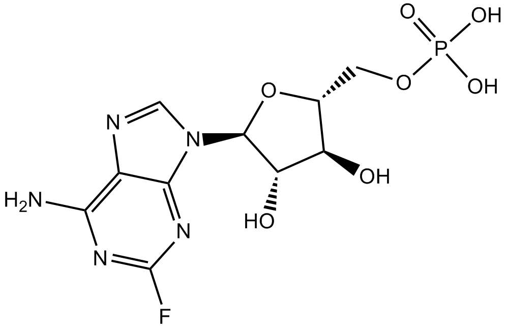
GC15134
Fludarabine Phosphate (Fludara)
フルダラビン (リン酸塩) はアデノシンとデオキシアデノシンの類似体であり、DNA への取り込みについて dATP と競合し、DNA 合成を阻害することができます。
-
GC48827
Flufenamic Acid-d4
フルフェナム酸の定量化のための内部標準

-
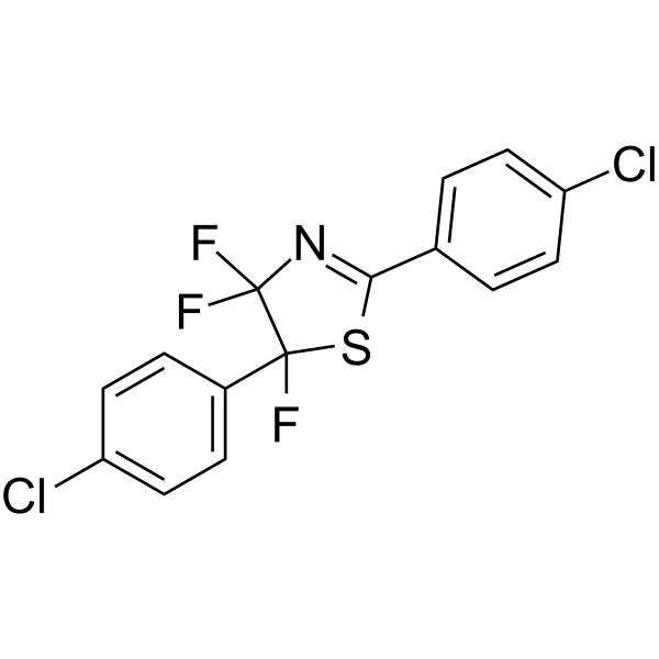
GC62167
Fluorizoline
フルオロゾリンは、プロヒビチン 1 (PHB1) および 2 (PHB2) に選択的かつ直接的に結合し、アポトーシスを誘導します。フルオロゾリンは、NOXA と BIM のアップレギュレーションを通じて、慢性リンパ性白血病 (CLL) 細胞の生存率を低下させます。フルオロゾリンは、p53 に依存しない方法で抗腫瘍作用を発揮します。
-
GC43689
Fluphenazine-N-2-chloroethane (hydrochloride)
フルペナジンは、ドーパミンD2受容体に強く結合する従来の抗精神病薬化合物であり(Ki = 0.55 nM)、マイクロモル濃度でカルモジュリンを可逆的に阻害します。

-
GC16880
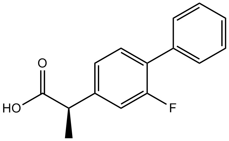
Flurbiprofen
フルルビプロフェン (dl-フルルビプロフェン) は、強力な経口活性非ステロイド性抗炎症剤 (NSAIA/NSAID) であり、解熱作用と鎮痛作用があります。
-
GC48831
Flutamide-d7
フルタミド-d7 は重水素標識フルタミドです。

-
GC49126
Folitixorin
フォリティキソリン (5,10-メチレンテトラヒドロ葉酸) はロイコボリンの補因子であり類似体です。 Folitixorin は、癌の補助研究における 5-FU 細胞毒性の調節に有望な薬剤です。

-
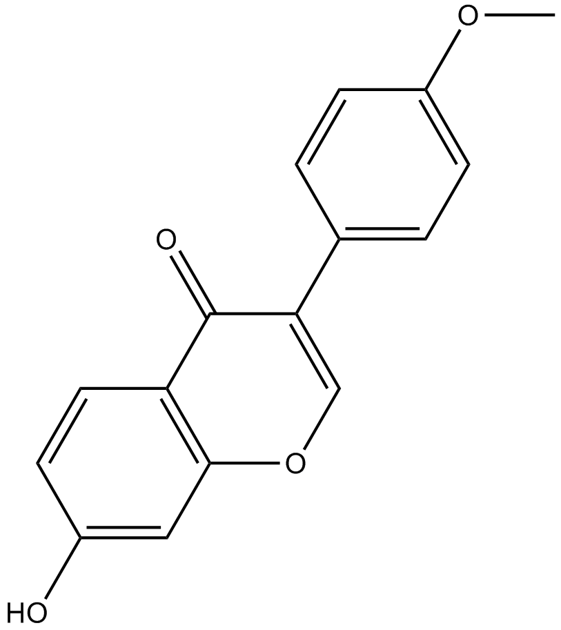
GN10527
Formononetin
-
GC36065
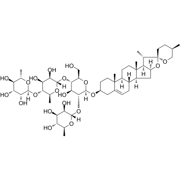
Formosanin C
フォルモサニン C は、パリフォルモサナ ハヤタから分離されたジオスゲニン サポニンであり、抗腫瘍活性を有する免疫調節物質です。フォルモサニン C はアポトーシスを誘導します。
-
GC33029
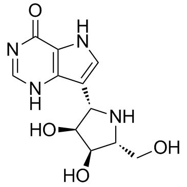
Forodesine (BCX-1777 freebase)
フォロデシン (BCX-1777 フリーベース) (BCX-1777) は、ヒト、マウス、ラット、サル、イヌの PNP に対して 0.48 ~ 1.57 nM の範囲の IC50 値を持つ、非常に強力で経口的に活性なプリン ヌクレオシド ホスホリラーゼ (PNP) 阻害剤です。 Forodesine (BCX-1777 フリーベース) は、強力なヒトリンパ球増殖阻害剤です。フォロデシン (BCX-1777 フリーベース) は、dGTP レベルを上昇させることにより、白血病細胞のアポトーシスを誘導する可能性があります。
-
GC32708
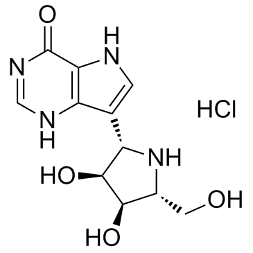
Forodesine hydrochloride (BCX-1777)
フォロデシン塩酸塩 (BCX-1777) (BCX-1777 塩酸塩) は、ヒト、マウス、ラット、サル、イヌの PNP に対して 0.48 ~ 1.57 nM の範囲の IC50 値を持つ、非常に強力で経口的に活性なプリン ヌクレオシド ホスホリラーゼ (PNP) 阻害剤です。フォロデシン塩酸塩 (BCX-1777) は、強力なヒトリンパ球増殖阻害剤です。フォロデシン塩酸塩 (BCX-1777) は、dGTP レベルを増加させることにより、白血病細胞のアポトーシスを誘導する可能性があります。
-
GN10727
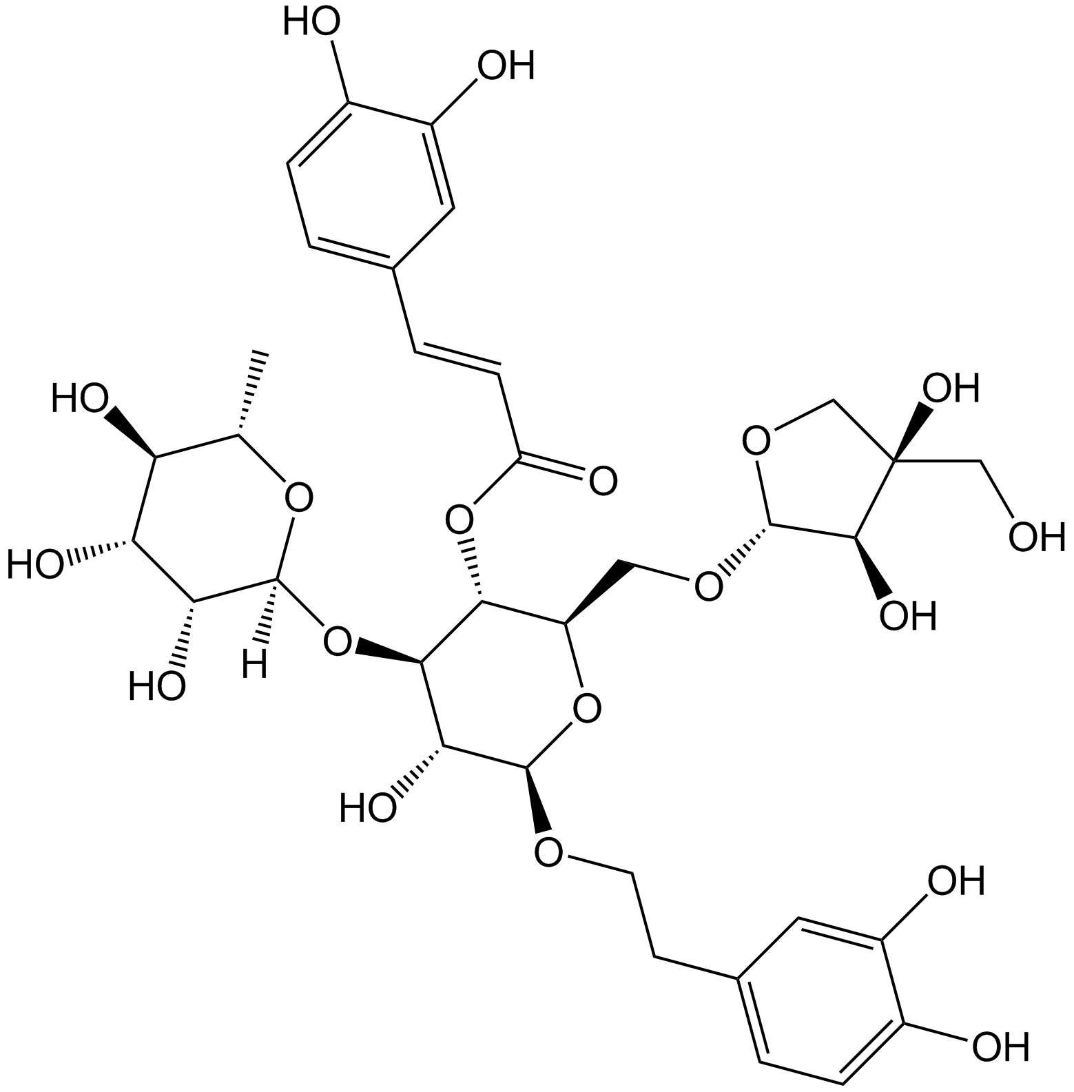
Forsythoside B
-
GC12308
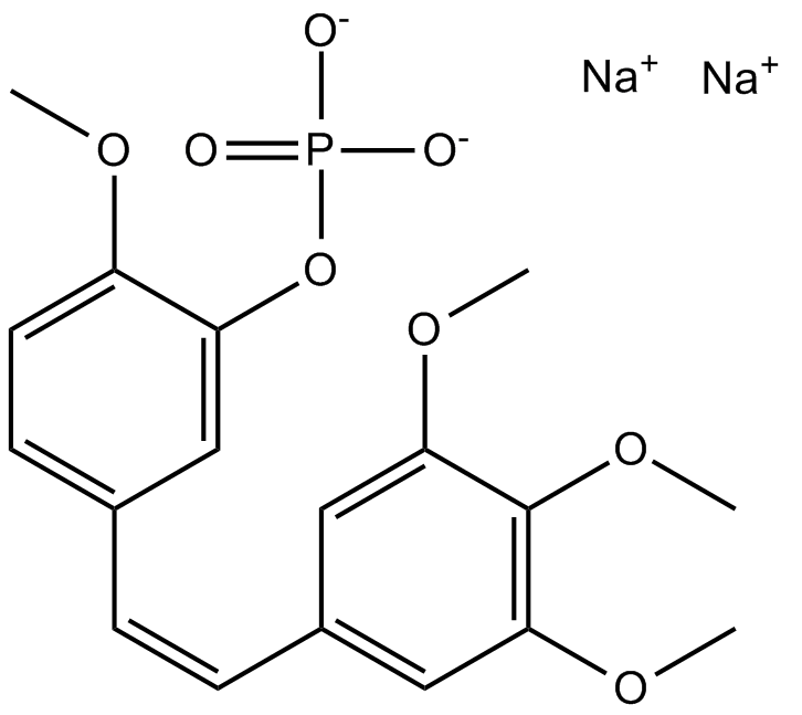
Fosbretabulin (Combretastatin A4 Phosphate (CA4P)) Disodium
Fosbretabulin (Combretastatin A4 Phosphate (CA4P)) 二ナトリウム (CA 4DP) は、チューブリン不安定化剤です。フォスブレタブリン (コンブレタスタチン A4 リン酸塩 (CA4P)) 二ナトリウムは、内皮細胞を選択的に標的とし、発生期の腫瘍新生血管の退縮を誘導し、腫瘍血流を減少させ、中心腫瘍壊死を引き起こすコンブレタスタチン A4 プロドラッグです。
-
GC25428
Foscenvivint (ICG-001)
Foscenvivint(ICG-001)は、Wnt / β-catenin / TCF媒介転写を拮抗し、CREB結合タンパク質(CBP)に特異的に結合するが、関連する転写共同活性化因子p300ではありません。 ICG-001はアポトーシスを誘導します。

-
GC62645
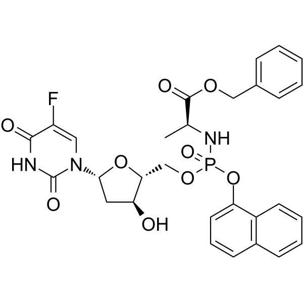
Fosifloxuridine nafalbenamide
フォシフロクスウリジン ナファルベンアミド (NUC-3373) は、ピリミジン ヌクレオチド アナログであり、チミジル酸シンターゼ阻害剤です。フォシフロクスウリジン ナファルベンアミドには、抗がん作用があります。フォシフロクスウリジン ナファルベンアミドは、宿主の免疫応答を誘発し、免疫療法を強化する可能性があります。
-
GC38551
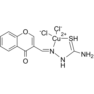
FPA-124
-
GC18652
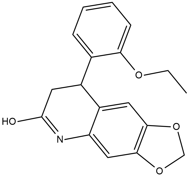
FQI 1
「Late SV40 Factorの阻害剤」
-
GC10647
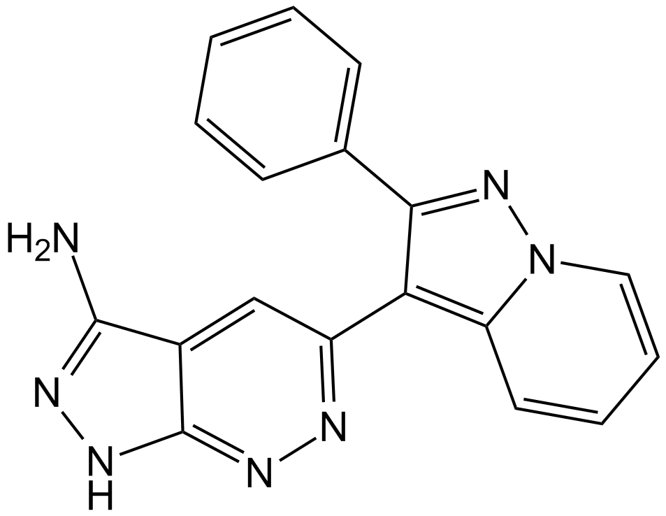
FR 180204
FR 180204 は、ATP 競合的かつ選択的な ERK 阻害剤です。 FR 180204 は、ERK1 と ERK2 をそれぞれ 0.51 μM (Ki=0.31 μM) と 0.33 μM (Ki=0.14 μM) の IC50 で阻害します。
-
GC38044
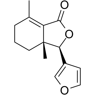
Fraxinellone
-
GC16310
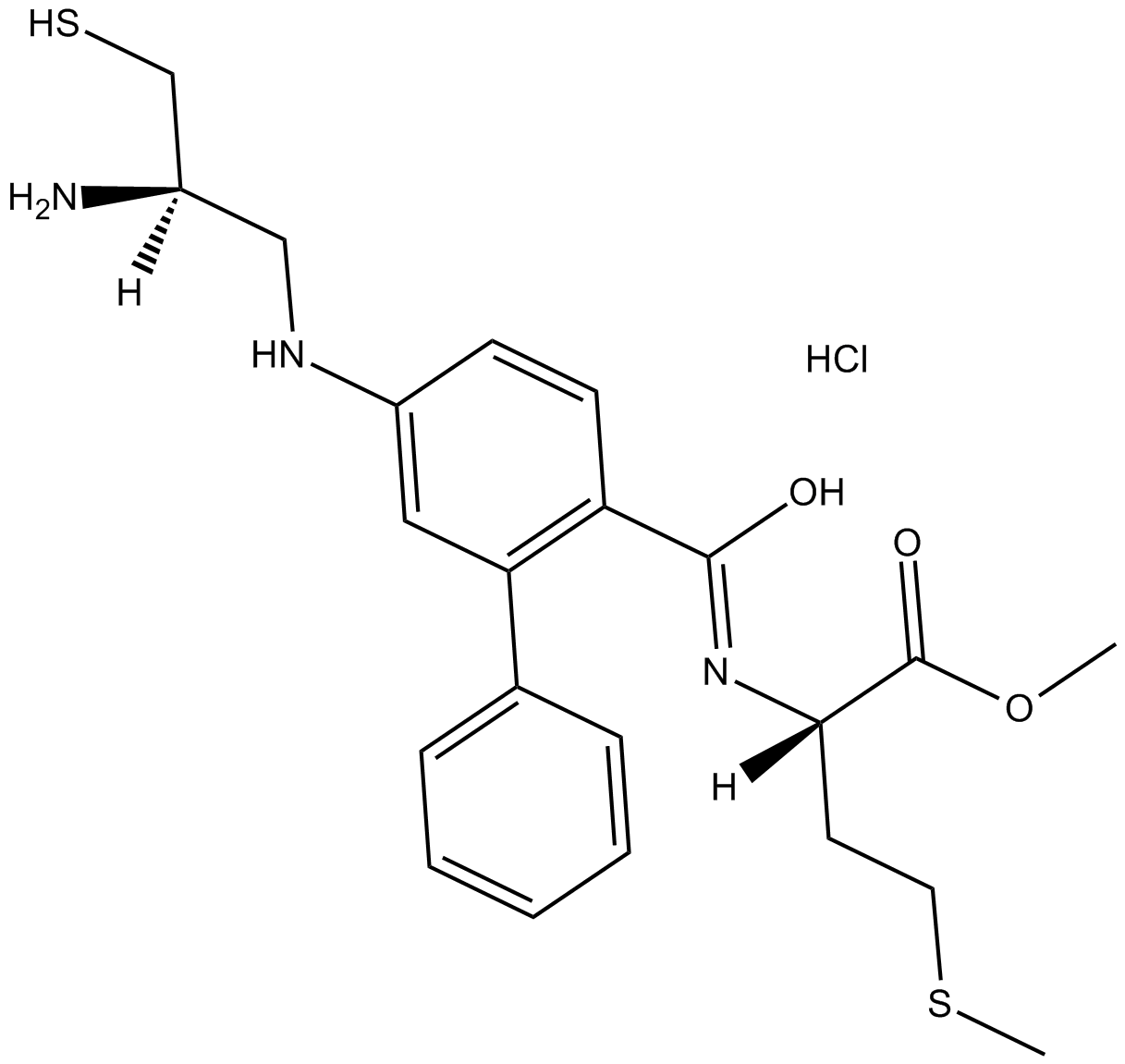
FTI 277 HCl
FTI 277 HCl は、ファルネシルトランスフェラーゼ (FTase) の阻害剤です。 H-およびK-Ras発癌性シグナル伝達の両方に拮抗する非常に強力なRas CAAXペプチドミメティック。
-
GC52288
Fumonisin B1-13C34
フモニシンB1の定量化のための内部標準

-
GC62981
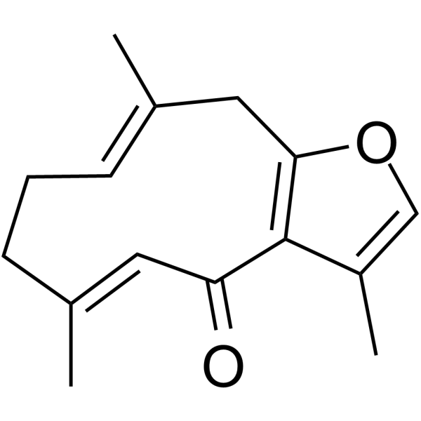
Furanodienone
フラノジエノンは、ウコン根茎由来の主要な生物活性成分の 1 つです。フラノジエノン誘導アポトーシス。
-
GC32138
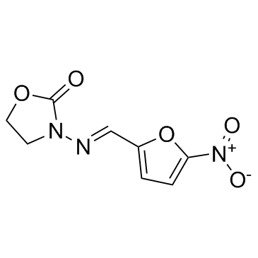
Furazolidone
フラゾリドンは、抗原虫および抗菌活性を有するニトロフラン誘導体であり、AML1-ETO 形質転換細胞を 12.7 μM の IC50 値で阻害します。
-
GC18611
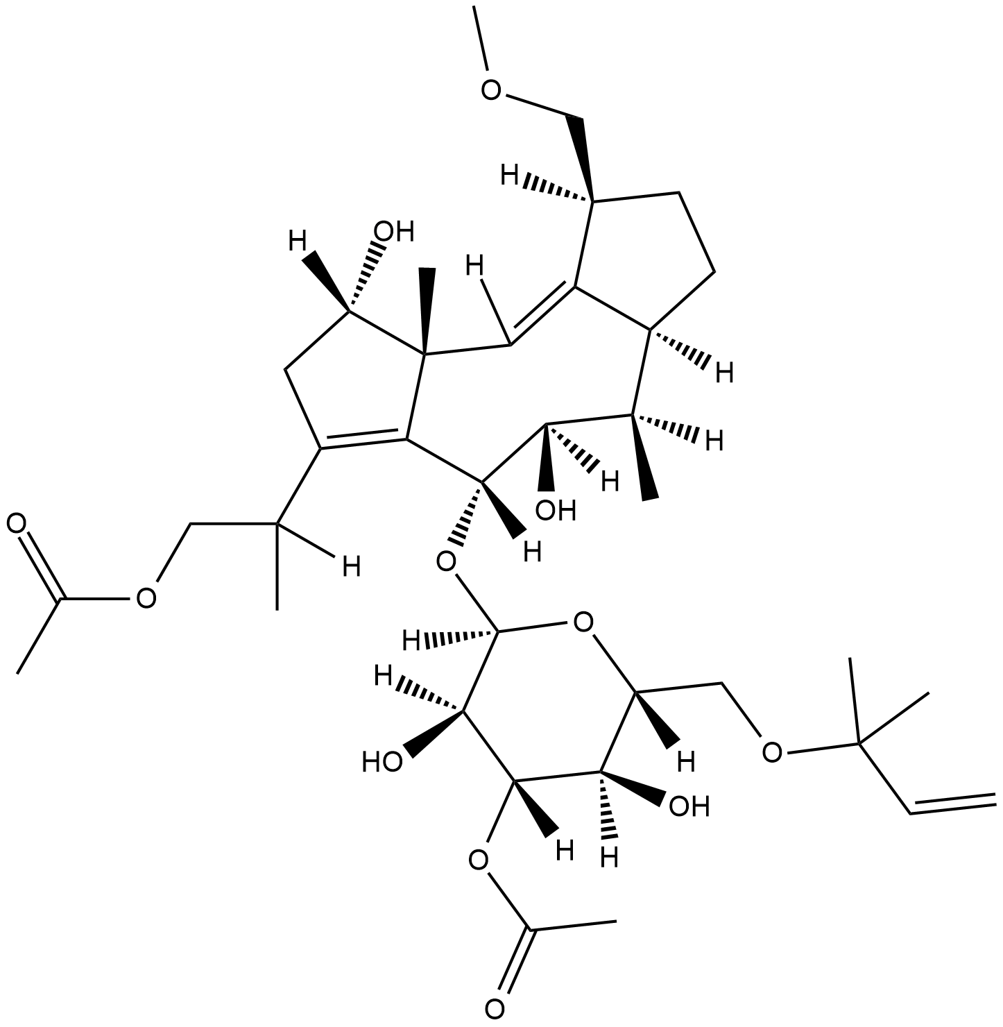
Fusicoccin
真菌のピトトキシンであるフシコクシン (Fusicoccin A) は、特定の 14-3-3 タンパク質間相互作用の安定剤です。フシコクシンは、ズボンの H+-ATPase/14-3-3 cmplex を安定化し、酵素を活性化状態に維持します。また、フシコクシンは、ERα、GPIbα、TASK3、CTFR、p53 などの C 末端 14-3-3 認識モチーフ (モード 3 モチーフ) を含む結合パートナーとの 14-3-3 タンパク質相互作用を安定化します。フシコクシンは癌細胞のアポトーシスを誘導し、抗癌活性を持っています。
-
GC39382
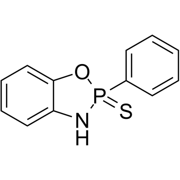
FW1256
FW1256 はフェニル類似体であり、徐放性の硫化水素 (H2S) 供与体です。
-
GC12178
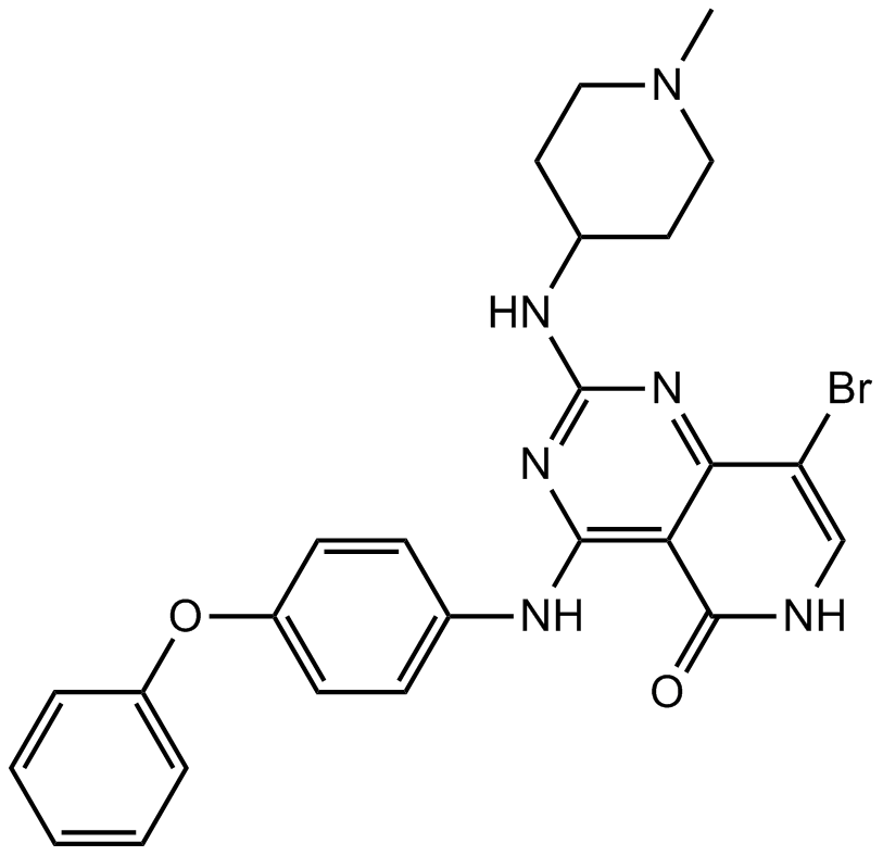
G-749
G-749 は、FLT3 野生型および FLT3-D835Y の IC50 がそれぞれ 0.4 nM および 0.6 nM である、強力な経口活性および ATP 競合 FLT3 阻害剤です。 G-749 は、急性骨髄性白血病 (AML) の薬剤耐性の研究に使用できます。
-
GC62246
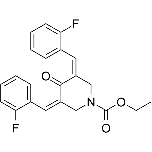
G5-7
G5-7 は経口活性型のアロステリックな JAK2 阻害剤であり、JAK2 に結合することにより、JAK2 を介したリン酸化と EGFR (Tyr1068) および STAT3 の活性化を選択的に阻害します。 G5-7 は、細胞周期の停止、アポトーシスを誘導し、抗血管新生効果を持っています。 G5-7 は神経膠腫研究の可能性を秘めています。
-
GC43723
Galactosylsphingosine (d18:1)
ガラクトセレブロシダーゼ (GALC) 酵素の基質であるガラクトシルスフィンゴシン (d18:1) (ガラクトシルスフィンゴシン) は、クラッベ病の潜在的なバイオマーカーです。

-
GC36103
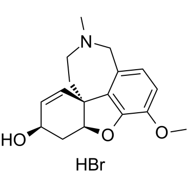
Galanthamine hydrobromide
-
GC38610
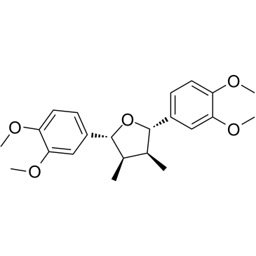
Galgravin
ガルグラビンはネクタンドラ メガポタミカに含まれる活性化合物で、抗炎症作用があります。ガルグラビンは in vitro で細胞傷害活性を示し、白血病細胞のアポトーシスを誘導します。
-
GN10388
Gallic acid

-
GC61436
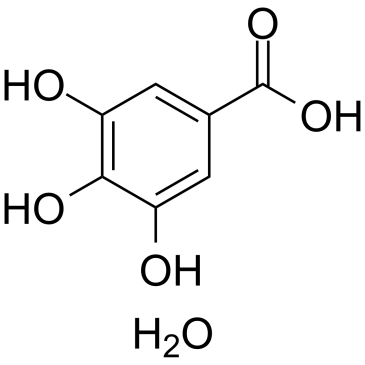
Gallic acid hydrate
没食子酸 (3,4,5-トリヒドロキシ安息香酸) 水和物は、天然のポリヒドロキシフェノール化合物であり、シクロオキシゲナーゼ-2 (COX-2) を阻害するフリーラジカルスカベンジャーです 。
-
GC12139
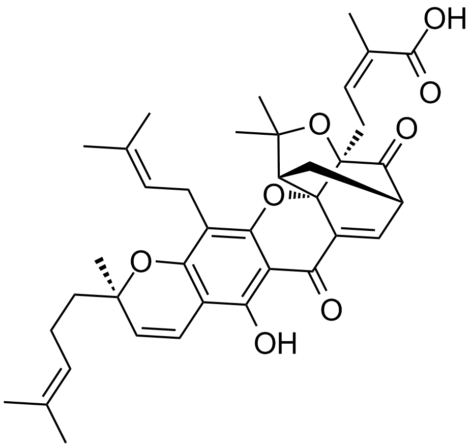
Gambogic Acid
抗がん作用を持つキサントノイド
-
GC36106
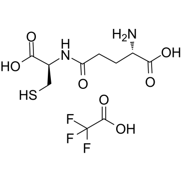
Gamma-glutamylcysteine (TFA)
γ-グルタミルシステイン (γ-グルタミルシステイン) グルタチオン (GSH) 合成の中間体である TFA は、抗酸化酵素グルタチオンペルオキシダーゼ (GPx) の必須補因子として機能するジペプチドです。
-
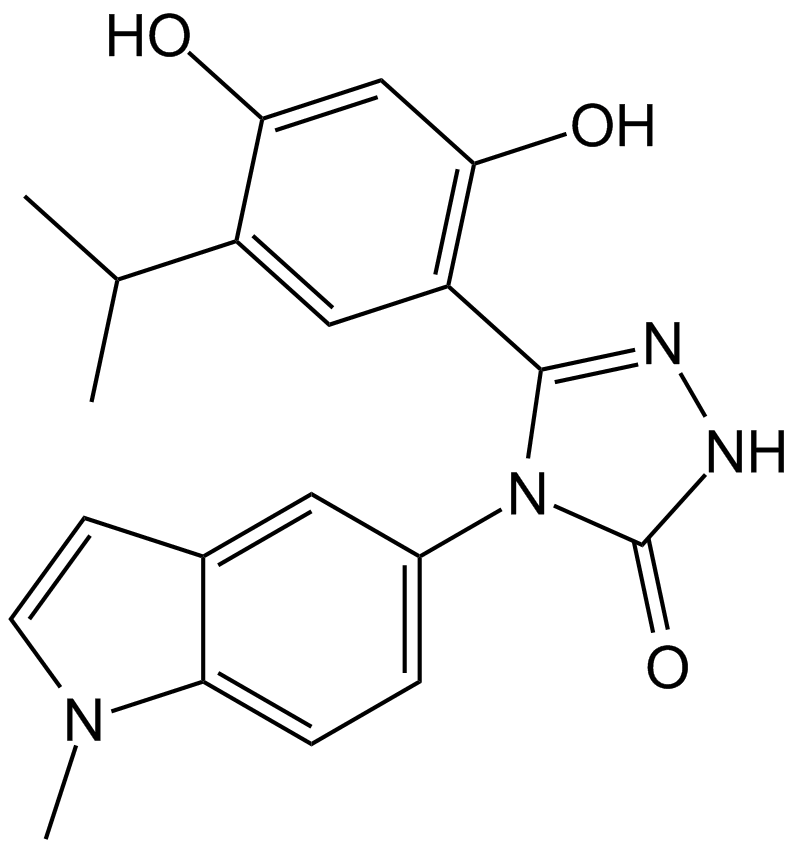
GC17655
Ganetespib (STA-9090)
-
GC43729
Ganglioside GD3 Mixture (sodium salt)
ガングリオシドGD3は、ラクトシルセラミドに2つのシアル酸残基を付加することで合成され、グリコシルおよびシアリルトランスフェラーゼの作用によってより複雑なガングリオシドの形成の前駆体として機能することができます。

-
GC47392
Ganglioside GM1 Mixture (ovine) (ammonium salt)
ガングリオシドGM1の混合物


