Apoptosis
As one of the cellular death mechanisms, apoptosis, also known as programmed cell death, can be defined as the process of a proper death of any cell under certain or necessary conditions. Apoptosis is controlled by the interactions between several molecules and responsible for the elimination of unwanted cells from the body.
Many biochemical events and a series of morphological changes occur at the early stage and increasingly continue till the end of apoptosis process. Morphological event cascade including cytoplasmic filament aggregation, nuclear condensation, cellular fragmentation, and plasma membrane blebbing finally results in the formation of apoptotic bodies. Several biochemical changes such as protein modifications/degradations, DNA and chromatin deteriorations, and synthesis of cell surface markers form morphological process during apoptosis.
Apoptosis can be stimulated by two different pathways: (1) intrinsic pathway (or mitochondria pathway) that mainly occurs via release of cytochrome c from the mitochondria and (2) extrinsic pathway when Fas death receptor is activated by a signal coming from the outside of the cell.
Different gene families such as caspases, inhibitor of apoptosis proteins, B cell lymphoma (Bcl)-2 family, tumor necrosis factor (TNF) receptor gene superfamily, or p53 gene are involved and/or collaborate in the process of apoptosis.
Caspase family comprises conserved cysteine aspartic-specific proteases, and members of caspase family are considerably crucial in the regulation of apoptosis. There are 14 different caspases in mammals, and they are basically classified as the initiators including caspase-2, -8, -9, and -10; and the effectors including caspase-3, -6, -7, and -14; and also the cytokine activators including caspase-1, -4, -5, -11, -12, and -13. In vertebrates, caspase-dependent apoptosis occurs through two main interconnected pathways which are intrinsic and extrinsic pathways. The intrinsic or mitochondrial apoptosis pathway can be activated through various cellular stresses that lead to cytochrome c release from the mitochondria and the formation of the apoptosome, comprised of APAF1, cytochrome c, ATP, and caspase-9, resulting in the activation of caspase-9. Active caspase-9 then initiates apoptosis by cleaving and thereby activating executioner caspases. The extrinsic apoptosis pathway is activated through the binding of a ligand to a death receptor, which in turn leads, with the help of the adapter proteins (FADD/TRADD), to recruitment, dimerization, and activation of caspase-8 (or 10). Active caspase-8 (or 10) then either initiates apoptosis directly by cleaving and thereby activating executioner caspase (-3, -6, -7), or activates the intrinsic apoptotic pathway through cleavage of BID to induce efficient cell death. In a heat shock-induced death, caspase-2 induces apoptosis via cleavage of Bid.
Bcl-2 family members are divided into three subfamilies including (i) pro-survival subfamily members (Bcl-2, Bcl-xl, Bcl-W, MCL1, and BFL1/A1), (ii) BH3-only subfamily members (Bad, Bim, Noxa, and Puma9), and (iii) pro-apoptotic mediator subfamily members (Bax and Bak). Following activation of the intrinsic pathway by cellular stress, pro‑apoptotic BCL‑2 homology 3 (BH3)‑only proteins inhibit the anti‑apoptotic proteins Bcl‑2, Bcl-xl, Bcl‑W and MCL1. The subsequent activation and oligomerization of the Bak and Bax result in mitochondrial outer membrane permeabilization (MOMP). This results in the release of cytochrome c and SMAC from the mitochondria. Cytochrome c forms a complex with caspase-9 and APAF1, which leads to the activation of caspase-9. Caspase-9 then activates caspase-3 and caspase-7, resulting in cell death. Inhibition of this process by anti‑apoptotic Bcl‑2 proteins occurs via sequestration of pro‑apoptotic proteins through binding to their BH3 motifs.
One of the most important ways of triggering apoptosis is mediated through death receptors (DRs), which are classified in TNF superfamily. There exist six DRs: DR1 (also called TNFR1); DR2 (also called Fas); DR3, to which VEGI binds; DR4 and DR5, to which TRAIL binds; and DR6, no ligand has yet been identified that binds to DR6. The induction of apoptosis by TNF ligands is initiated by binding to their specific DRs, such as TNFα/TNFR1, FasL /Fas (CD95, DR2), TRAIL (Apo2L)/DR4 (TRAIL-R1) or DR5 (TRAIL-R2). When TNF-α binds to TNFR1, it recruits a protein called TNFR-associated death domain (TRADD) through its death domain (DD). TRADD then recruits a protein called Fas-associated protein with death domain (FADD), which then sequentially activates caspase-8 and caspase-3, and thus apoptosis. Alternatively, TNF-α can activate mitochondria to sequentially release ROS, cytochrome c, and Bax, leading to activation of caspase-9 and caspase-3 and thus apoptosis. Some of the miRNAs can inhibit apoptosis by targeting the death-receptor pathway including miR-21, miR-24, and miR-200c.
p53 has the ability to activate intrinsic and extrinsic pathways of apoptosis by inducing transcription of several proteins like Puma, Bid, Bax, TRAIL-R2, and CD95.
Some inhibitors of apoptosis proteins (IAPs) can inhibit apoptosis indirectly (such as cIAP1/BIRC2, cIAP2/BIRC3) or inhibit caspase directly, such as XIAP/BIRC4 (inhibits caspase-3, -7, -9), and Bruce/BIRC6 (inhibits caspase-3, -6, -7, -8, -9).
Any alterations or abnormalities occurring in apoptotic processes contribute to development of human diseases and malignancies especially cancer.
References:
1.Yağmur Kiraz, Aysun Adan, Melis Kartal Yandim, et al. Major apoptotic mechanisms and genes involved in apoptosis[J]. Tumor Biology, 2016, 37(7):8471.
2.Aggarwal B B, Gupta S C, Kim J H. Historical perspectives on tumor necrosis factor and its superfamily: 25 years later, a golden journey.[J]. Blood, 2012, 119(3):651.
3.Ashkenazi A, Fairbrother W J, Leverson J D, et al. From basic apoptosis discoveries to advanced selective BCL-2 family inhibitors[J]. Nature Reviews Drug Discovery, 2017.
4.McIlwain D R, Berger T, Mak T W. Caspase functions in cell death and disease[J]. Cold Spring Harbor perspectives in biology, 2013, 5(4): a008656.
5.Ola M S, Nawaz M, Ahsan H. Role of Bcl-2 family proteins and caspases in the regulation of apoptosis[J]. Molecular and cellular biochemistry, 2011, 351(1-2): 41-58.
対象は Apoptosis
- Pyroptosis(15)
- Caspase(77)
- 14.3.3 Proteins(3)
- Apoptosis Inducers(71)
- Bax(15)
- Bcl-2 Family(136)
- Bcl-xL(13)
- c-RET(15)
- IAP(32)
- KEAP1-Nrf2(73)
- MDM2(21)
- p53(137)
- PC-PLC(6)
- PKD(8)
- RasGAP (Ras- P21)(2)
- Survivin(8)
- Thymidylate Synthase(12)
- TNF-α(141)
- Other Apoptosis(1145)
- Apoptosis Detection(0)
- Caspase Substrate(0)
- APC(6)
- PD-1/PD-L1 interaction(60)
- ASK1(4)
- PAR4(2)
- RIP kinase(47)
- FKBP(22)
製品は Apoptosis
- Cat.No. 商品名 インフォメーション
-
GC17448
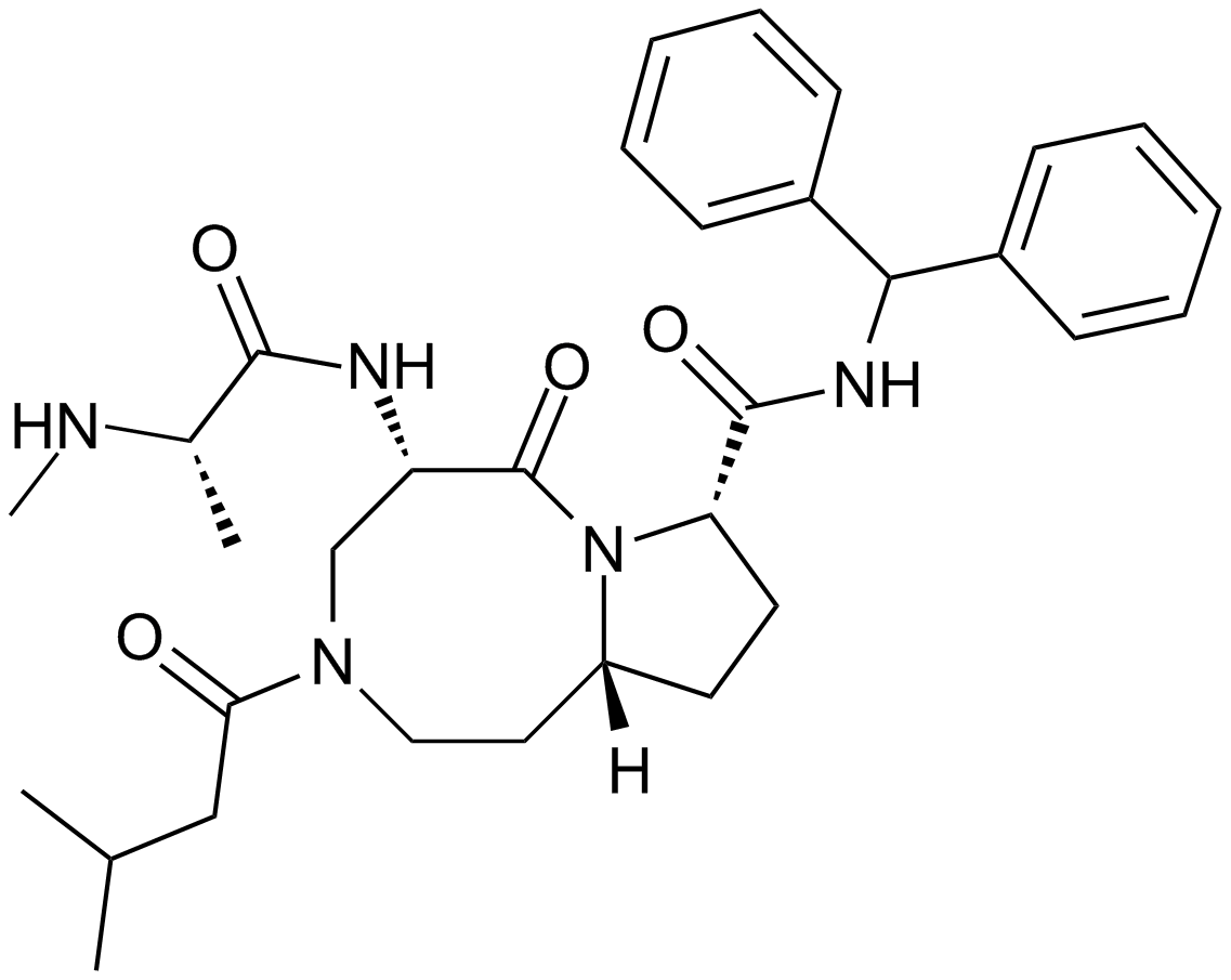
AT-406 (SM-406)
AT-406 (SM-406) (AT-406) は、経口で生物学的に利用可能な強力な Smac 模倣体であり、IAP のアンタゴニストであり、それぞれ 66.4、1.9、および 5.1 nM の Ki で XIAP、cIAP1、および cIAP2 タンパク質に結合します。 .
-
GC15870
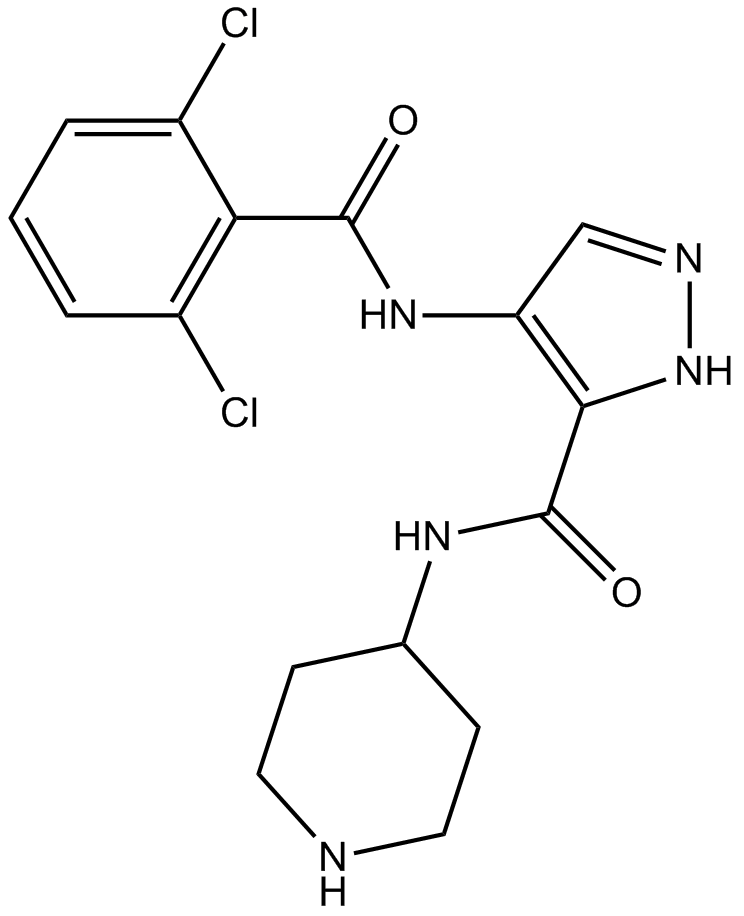
AT7519
AT7519 (AT7519M) は CDK の強力な阻害剤であり、CDK1、CDK2、CDK4 から CDK6、および CDK9 の IC50 はそれぞれ 210、47、100、13、170、および <10 nM です。
-
GC13998
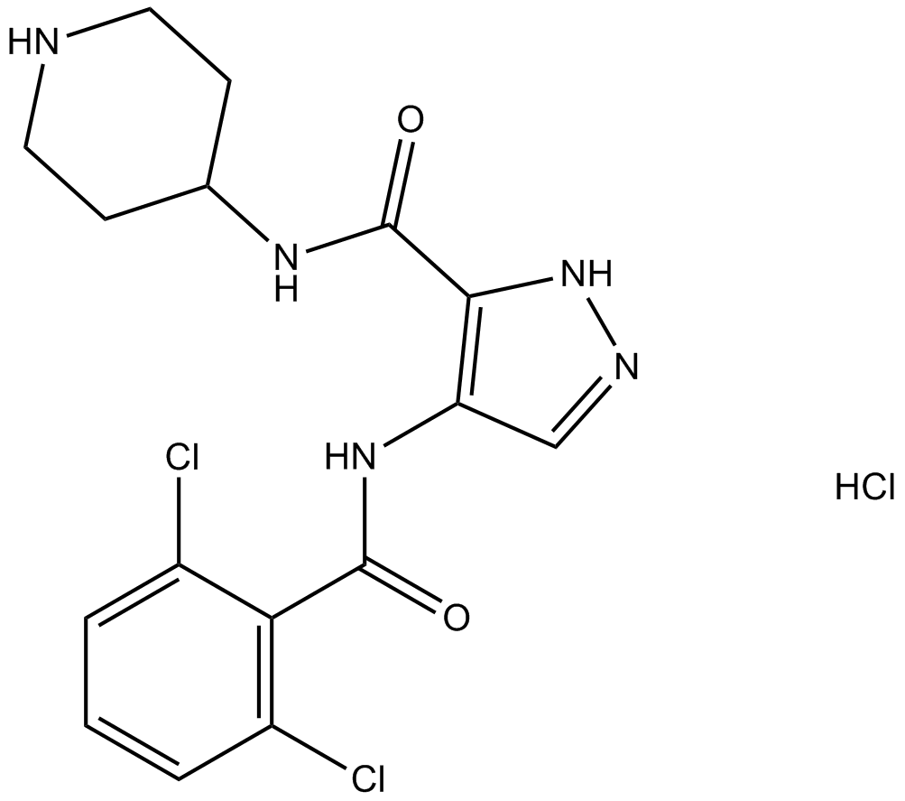
AT7519 Hydrochloride
Cdk阻害剤
-
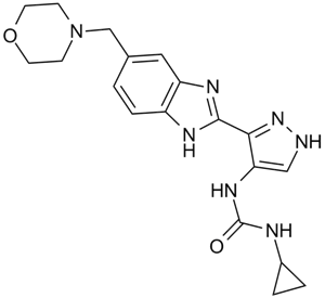
GC10638
AT9283
広範なスペクトルのキナーゼ阻害剤
-
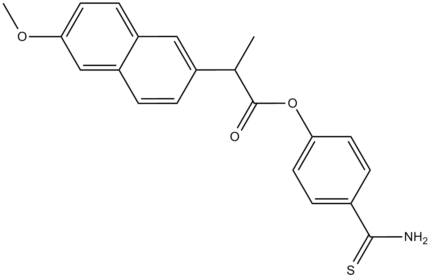
GC18133
ATB-346
ATB-346 (ATB-346)、経口活性非ステロイド性抗炎症薬 (NSAID) は、シクロオキシゲナーゼ-1 および 2 (COX-1 および 2) を阻害します。
-
GC32704
Atezolizumab (MPDL3280A)
アテゾリズマブ(MPDL3280A)は、がん研究に使用されるプログラム細胞死リガンド1(PD-L1)に対する選択的なヒト化モノクローナルIgG1抗体です。

-
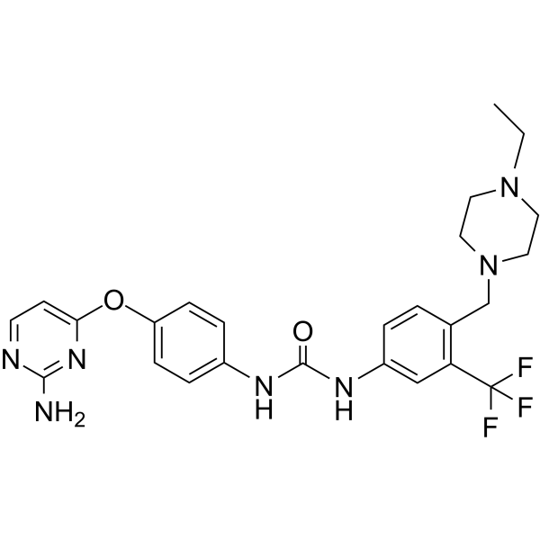
GC62499
ATH686
ATH686 は、強力な選択的 ATP 競合 FLT3 阻害剤です。 ATH686 は変異体 FLT3 プロテインキナーゼ活性を標的とし、アポトーシスの誘導と細胞周期阻害を介して FLT3 変異体を含む細胞の増殖を阻害します。 ATH686 には抗白血病効果があります。
-
GC46892
ATRA-BA Hybrid
オールトランスレチノイン酸とブチル酸のプロドラッグ形態

-
GN10394
Atractylenolide III

-
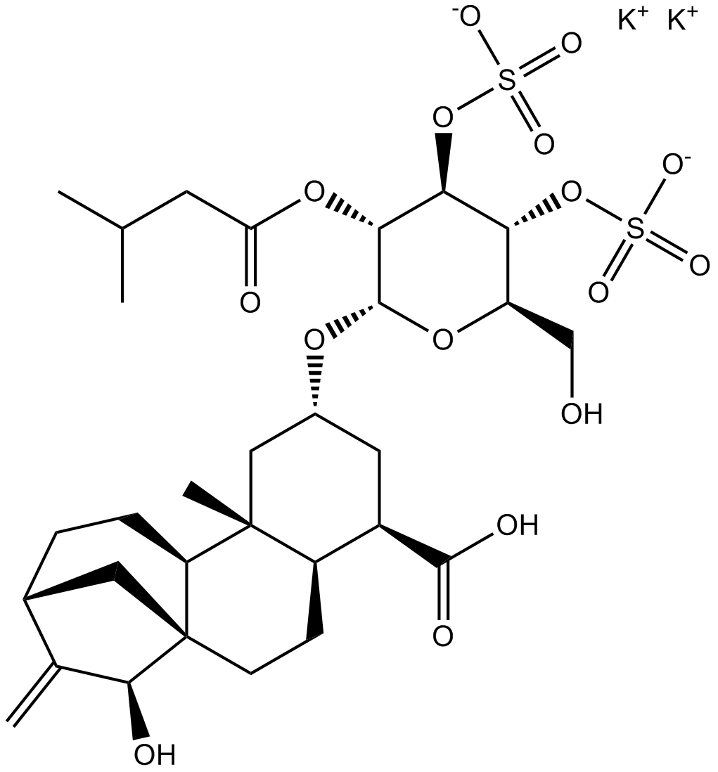
GC15878
Atractyloside Dipotassium Salt
ADP/ATPトランスロケーゼの阻害剤
-
GC39699
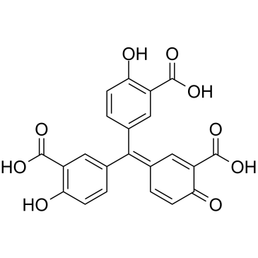
Aurintricarboxylic acid
アウリントリカルボン酸は、αβ-メチレン-ATP 感受性の P2X1R および P2X3R に対する選択性を備えたナノモル強度のアロステリック アンタゴニストであり、rP2X1R および rP2X3R の IC50 はそれぞれ 8.6 nM および 72.9 nM です 。
-
GC46895
Aurintricarboxylic Acid (ammonium salt)
多様な生物学的活性を持つタンパク質合成阻害剤

-
GC13332
Aurora A Inhibitor I
オーロラAキナーゼの強力で選択的な阻害剤

-
GC15295
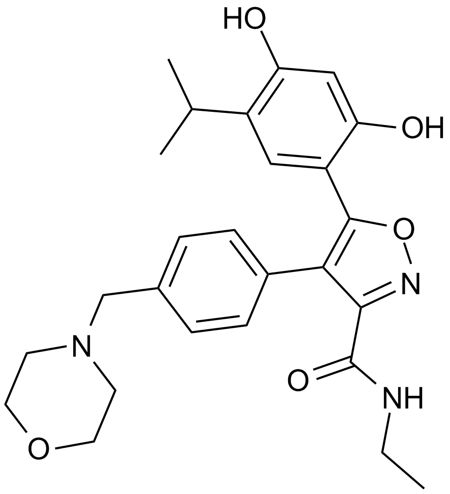
AUY922 (NVP-AUY922)
Hsp90阻害剤
-
GC31719
Avelumab (Anti-Human PD-L1, Human Antibody)
アベルマブ (Anti-Human PD-L1, Human Antibody) は、潜在的な抗体依存性細胞媒介性細胞傷害を有する完全ヒト IgG1 抗 PD-L1 モノクローナル抗体です。

-
GC42880
Avenanthramide-C methyl ester
アベナントラミドCメチルエステルは、IKKおよびIκBのリン酸化を阻害することによってNF-κB活性化の阻害剤であり(IC50〜40μM)、作用します。

-
GC35440
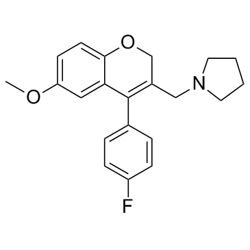
AX-024
AX-024 は、TCR によって誘発される T 細胞の活性化を IC50 ~1 nM で選択的に阻害する、TCR-Nck 相互作用の経口投与可能なクラス初の阻害剤です。
-
GC19046
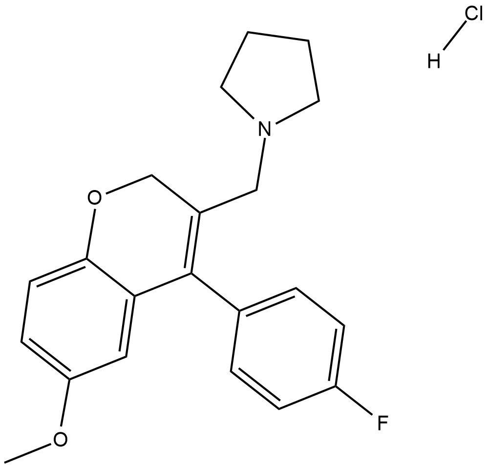
AX-024 hydrochloride
AX-024 塩酸塩は、TCR によって誘発される T 細胞の活性化を IC50 ~1 nM で選択的に阻害する、TCR-Nck 相互作用の経口投与可能なクラス初の阻害剤です。
-
GC17045
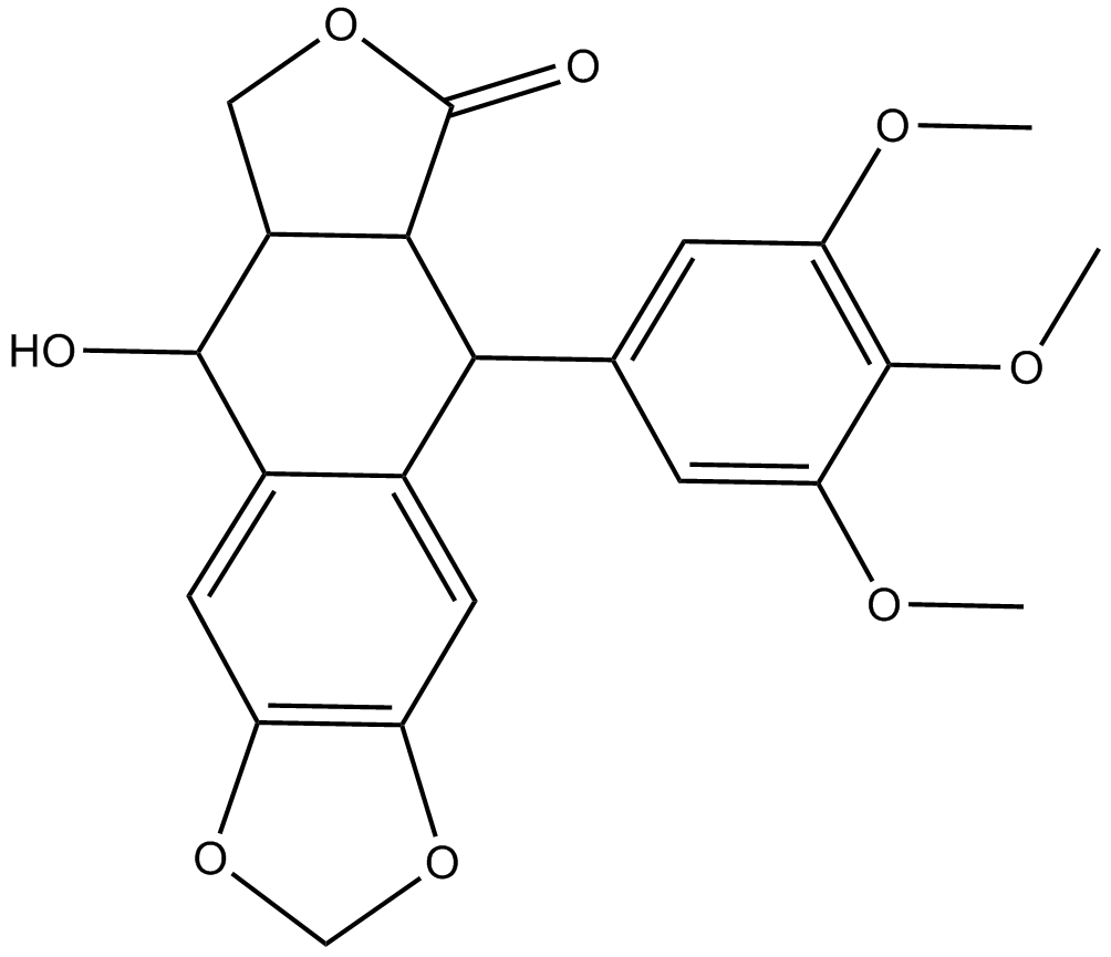
AXL1717
IGF-1Rの強力で選択的な阻害剤
-
GC15055
AZ 628
AZ 628 は、B-Raf、B-RafV600E、および c-Raf-1 に対してそれぞれ 105、34、および 29 nM の IC50 を持つ汎 Raf キナーゼ阻害剤です。

-
GC13433
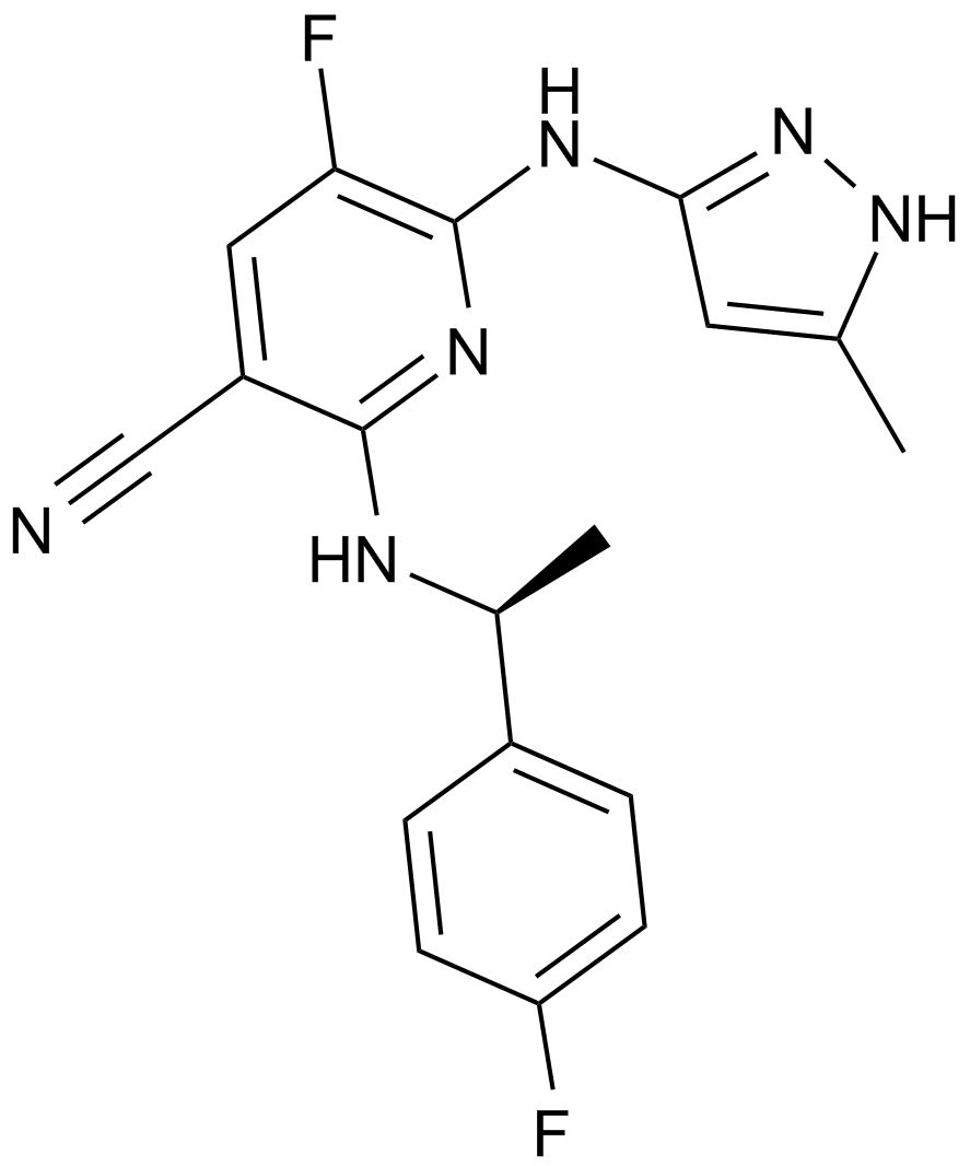
AZ 960
JAK2阻害剤
-
GC46901
Azadirachtin
最も有望な植物性殺虫剤の 1 つであるアザディラクチンは、害虫駆除に広く使用されています。

-
GC15033
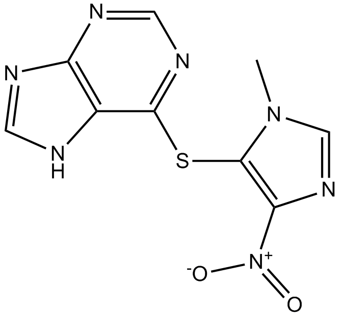
Azathioprine
アザチオプリン (BW 57-322) は、経口的に活性な免疫抑制剤です。
-
GC48971
AZD 1152 (hydrochloride)
強力なオーロラB阻害剤のプロドラッグ

-
GC18566
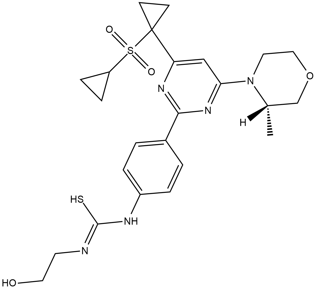
AZD 3147
AZD 3147 は、1.5 nM の IC50 値を持つ、mTORC1 および mTORC2 の強力な経口活性選択的二重阻害剤です。 AZD 3147 は、PI3K に対しても選択的な効果があります。
-
GC50109
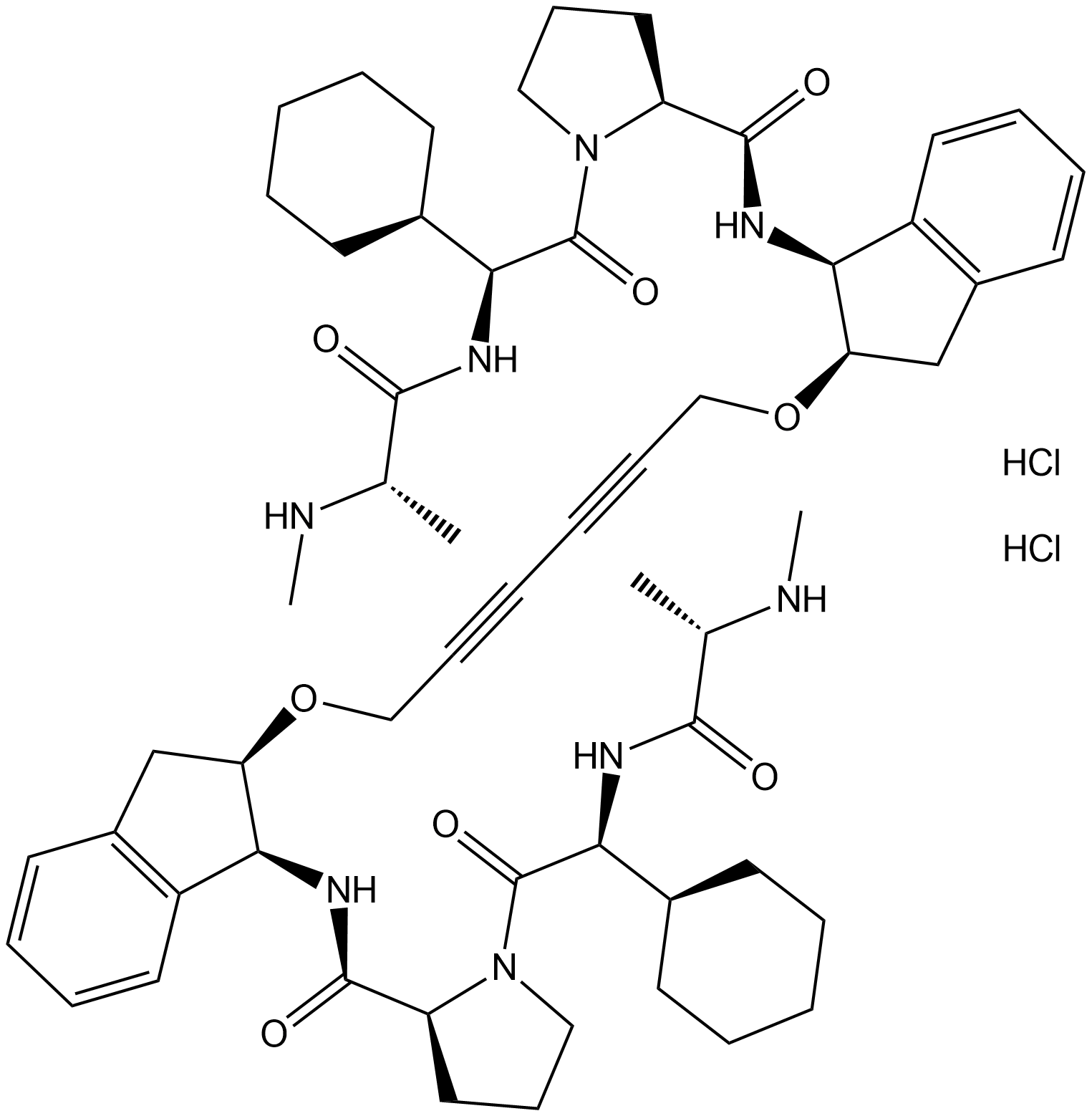
AZD 5582 dihydrochloride
AZD 5582 二塩酸塩は、BIR3 ドメイン cIAP1、cIAP2、および XIAP にそれぞれ 15、21、および 15 nM の IC50 で結合するアポトーシス タンパク質 (IAP) の阻害剤のアンタゴニストです。 AZD5582 はアポトーシスを誘導します。
-
GC33247
AZD-5991
AZD-5991 は、FRET アッセイで 0.7 nM の IC50、表面プラズモン共鳴 (SPR) アッセイで 0.17 nM の Kd を持つ、強力かつ選択的な Mcl-1 阻害剤です。

-
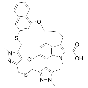
GC33283
AZD-5991 Racemate
AZD-5991 ラセミ体は AZD-5991 のラセミ体です。 AZD-5991 ラセミ体は、FRET アッセイでの IC50 が 3 nM 未満の Mcl-1 阻害剤です。
-
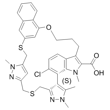
GC33239
AZD-5991 S-enantiomer
AZD-5991 S-エナンチオマーは、AZD-5991 の活性の低いエナンチオマーです。 AZD-5991 S-エナンチオマーは、FRET アッセイで 6.3 μM の IC50、表面プラズモン共鳴 (SPR) アッセイで 0.98 μM の Kd を持つ Mcl-1 阻害剤です。
-
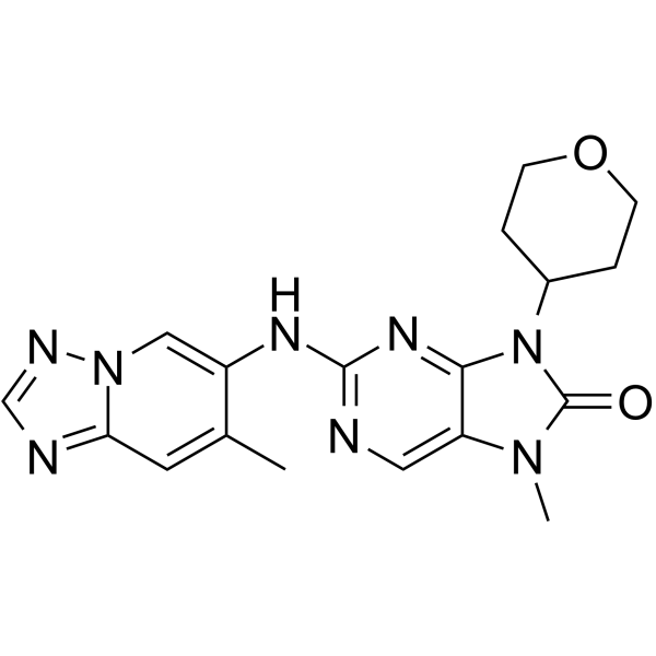
GC64938
AZD-7648
AZD-7648 は、0.6 nM の IC50 を持つ強力な経口活性の選択的 DNA-PK 阻害剤です。 AZD-7648 はアポトーシスを誘導し、抗腫瘍活性を示します。
-
GC12660
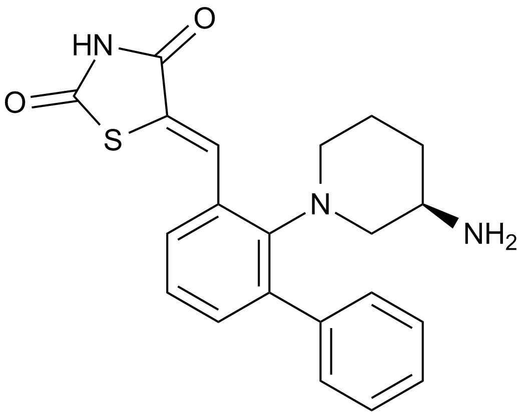
AZD1208
パン-Pimキナーゼ阻害剤
-
GC13029
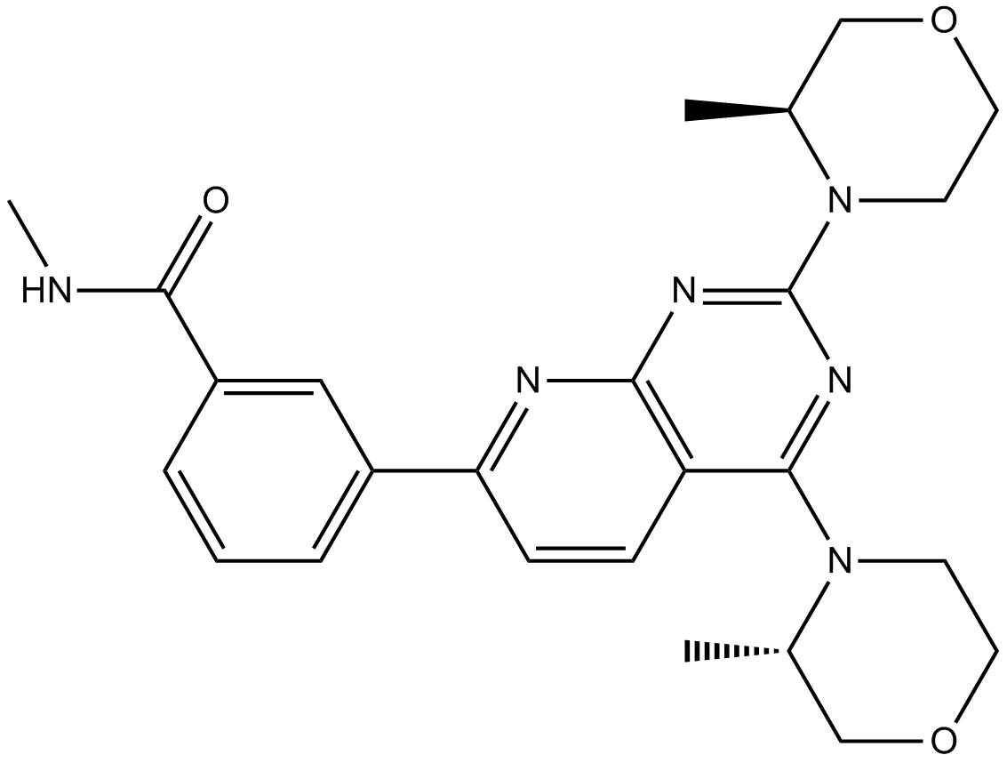
AZD2014
AZD2014 (AZD2014) は、IC50 が 2.81 nM の ATP 競合 mTOR 阻害剤です。 AZD2014 は、mTORC1 複合体と mTORC2 複合体の両方を阻害します。
-
GC33255
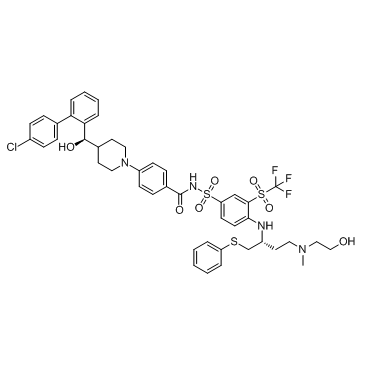
AZD4320
AZD4320 は、KPUM-MS3、KPUM-UH1、および STR-428 細胞に対してそれぞれ 26 nM、17 nM、および 170 nM の IC50 を持つ、新規の BH3 模倣デュアル BCL2/BCLxL 阻害剤です。
-
GC19050
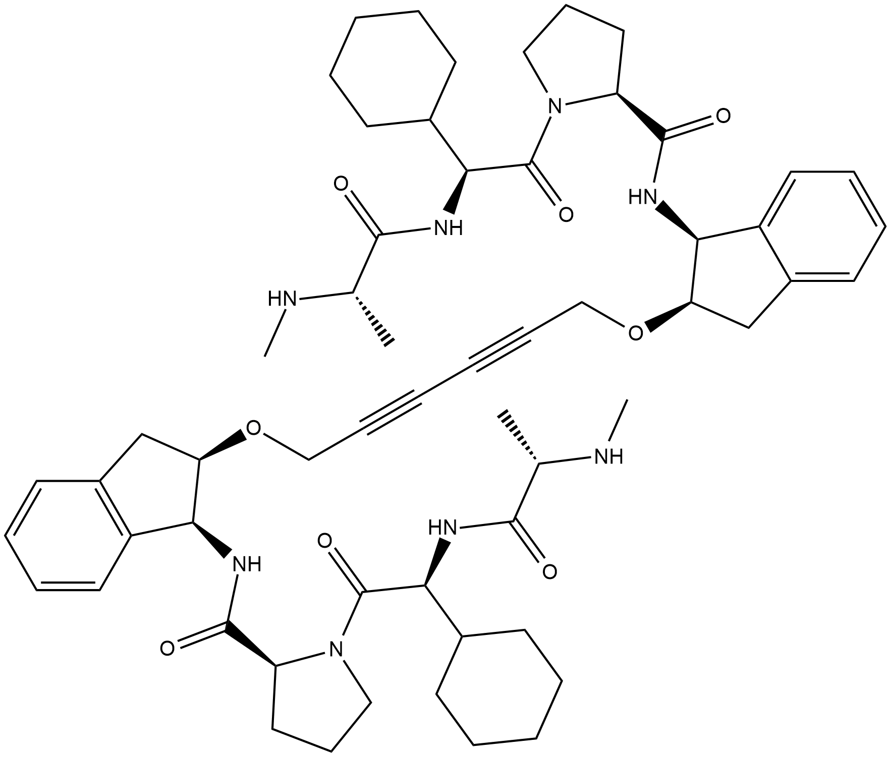
AZD5582
AZD5582 は、それぞれ 15、21、および 15 nM の IC50 で BIR3 ドメイン cIAP1、cIAP2、および XIAP に結合するアポトーシスタンパク質 (IAP) の阻害剤のアンタゴニストです。 AZD5582 はアポトーシスを誘導します。
-
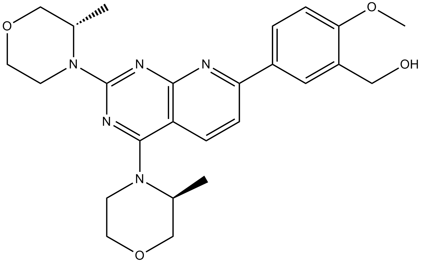
GC16380
AZD8055
MTOR阻害剤
-
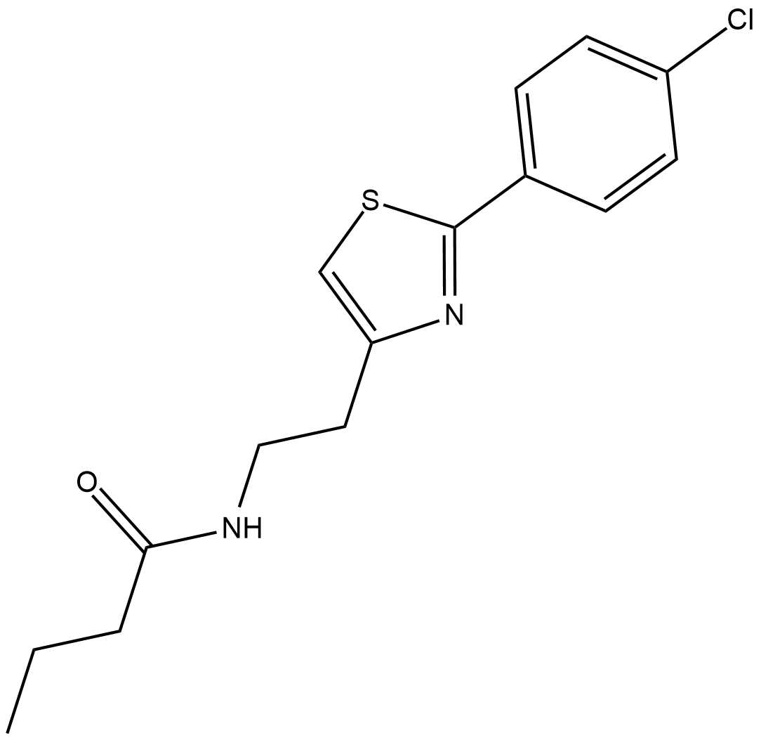
GC19054
Azoramide
アゾラミドは、展開タンパク質応答 (UPR) の強力な経口活性低分子モジュレーターです。
-
GC46904
Azoxystrobin
アゾキシストロビンは、広域スペクトルのβ-メトキシアクリレート殺菌剤です。

-
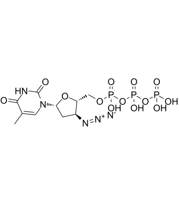
GC60616
AZT triphosphate
AZT 三リン酸 (3'-アジド-3'-デオキシチミジン-5'-三リン酸) は、ジドブジン (AZT) の活性な三リン酸代謝物です。
-
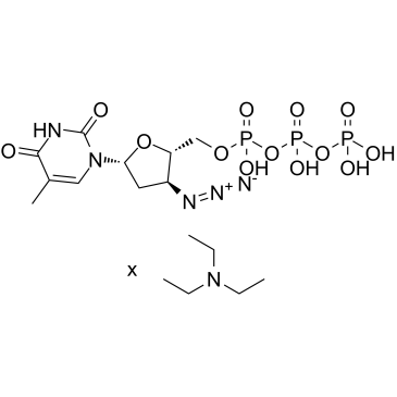
GC60617
AZT triphosphate TEA
AZT 三リン酸 TEA (3'-アジド-3'-デオキシチミジン-5'-三リン酸 TEA) は、ジドブジン (AZT) の活性三リン酸代謝物です。
-
GC35458
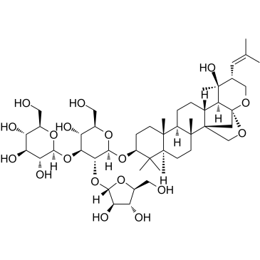
Bacopaside II
Bacopaside II は、薬草 Bacopa monnieri からの抽出物で、Aquaporin-1 (AQP1) 水チャネルを遮断し、AQP1 を発現する細胞の移動を阻害します。バコパシド II は、細胞周期の停止とアポトーシスを誘導します。
-
GC34263
Bak BH3
Bak BH3 は、Bak の BH3 ドメインに由来し、細胞内の Bcl-xL の機能に拮抗することができます。

-
GC52344
Bak BH3 (72-87) (human) (trifluoroacetate salt)
バク由来のペプチド

-
GC12053
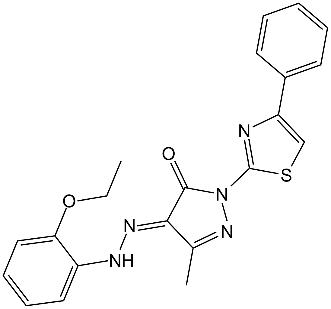
BAM7
Baxの直接活性化剤
-
GN10507
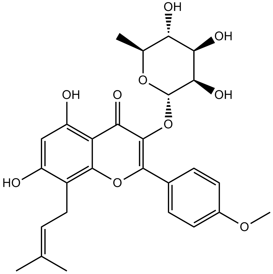
Baohuoside I
Baohuoside I, a flavonoid isolated from Epimedium koreanum Nakai, acts as an inhibitor of CXCR4, downregulates CXCR4 expression, induces apoptosis and shows anti-tumor activity.
-
GC15371
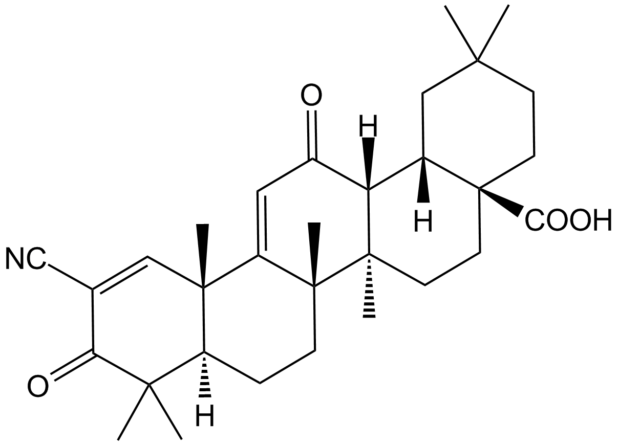
Bardoxolone
Nrf2/AREシグナルを活性化する抗炎症化合物
-
GC11572
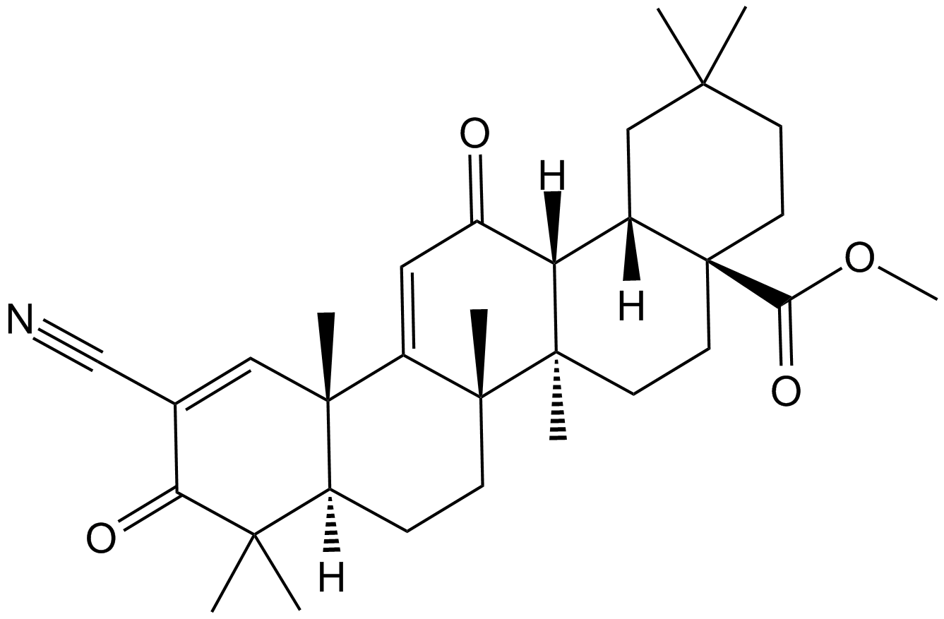
Bardoxolone methyl
がんと糖尿病に対する強力な抗癌作用および抗糖尿病作用を持つ合成トリテルペノイド
-
GC60620
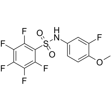
Batabulin
バタブリン (T138067) は抗腫瘍剤であり、β-チューブリン アイソタイプのサブセットに共有結合および選択的に結合し、それによって微小管重合を妨害します。バタブリンは細胞の形態に影響を与え、細胞周期の停止を引き起こし、最終的にアポトーシス細胞死を誘導します。
-
GC60621
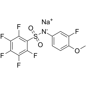
Batabulin sodium
バタブリン ナトリウム (T138067 ナトリウム) は抗腫瘍剤であり、β-チューブリン アイソタイプのサブセットに共有結合および選択的に結合し、それによって微小管重合を妨害します。バタブリンナトリウムは細胞の形態に影響を与え、細胞周期の停止を引き起こし、最終的にアポトーシス細胞死を誘導します。
-
GC12763
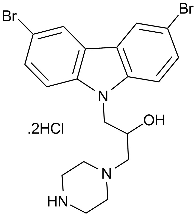
Bax channel blocker
-
GC16023
Bax inhibitor peptide P5
Bax阻害剤

-
GC17195
Bax inhibitor peptide V5
バックス阻害剤

-
GC52476
Bax Inhibitor Peptide V5 (trifluoroacetate salt)
バックス阻害剤

-
GC16695
Bax inhibitor peptide, negative control
ペプチドはBaxのミトコンドリアへの移行を抑制する。

-
GC10345
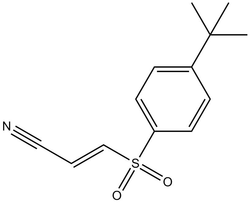
Bay 11-7085
BAY 11-7085 (BAY 11-7083) は、NF-κB の活性化と IκBα のリン酸化の阻害剤です。 10 μM の IC50 で IκBα を安定化します。
-
GC13035
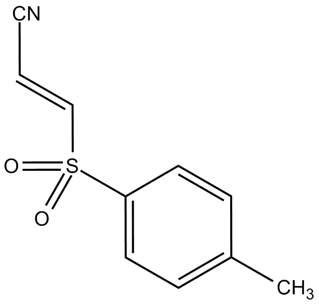
Bay 11-7821
選択的かつ不可逆的なNF-κB阻害剤
-
GC16389
BAY 61-3606
Syk阻害剤

-
GC42897
BAY 61-3606 (hydrochloride)
BAY 61-3606は、細胞膜透過性のある可逆的なスプレンチロシンキナーゼ(Syk)阻害剤であり(Ki = 7.5 nM; IC50 = 10 nM)、その効果は細胞内に及ぶ。

-
GC12136
BAY 61-3606 dihydrochloride
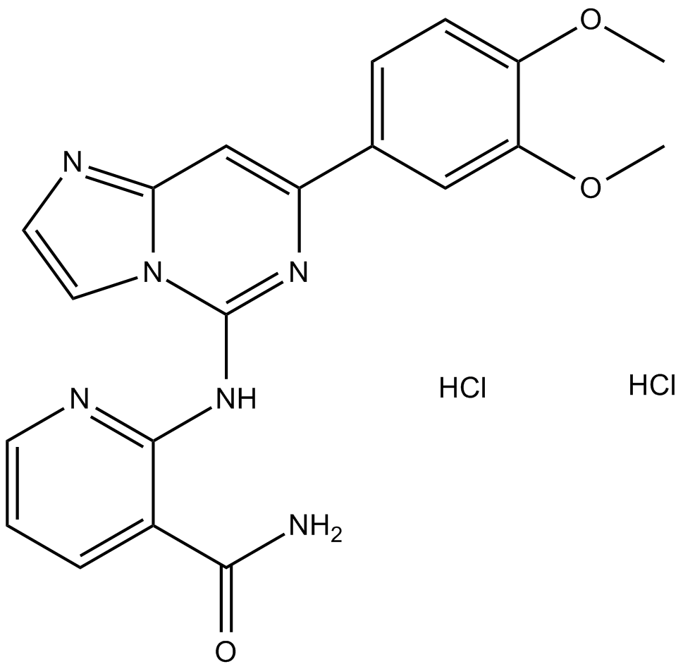
-
GC62164
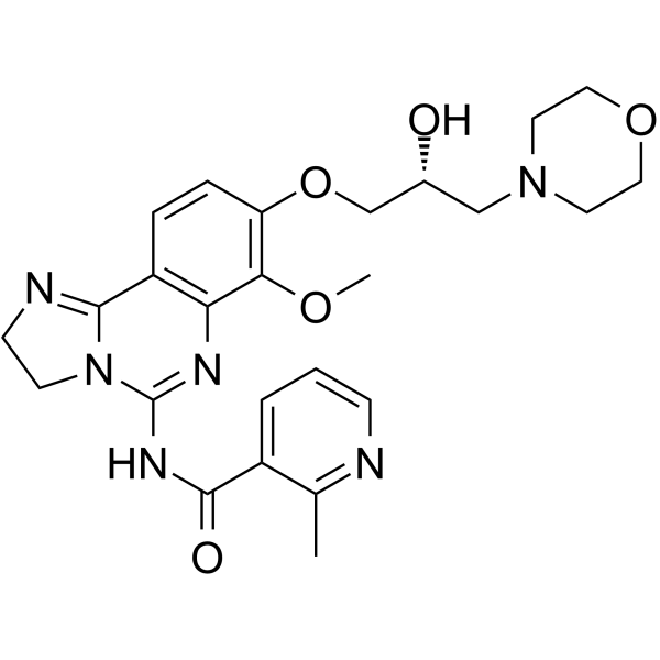
BAY1082439
BAY1082439 は、経口で生物学的に利用可能な選択的 PI3Kα/β/δ 阻害剤です。 BAY1082439 は、PIK3CA の変異型も阻害します。 BAY1082439 は、Pten-null 前立腺癌の増殖を阻害するのに非常に効果的です。
-
GC16516
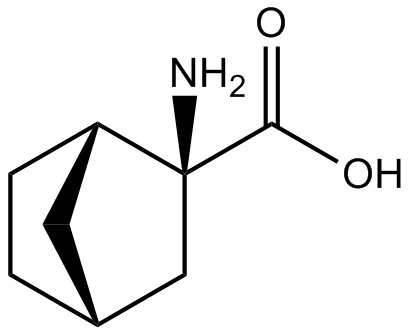
BCH
BCH (BCH) は、大型中性アミノ酸輸送体 1 (LAT1) の選択的かつ競合的な阻害剤であり、アミノ酸の細胞取り込みと mTOR リン酸化を大幅に阻害し、癌の増殖とアポトーシスの抑制を誘導します。
-
GC63325
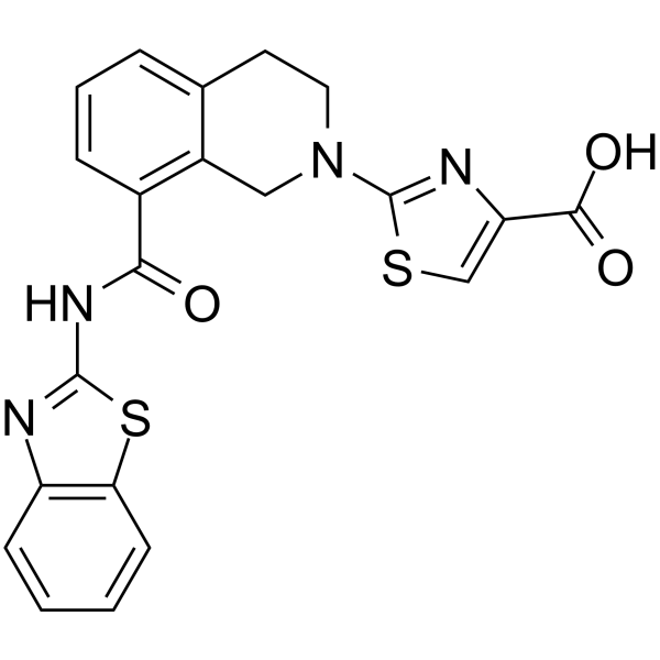
Bcl-xL antagonist 2
Bcl-xL アンタゴニスト 2 は、BCL-XL の強力で選択的な経口活性アンタゴニストであり、IC50 と Ki はそれぞれ 0.091 μM と 65 nM です。 Bcl-xL アンタゴニスト 2 は、がん細胞のアポトーシスを促進します。 Bcl-xL アンタゴニスト 2 は、慢性リンパ性白血病 (CLL) および非ホジキンリンパ腫 (NHL) の研究の可能性を秘めています。
-
GC62599
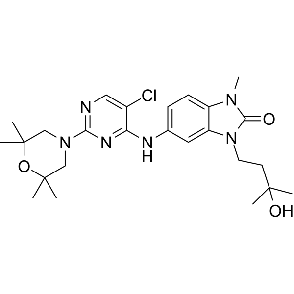
BCL6-IN-4
BCL6-IN-4 は、97 nM の IC50 を持つ強力な B 細胞リンパ腫 6 (BCL6) 阻害剤です。 BCL6-IN-4 には抗腫瘍作用があります。
-
GC68012
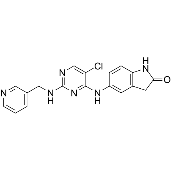
BCL6-IN-7
-
GC10721
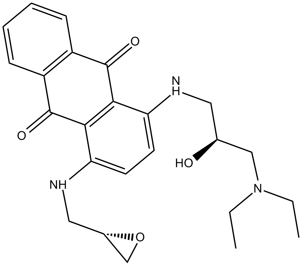
BDA-366
BDA-366 は強力な Bcl2 アンタゴニスト (Ki = 3.3 nM) であり、高い親和性と選択性で Bcl2-BH4 ドメインに結合します。 BDA-366 は、Bcl2 のコンフォメーション変化を誘発し、その抗アポトーシス機能を無効にし、生存分子から細胞死誘導因子に変換します。 BDA-366は肺がん細胞の増殖を抑制します。
-
GC42912
Becatecarin
ベカテカリンは、抗腫瘍効果を持つレベッカマイシン類似体です。ベカテカリンは DNA にインターカレートし、トポイソメラーゼ I/II の触媒活性を阻害します。

-
GC68369
Belantamab

-
GC65031
Belimumab
ベリムマブ (LymphoStat B) は、B 細胞活性化因子 (BAFF) を阻害するヒト IgG1Λ モノクローナル抗体です。

-
GC49042
Benastatin A
多様な生物学的活性を持つ細菌代謝産物

-
GC64354
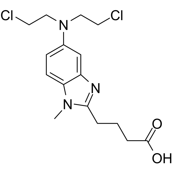
Bendamustine
プリン類似体であるベンダムスチン (SDX-105 遊離塩基) は、DNA 架橋剤です。ベンダムスチンは DNA 損傷ストレス応答とアポトーシスを活性化します。ベンダムスチンには、強力なアルキル化、抗がん、および代謝拮抗作用があります。
-
GC10744
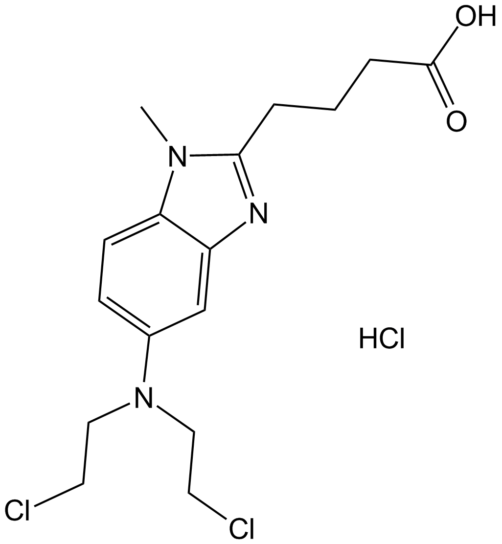
Bendamustine HCl
プリン類似体であるベンダムスチン HCl (SDX-105) は、DNA 架橋剤です。ベンダムスチン HCl は、DNA 損傷ストレス応答とアポトーシスを活性化します。ベンダムスチン HCl には、強力なアルキル化、抗がん、および代謝拮抗作用があります。
-
GC49781
Benomyl
カルバメート系農薬

-
GC62451
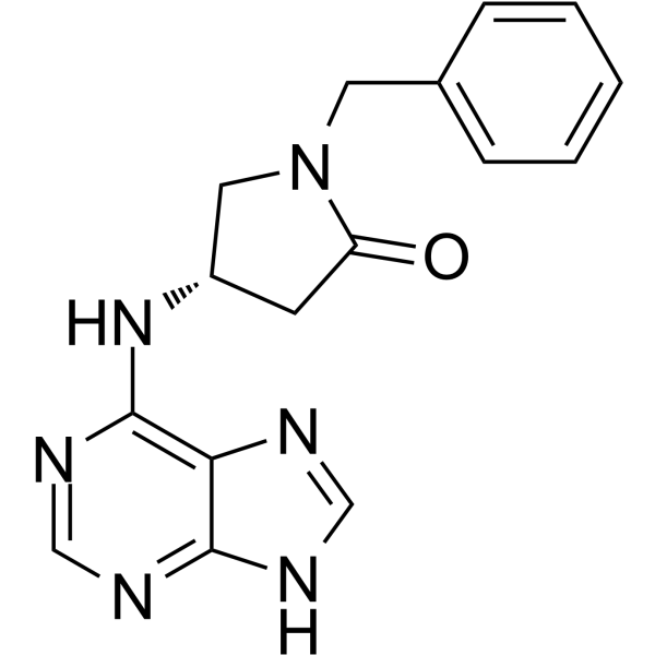
Benpyrine
ベンピリンは、82.1 μM の KD 値を持つ、非常に特異的で経口的に活性な TNF-α 阻害剤です。
-
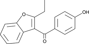
GC49403
Benzarone
ベンザロン (Fragivix) は強力なヒト尿酸トランスポーター 1 (hURAT1) 阻害剤であり、卵母細胞での IC50 は 2.8 μM です。
-
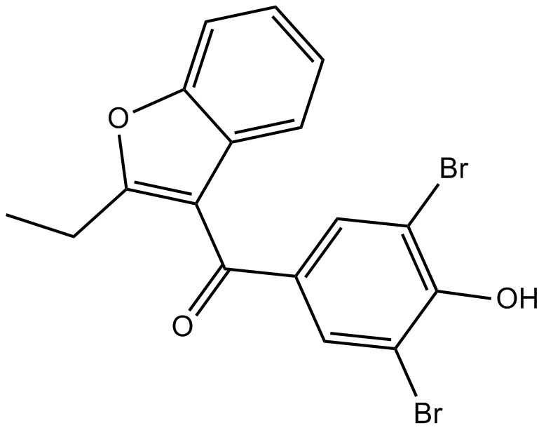
GC14930
Benzbromarone
ベンズブロマロンは、キサンチンオキシダーゼの非常に効果的で忍容性の高い非競合的阻害剤であり、痛風の治療に使用される尿酸排泄抑制剤として使用されます。
-
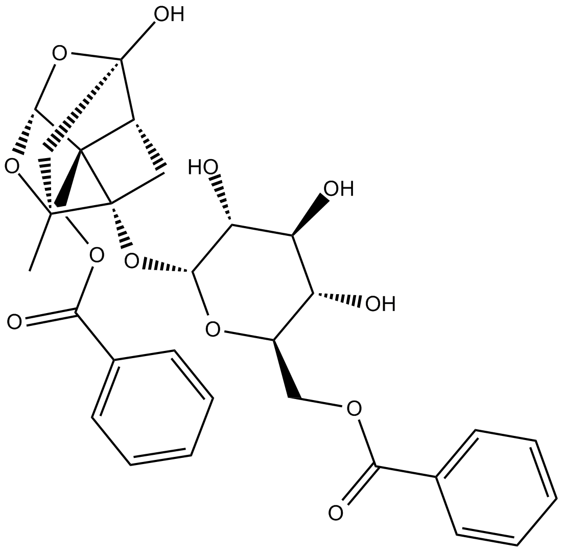
GN10520
Benzoylpaeoniflorin
-
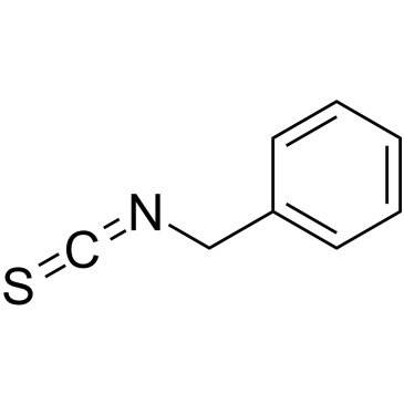
GC38683
Benzyl isothiocyanate
ベンジルイソチオシアネートは、抗菌活性を持つ天然イソチオシアネートのメンバーです。
-
GN10358
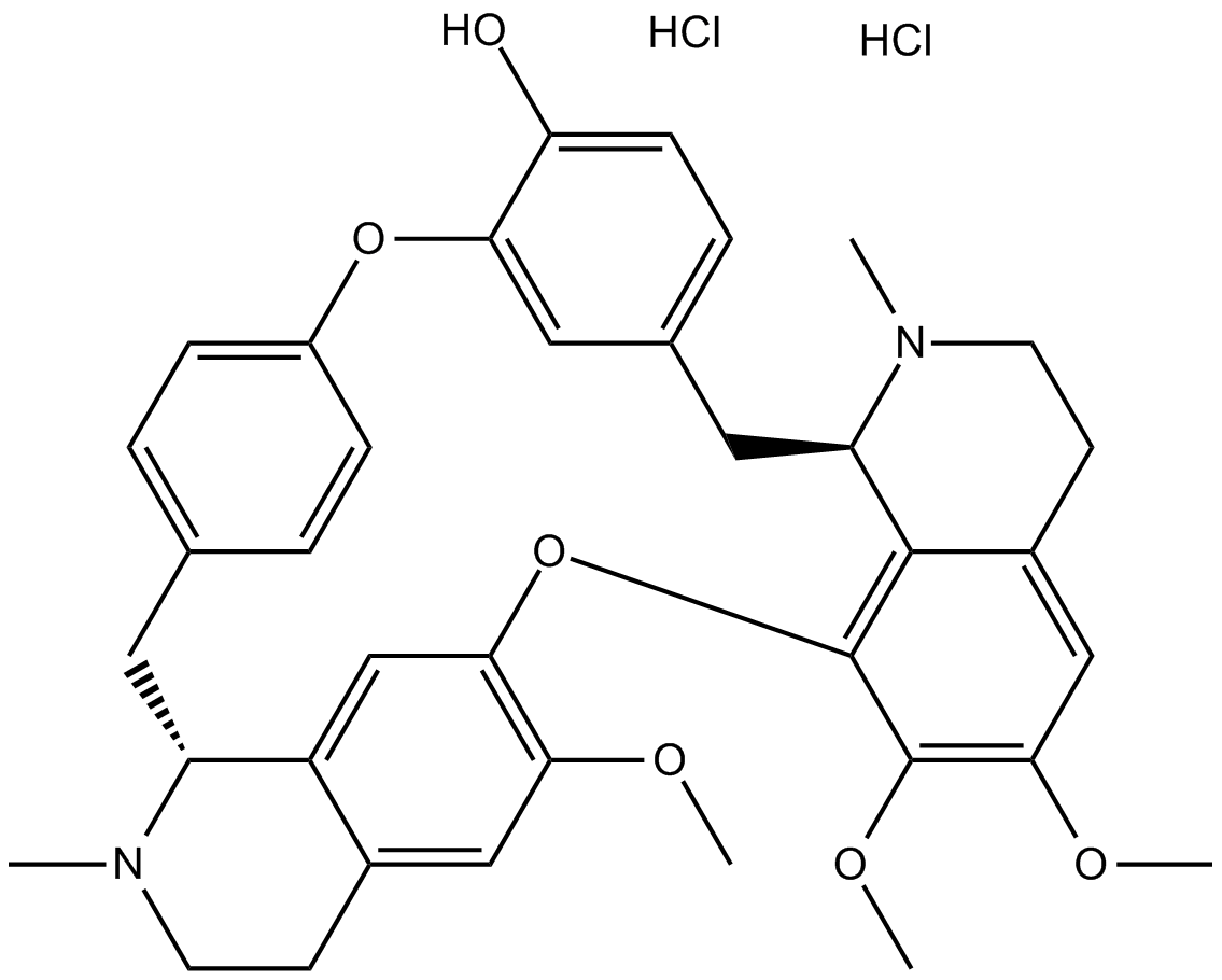
Berbamine hydrochloride
-
GN10539
Bergenin

-
GC42925
Berteroin
天然のスルフォラファン類似体であるベルテロインは、抗転移剤です。

-
GC10734
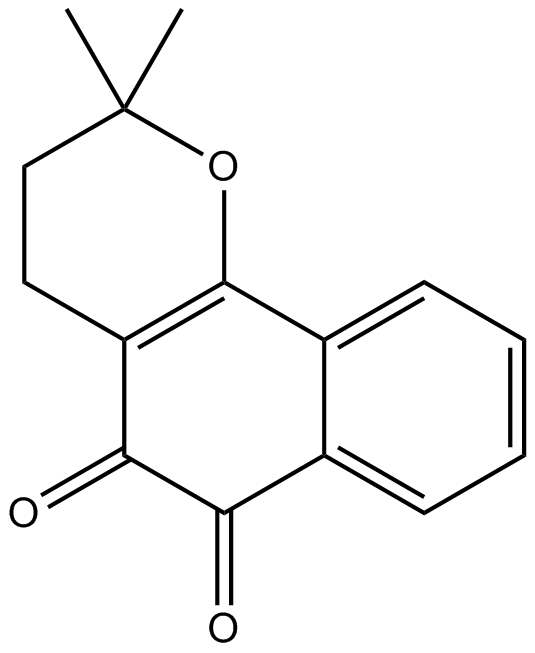
Beta-Lapachone
β-ラパコン (ARQ-501; NSC-26326) は、天然に存在する O-ナフトキノンであり、トポイソメラーゼ I 阻害剤として作用し、細胞周期の進行を阻害することによってアポトーシスを誘導します。
-
GC35504
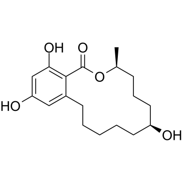
Beta-Zearalanol
Beta-Zearalenol は、Fusarium spp によって産生されるマイコトキシンであり、哺乳動物の生殖細胞にアポトーシスと酸化ストレスを引き起こします 。
-
GN10632
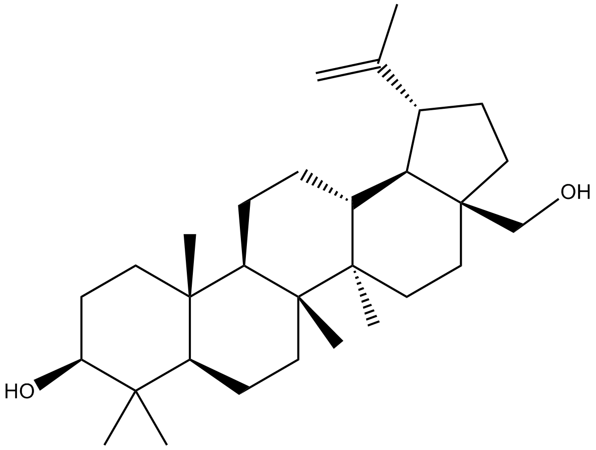
Betulin
-
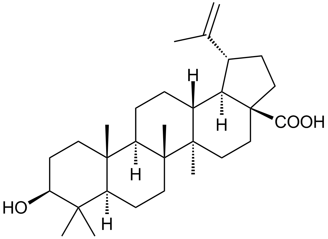
GC10480
Betulinic acid
胆汁酸に似た植物トリテルペノイド
-
GC48477
Betulinic Acid propargyl ester
ベチュリン酸のアルキン誘導体

-
GC48504
Betulinic Aldehyde oxime
ベチュリンの誘導体

-
GC48520
Betulonaldehyde
五環式トリテルペノイド

-
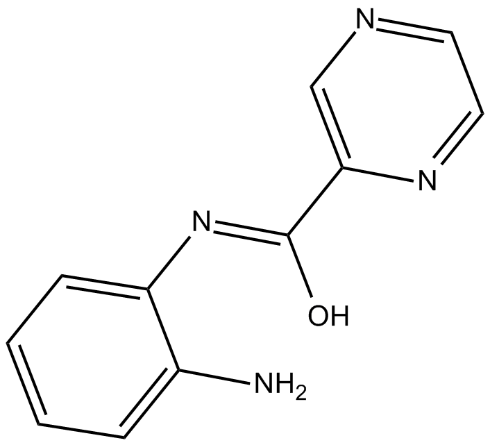
GC12074
BG45
BG45 は、HDAC3 に対する選択性を持つ HDAC クラス I 阻害剤です (IC50 = 289 nM)。
-
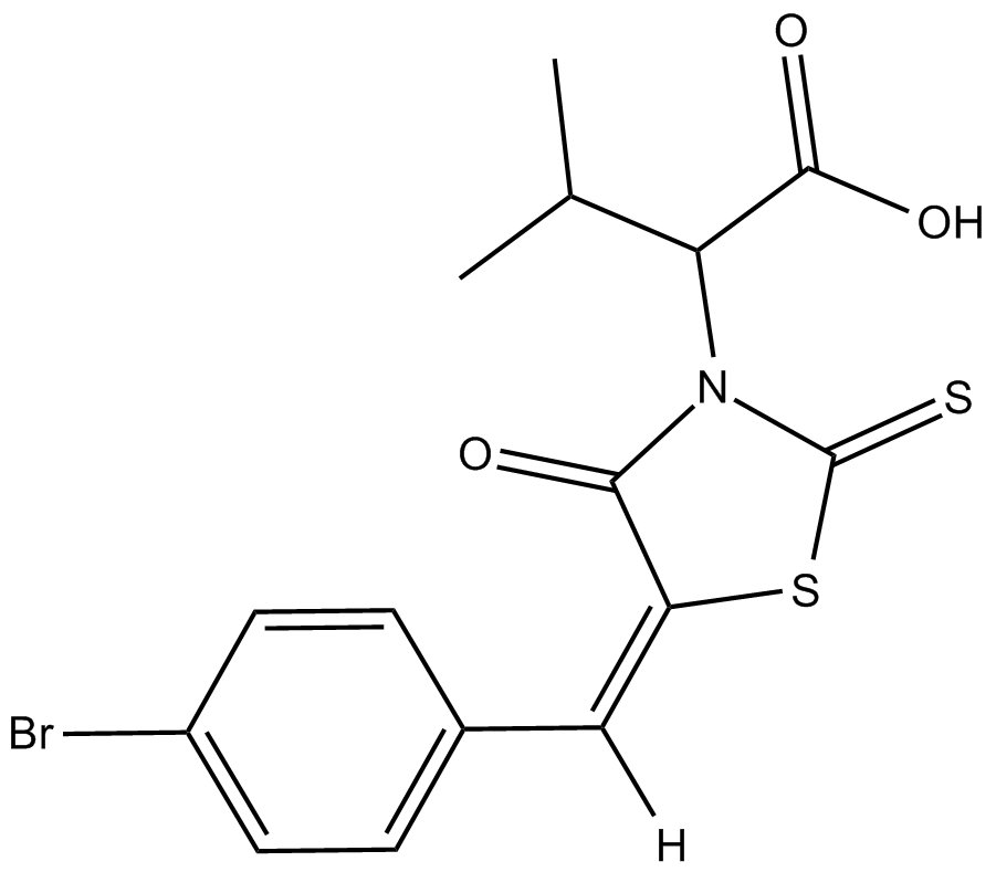
GC18136
BH3I-1
BH3I-1 は Bcl-2 ファミリーのアンタゴニストであり、FP アッセイで 2.4±0.2 μM の Ki で Bak BH3 ペプチドの Bcl-xL への結合を阻害します。 BH3I-1 は、p53/MDM2 ペアに対して 5.3 μM の Kd を持っています。
-
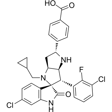
GC35511
BI-0252
BI-0252 は、4 nM の IC50 を持つ経口活性の選択的 MDM2-p53 阻害剤です。 BI-0252 は、マウス SJSA-1 異種移植片のすべての動物で腫瘍退行を誘発し、同時に腫瘍タンパク質 p53 (TP53) 標的遺伝子とアポトーシスのマーカーを誘発します。
-
GC17828
BI-847325
BI-847325 は、MEK および aurora キナーゼ (AK) の ATP 競合二重阻害剤であり、IC50 値は、ヒト MEK2 および AK-C に対してそれぞれ 4 および 15 nM です。

-
GC11224
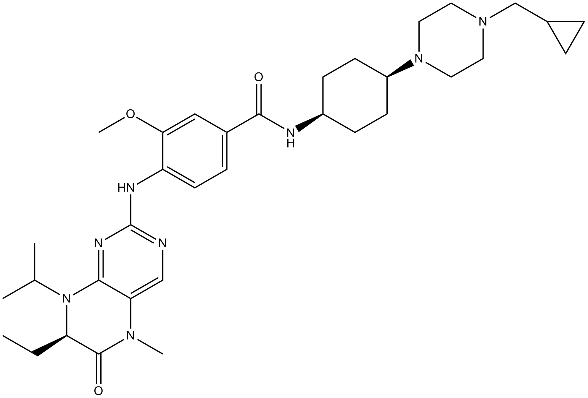
BI6727(Volasertib)
BI6727(Volasertib) (BI 6727) は、0.87 nM の IC50 を持つ、経口で活性があり、非常に強力で ATP 競合性の Polo 様キナーゼ 1 (PLK1) 阻害剤です。 BI6727(Volasertib) は、それぞれ 5 および 56 nM の IC50 で PLK2 および PLK3 を阻害します。 BI6727(Volasertib) は、有糸分裂停止とアポトーシスを誘導します。ジヒドロプテリジノン誘導体である BI6727(Volasertib) は、複数の癌モデルで顕著な抗腫瘍活性を示します。
-
GC13636
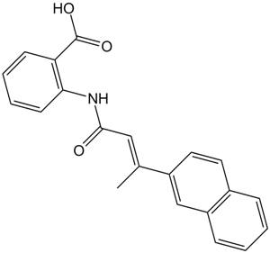
BIBR 1532
BIBR 1532 は、無細胞アッセイで IC50 が 100 nM の、強力で選択的かつ非競合的なテロメラーゼ阻害剤です。
-
GC60076
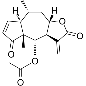
Bigelovin
Inula helianthus-aquatica から分離されたセスキテルペンラクトンであるビゲロビンは、選択的なレチノイド X 受容体 α アゴニストです。ビゲロビンは、ROS 生成によって調節される mTOR 経路の阻害を介してアポトーシスとオートファジーを誘導することにより、腫瘍の成長を抑制します。
-
GC15987
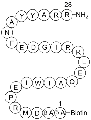
BIM, Biotinylated
バイオチン基が付加されたBimペプチドフラグメント
-
GC49513
Bim/BOD (IN) Polyclonal Antibody
Bim関連タンパク質の免疫検出には、以下のような方法があります。

-
GC52355
BimS BH3 (51-76) (human) (trifluoroacetate salt)
「Bim由来ペプチド」

-
GC14233
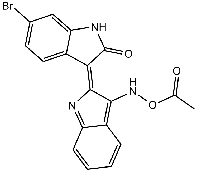
BIO-acetoxime
BIO-アセトキシム (BIA) は強力かつ選択的な GSK-3 阻害剤であり、GSK-3α/β の IC50 は両方とも 10 nM です。
-
GC67680
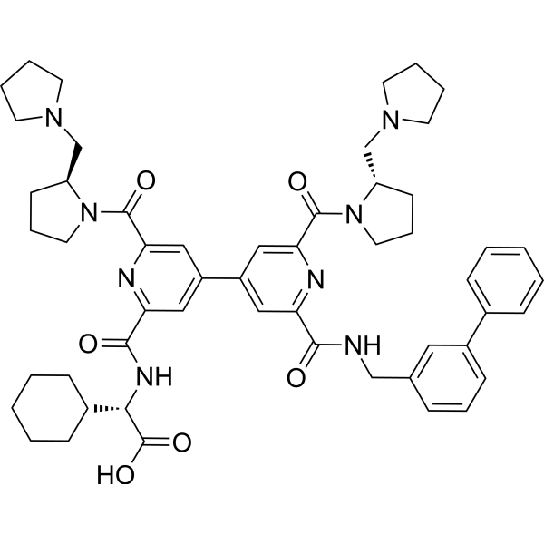
BIO8898
-
GC18476
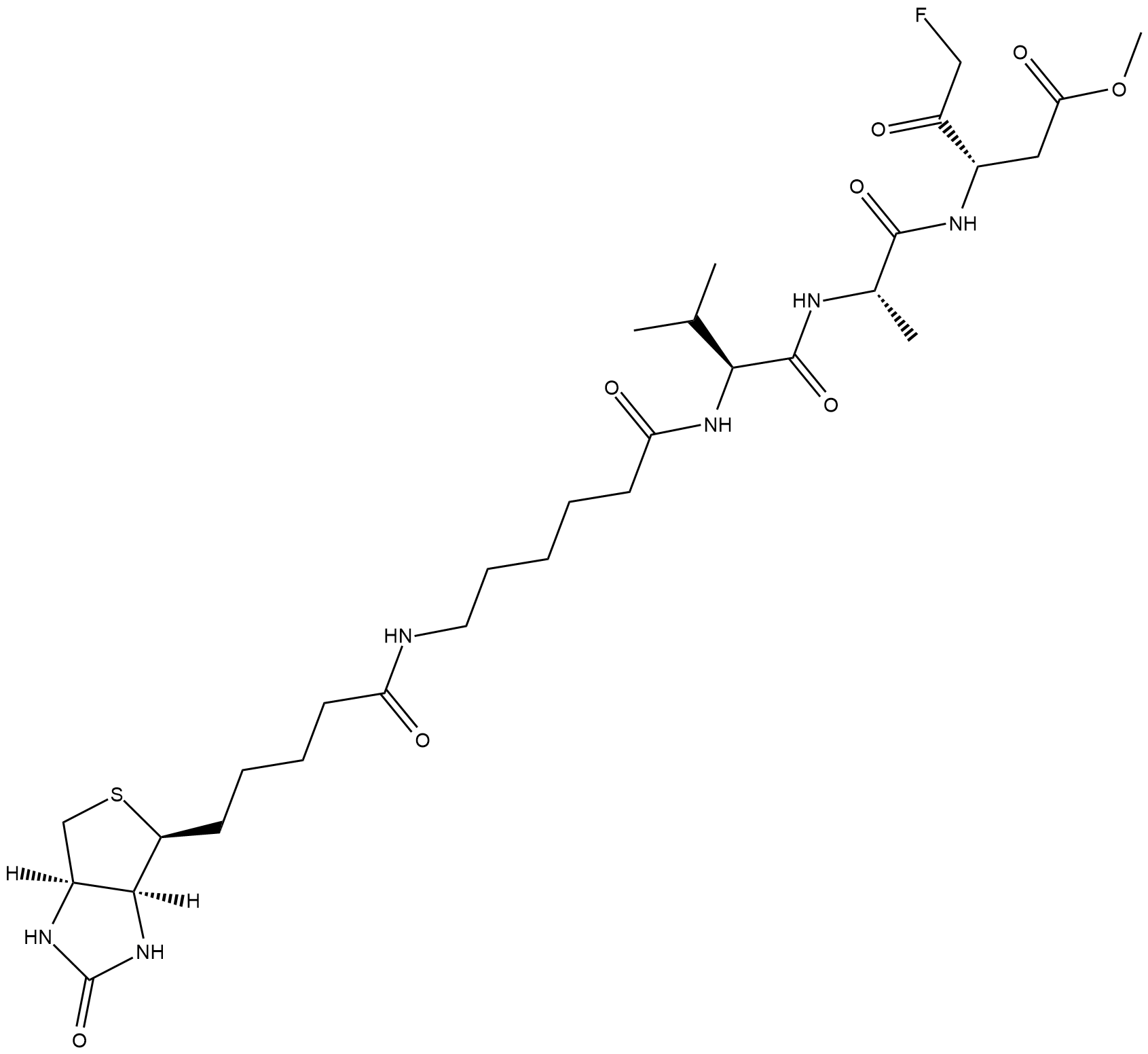
Biotin-VAD-FMK
ビオチン-VAD-FMK は細胞透過性で不可逆的なビオチン標識カスパーゼ阻害剤で、細胞ライセート中の活性カスパーゼを同定するために使用されます。
-
GC35523
Bioymifi
強力な TRAIL 受容体 DR5 アクチベーターである Bioymifi (DR5 Activator) は、1.2 μM の Kd で DR5 の細胞外ドメイン (ECD) に結合します。 Bioymifi は、DR5 のクラスタリングと凝集を誘導する単一の薬剤として作用し、アポトーシスにつながります。
