Apoptosis
As one of the cellular death mechanisms, apoptosis, also known as programmed cell death, can be defined as the process of a proper death of any cell under certain or necessary conditions. Apoptosis is controlled by the interactions between several molecules and responsible for the elimination of unwanted cells from the body.
Many biochemical events and a series of morphological changes occur at the early stage and increasingly continue till the end of apoptosis process. Morphological event cascade including cytoplasmic filament aggregation, nuclear condensation, cellular fragmentation, and plasma membrane blebbing finally results in the formation of apoptotic bodies. Several biochemical changes such as protein modifications/degradations, DNA and chromatin deteriorations, and synthesis of cell surface markers form morphological process during apoptosis.
Apoptosis can be stimulated by two different pathways: (1) intrinsic pathway (or mitochondria pathway) that mainly occurs via release of cytochrome c from the mitochondria and (2) extrinsic pathway when Fas death receptor is activated by a signal coming from the outside of the cell.
Different gene families such as caspases, inhibitor of apoptosis proteins, B cell lymphoma (Bcl)-2 family, tumor necrosis factor (TNF) receptor gene superfamily, or p53 gene are involved and/or collaborate in the process of apoptosis.
Caspase family comprises conserved cysteine aspartic-specific proteases, and members of caspase family are considerably crucial in the regulation of apoptosis. There are 14 different caspases in mammals, and they are basically classified as the initiators including caspase-2, -8, -9, and -10; and the effectors including caspase-3, -6, -7, and -14; and also the cytokine activators including caspase-1, -4, -5, -11, -12, and -13. In vertebrates, caspase-dependent apoptosis occurs through two main interconnected pathways which are intrinsic and extrinsic pathways. The intrinsic or mitochondrial apoptosis pathway can be activated through various cellular stresses that lead to cytochrome c release from the mitochondria and the formation of the apoptosome, comprised of APAF1, cytochrome c, ATP, and caspase-9, resulting in the activation of caspase-9. Active caspase-9 then initiates apoptosis by cleaving and thereby activating executioner caspases. The extrinsic apoptosis pathway is activated through the binding of a ligand to a death receptor, which in turn leads, with the help of the adapter proteins (FADD/TRADD), to recruitment, dimerization, and activation of caspase-8 (or 10). Active caspase-8 (or 10) then either initiates apoptosis directly by cleaving and thereby activating executioner caspase (-3, -6, -7), or activates the intrinsic apoptotic pathway through cleavage of BID to induce efficient cell death. In a heat shock-induced death, caspase-2 induces apoptosis via cleavage of Bid.
Bcl-2 family members are divided into three subfamilies including (i) pro-survival subfamily members (Bcl-2, Bcl-xl, Bcl-W, MCL1, and BFL1/A1), (ii) BH3-only subfamily members (Bad, Bim, Noxa, and Puma9), and (iii) pro-apoptotic mediator subfamily members (Bax and Bak). Following activation of the intrinsic pathway by cellular stress, pro‑apoptotic BCL‑2 homology 3 (BH3)‑only proteins inhibit the anti‑apoptotic proteins Bcl‑2, Bcl-xl, Bcl‑W and MCL1. The subsequent activation and oligomerization of the Bak and Bax result in mitochondrial outer membrane permeabilization (MOMP). This results in the release of cytochrome c and SMAC from the mitochondria. Cytochrome c forms a complex with caspase-9 and APAF1, which leads to the activation of caspase-9. Caspase-9 then activates caspase-3 and caspase-7, resulting in cell death. Inhibition of this process by anti‑apoptotic Bcl‑2 proteins occurs via sequestration of pro‑apoptotic proteins through binding to their BH3 motifs.
One of the most important ways of triggering apoptosis is mediated through death receptors (DRs), which are classified in TNF superfamily. There exist six DRs: DR1 (also called TNFR1); DR2 (also called Fas); DR3, to which VEGI binds; DR4 and DR5, to which TRAIL binds; and DR6, no ligand has yet been identified that binds to DR6. The induction of apoptosis by TNF ligands is initiated by binding to their specific DRs, such as TNFα/TNFR1, FasL /Fas (CD95, DR2), TRAIL (Apo2L)/DR4 (TRAIL-R1) or DR5 (TRAIL-R2). When TNF-α binds to TNFR1, it recruits a protein called TNFR-associated death domain (TRADD) through its death domain (DD). TRADD then recruits a protein called Fas-associated protein with death domain (FADD), which then sequentially activates caspase-8 and caspase-3, and thus apoptosis. Alternatively, TNF-α can activate mitochondria to sequentially release ROS, cytochrome c, and Bax, leading to activation of caspase-9 and caspase-3 and thus apoptosis. Some of the miRNAs can inhibit apoptosis by targeting the death-receptor pathway including miR-21, miR-24, and miR-200c.
p53 has the ability to activate intrinsic and extrinsic pathways of apoptosis by inducing transcription of several proteins like Puma, Bid, Bax, TRAIL-R2, and CD95.
Some inhibitors of apoptosis proteins (IAPs) can inhibit apoptosis indirectly (such as cIAP1/BIRC2, cIAP2/BIRC3) or inhibit caspase directly, such as XIAP/BIRC4 (inhibits caspase-3, -7, -9), and Bruce/BIRC6 (inhibits caspase-3, -6, -7, -8, -9).
Any alterations or abnormalities occurring in apoptotic processes contribute to development of human diseases and malignancies especially cancer.
References:
1.Yağmur Kiraz, Aysun Adan, Melis Kartal Yandim, et al. Major apoptotic mechanisms and genes involved in apoptosis[J]. Tumor Biology, 2016, 37(7):8471.
2.Aggarwal B B, Gupta S C, Kim J H. Historical perspectives on tumor necrosis factor and its superfamily: 25 years later, a golden journey.[J]. Blood, 2012, 119(3):651.
3.Ashkenazi A, Fairbrother W J, Leverson J D, et al. From basic apoptosis discoveries to advanced selective BCL-2 family inhibitors[J]. Nature Reviews Drug Discovery, 2017.
4.McIlwain D R, Berger T, Mak T W. Caspase functions in cell death and disease[J]. Cold Spring Harbor perspectives in biology, 2013, 5(4): a008656.
5.Ola M S, Nawaz M, Ahsan H. Role of Bcl-2 family proteins and caspases in the regulation of apoptosis[J]. Molecular and cellular biochemistry, 2011, 351(1-2): 41-58.
対象は Apoptosis
- Pyroptosis(15)
- Caspase(77)
- 14.3.3 Proteins(3)
- Apoptosis Inducers(71)
- Bax(15)
- Bcl-2 Family(136)
- Bcl-xL(13)
- c-RET(15)
- IAP(32)
- KEAP1-Nrf2(73)
- MDM2(21)
- p53(137)
- PC-PLC(6)
- PKD(8)
- RasGAP (Ras- P21)(2)
- Survivin(8)
- Thymidylate Synthase(12)
- TNF-α(141)
- Other Apoptosis(1145)
- Apoptosis Detection(0)
- Caspase Substrate(0)
- APC(6)
- PD-1/PD-L1 interaction(60)
- ASK1(4)
- PAR4(2)
- RIP kinase(47)
- FKBP(22)
製品は Apoptosis
- Cat.No. 商品名 インフォメーション
-
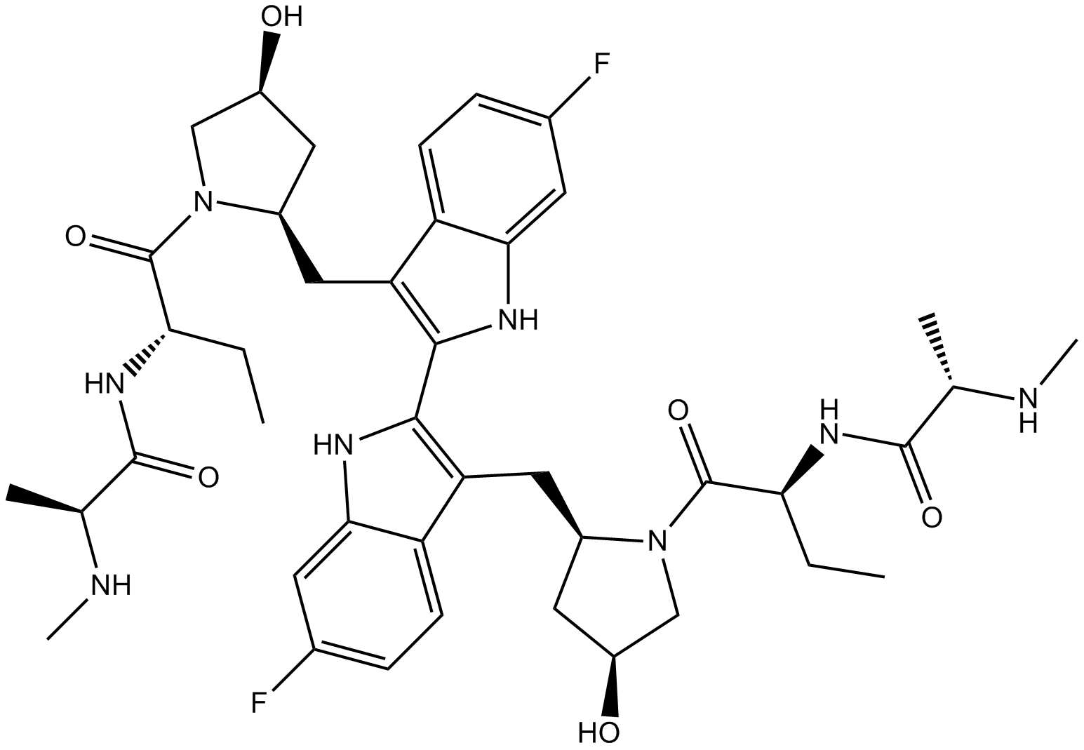
GC12426
Birinapant (TL32711)
cIAP1、cIAP2、およびXIAPの拮抗剤
-
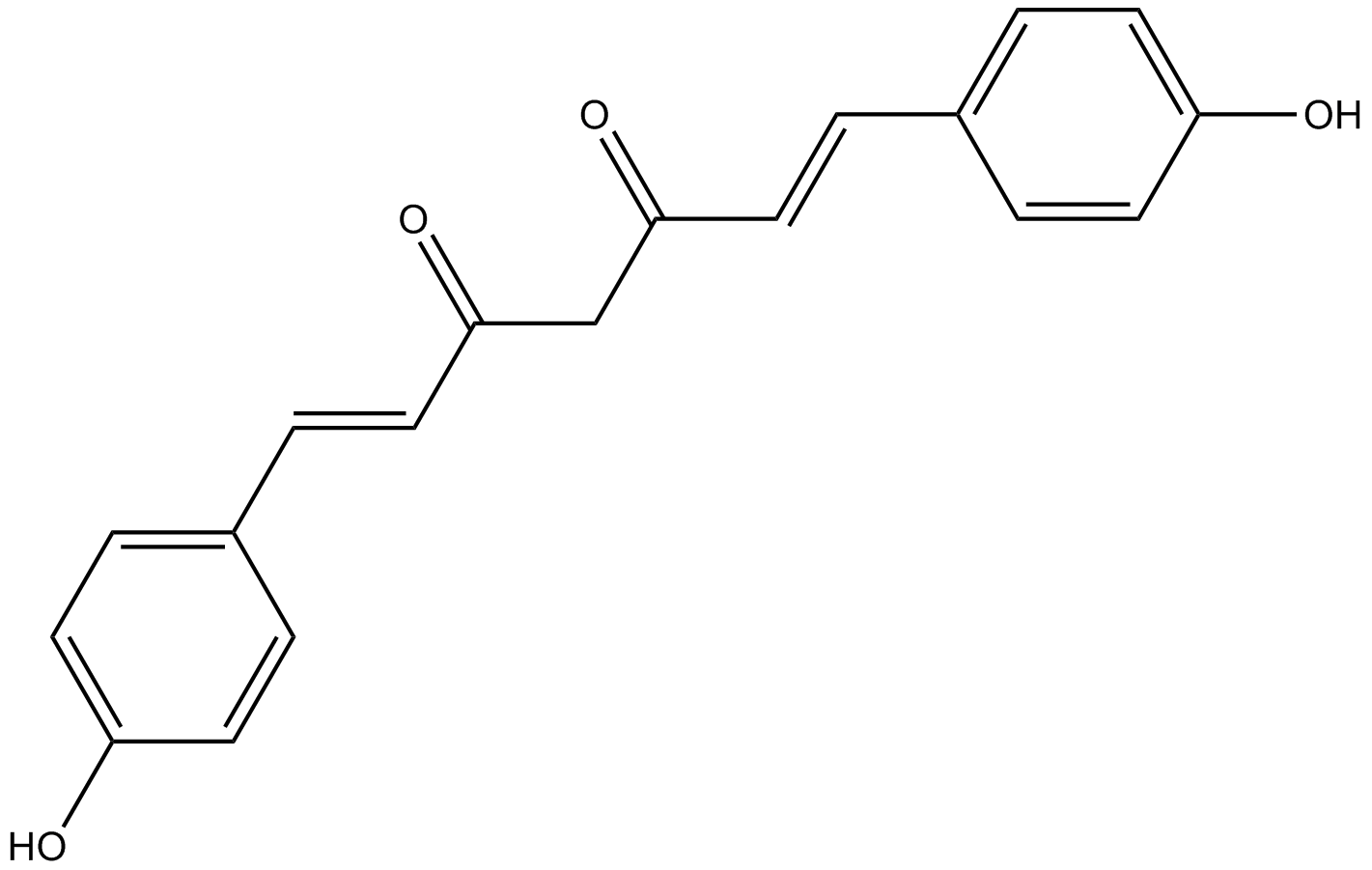
GN10037
Bisdemethoxycurcumin
-
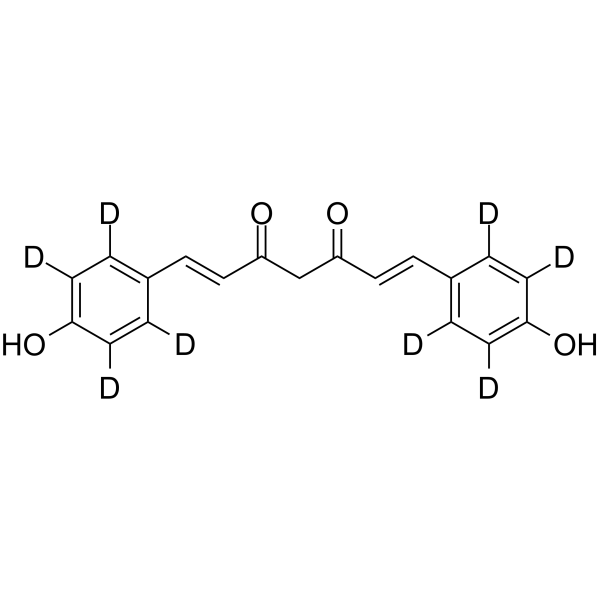
GC68308
Bisdemethoxycurcumin-d8
-
GC35530
BJE6-106
BJE6-106 (B106) は、IC50 が 0.05 μM の強力な選択的第 3 世代 PKCδ 阻害剤であり、古典的な PKC アイソザイム PKCα (IC50 = 50 μM) よりも選択的にターゲットを絞っています。

-
GC11931
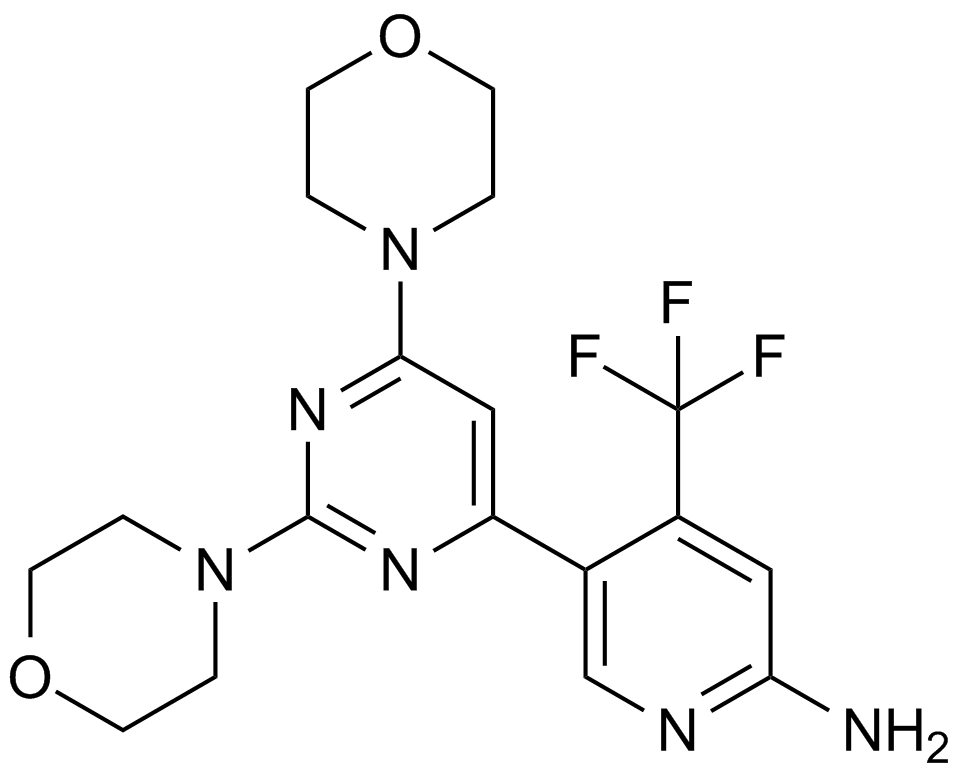
BKM120
クラスI PI3Kアイソフォームの阻害剤
-
GC65428
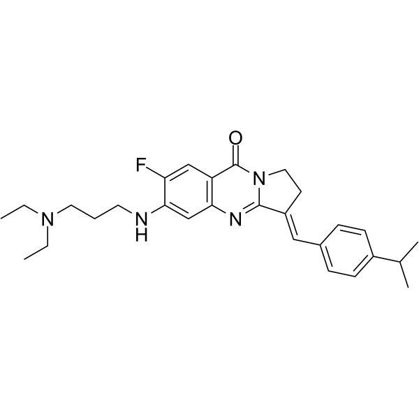
BLM-IN-1
BLM-IN-1(化合物29)は、効果的なブルーム症候群タンパク質(BLM)阻害剤であり、BLMに対して1.81μMの強力なBLM結合KDおよび0.95μMのIC50を有する。 DNA 損傷応答、ならびに癌細胞のアポトーシスおよび増殖停止を誘導します。
-
GC33407
BM 957
BM 957 は強力な Bcl-2 および Bcl-xL 阻害剤であり、Kis はそれぞれ 1.2、<1 nM、IC50 は 5.4、6.0 nM です。
-
GC13498
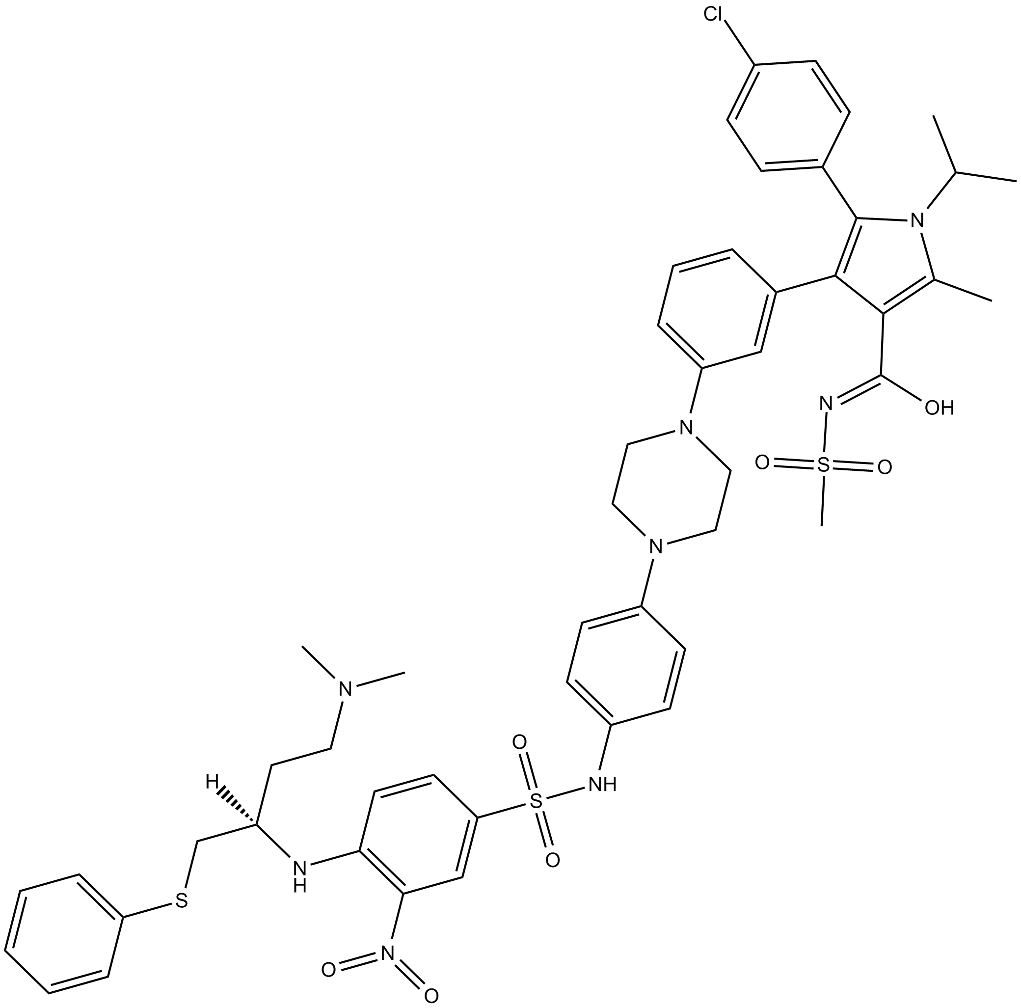
BM-1074
-
GC62871
BM-1244
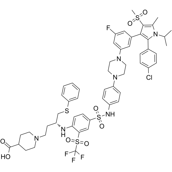
BM-1244 (APG-1252-M1) は強力な Bcl-xL/Bcl-2 阻害剤であり、Bcl-xL と Bcl-2 の Kis はそれぞれ 134 nM と 450 nM です。 BM-1244 は、5 nM の EC50 で老化線維芽細胞 (SnC) を阻害します。 (特許 WO2019033119A1 より)。
-
GC12822
BML-210(CAY10433)
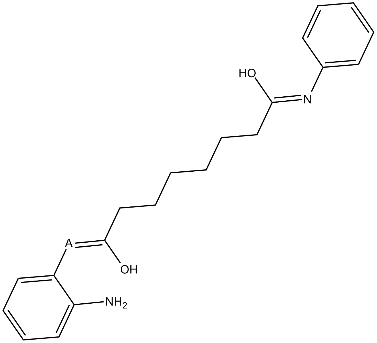
BML-210(CAY10433) は新規の HDAC 阻害剤であり、その作用機序は解明されていません。
-
GC11648
BML-277
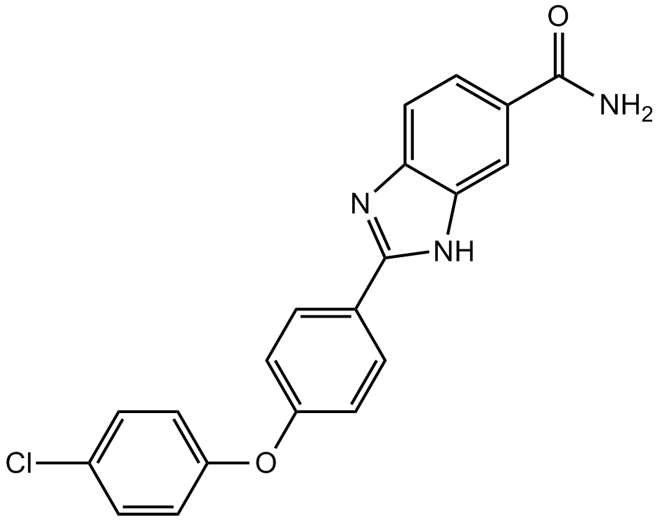
BML-277 は、IC50 が 15 nM の選択的チェックポイントキナーゼ 2 (Chk2) 阻害剤です。
-
GC42953
BMS 345541 (trifluoroacetate salt)
BMS 345541は、IκBキナーゼIKKαおよびIKKβの細胞浸透性阻害剤です(IC50値はそれぞれ4および0.3μM)。

-
GC25160
BMS-1001
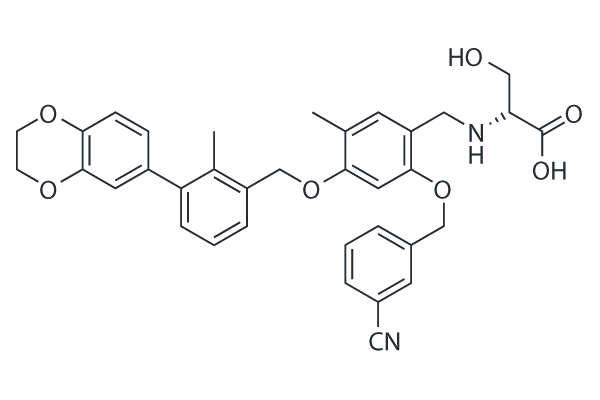
BMS-1001は、PD-1/PD-L1相互作用の強力な阻害剤であり、EC50値は253 nMです。BMS-1001は、可溶性PD-L1の抑制効果を軽減し、T細胞受容体介在性活性化によるTリンパ球の活性化を促進します。
-
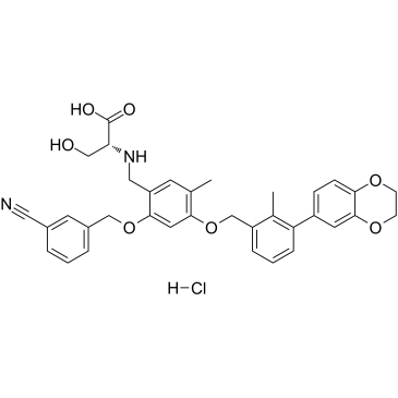
GC38740
BMS-1001 hydrochloride
BMS-1001 塩酸塩は、経口で活性なヒト PD-L1/PD-1 免疫チェックポイント阻害剤です。
-
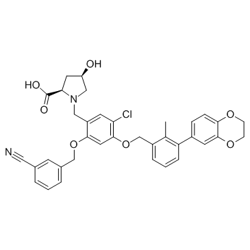
GC31753
BMS-1166 (PD-1/PD-L1-IN1)
BMS-1166 (PD-1/PD-L1-IN1) は、強力な PD-1/PD-L1 免疫チェックポイント阻害剤です。
-
GC38131
BMS-1166 hydrochloride
BMS-1166 塩酸塩は、強力な PD-1/PD-L1 免疫チェックポイント阻害剤です。

-
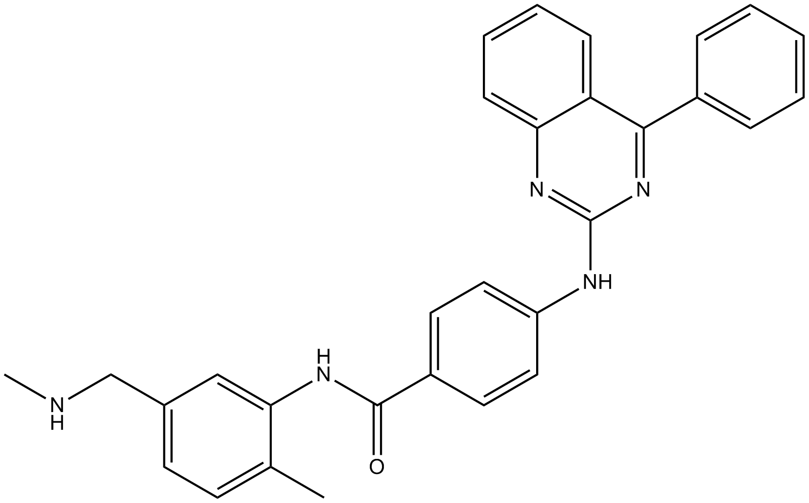
GC13628
BMS-833923
経口投与可能なSmo阻害剤
-
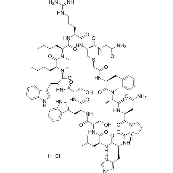
GC62682
BMSpep-57 hydrochloride
BMSpep-57 塩酸塩は、7.68 nM の IC50 を持つ PD-1/PD-L1 相互作用の強力で競合的な大環状ペプチド阻害剤です。 BMSpep-57 塩酸塩は、MST および SPR アッセイでそれぞれ 19 nM および 19.88 nM の Kds で PD-L1 に結合します。 BMSpep-57 塩酸塩は、PBMC での IL-2 産生を増加させることにより、T 細胞機能を促進します。
-
GA20897
Boc-Arg(Boc)₂-OH
アミノ酸の構成要素
-
GC16774
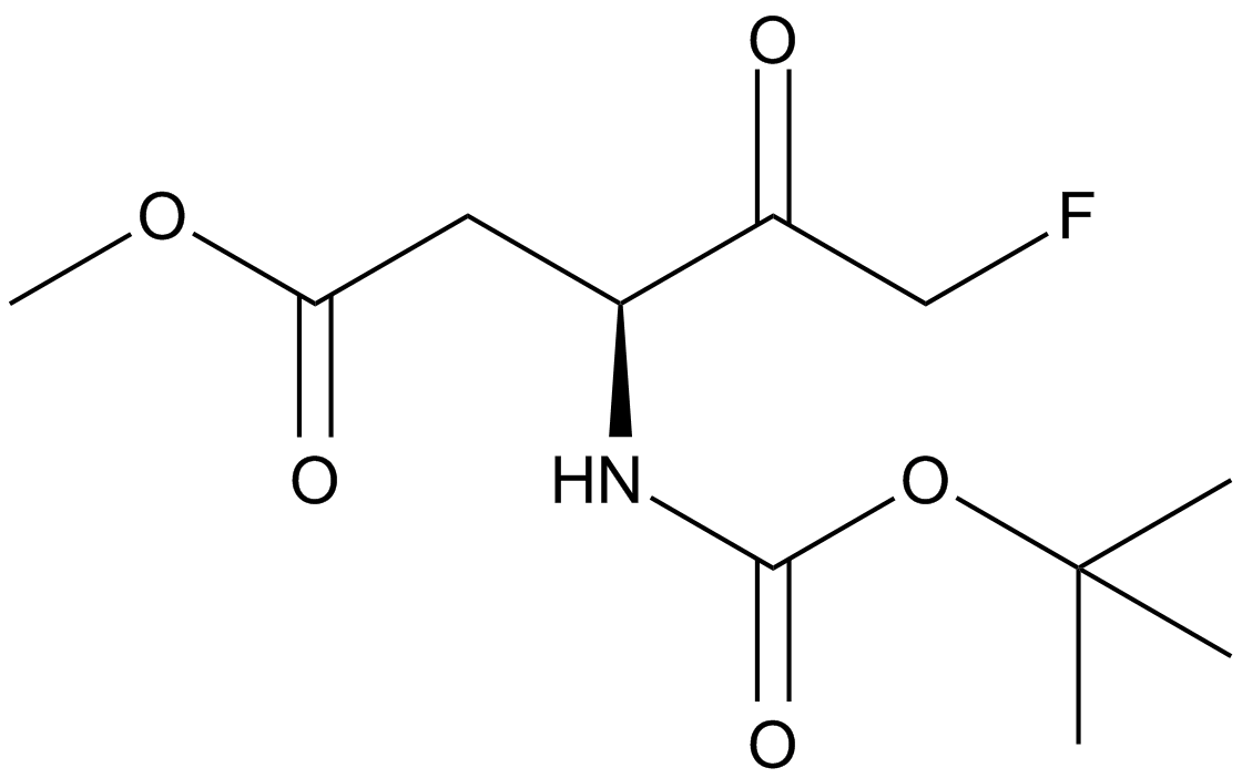
Boc-D-FMK
不可逆的なパンカスパーゼ阻害剤
-
GC33501
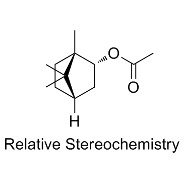
Bornyl acetate
酢酸ボルニルは強力な臭気物質であり、最高のフレーバー希釈係数 (FD 係数) の 1 つを示します。酢酸ボルニルには抗がん作用があります。
-
GC11040
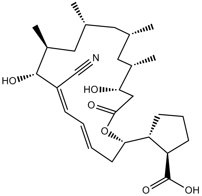
Borrelidin
ボレリジン (トレポネマイシン) は、Streptomyces rochei から分離されたニトリル含有マクロライド系抗生物質である、細菌および真核生物のスレオニル tRNA 合成酵素阻害剤です 。
-
GC17644
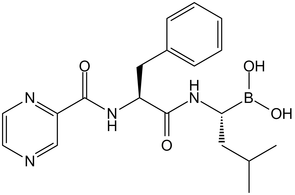
Bortezomib (PS-341)
強力で可逆的な20Sプロテアソーム阻害剤
-
GC65010
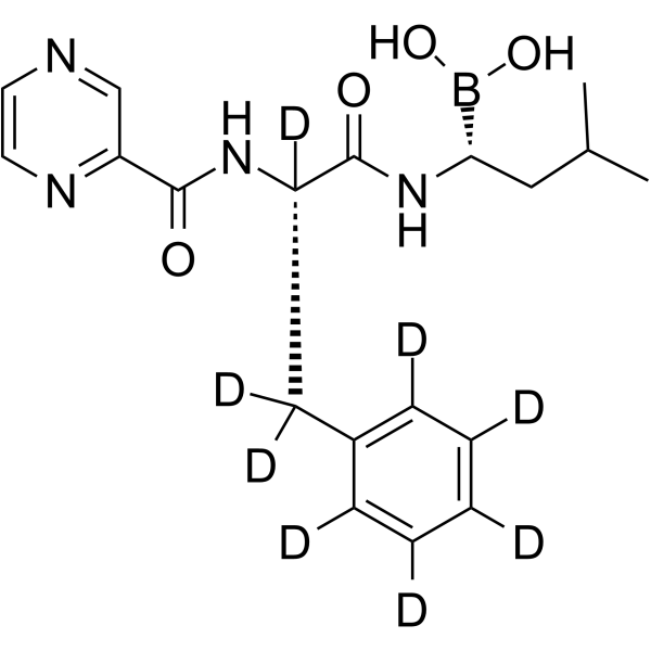
Bortezomib-d8
ボルテゾミブ-d8 (PS-341-d8) は、ボルテゾミブとラベル付けされた重水素です。ボルテゾミブ (PS-341) は、可逆的かつ選択的なプロテアソーム阻害剤であり、スレオニン残基を標的とすることで 20S プロテアソーム (Ki = 0.6 nM) を強力に阻害します。ボルテゾミブは細胞周期を乱し、アポトーシスを誘導し、NF-κB を阻害します。ボルテゾミブは、最初のプロテアソーム阻害抗がん剤です。抗がん作用。
-
GC40009
Bostrycin
ボストリシンは、もともとBから分離されたアントラキノンです。

-
GC42969
bpV(phen) (potassium hydrate)
インスリン模倣剤である bpV(phen) (水和カリウム) は、強力なプロテイン チロシン ホスファターゼ (PTP) および PTEN 阻害剤であり、PTEN の IC50 は 38 nM、343 nM、および 920 nM、PTP-β です。それぞれ、PTP-1B。

-
GC42974
Brassinin
ブラシニンは、アブラナ属の植物アレキシンの代謝です。

-
GC11632
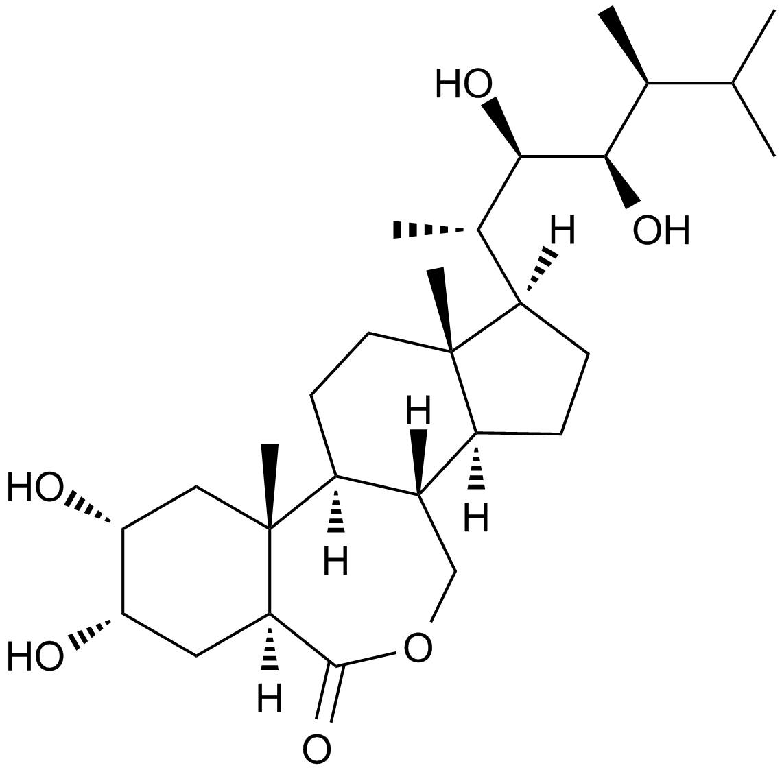
Brassinolide
植物成長調節剤
-
GC52101
Brazilein
ブラジレインは、Caesalpinia sappan L. から分離された重要な免疫抑制成分です。

-
GN10802
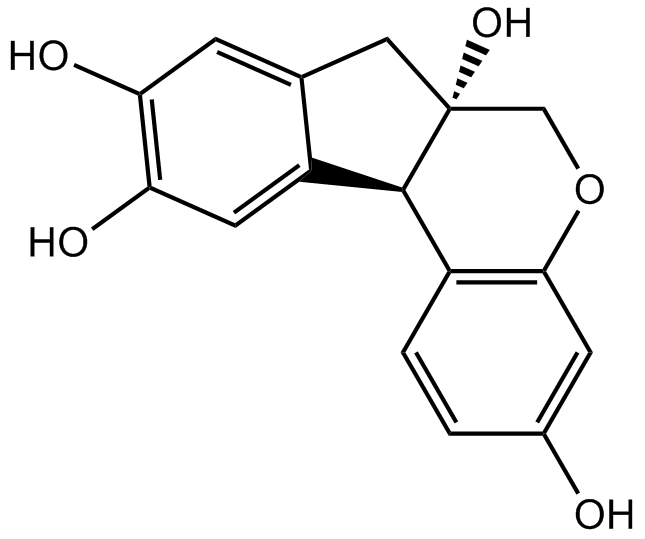
Brazilin
-
GC68288
Brentuximab

-
GC35554
Brevilin A
Brevilin A は、経口で活性な STAT3/JAK 阻害剤です (STAT3 IC50= 10.6 μM)。ブレビリン A は、抗腫瘍活性、癌細胞に対する抗増殖活性を示し、アポトーシスとオートファジーを誘導することができます。

-
GC35555
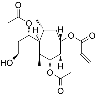
Britannin
Inula aucheriana から分離されたブリタニンは、セスキテルペンラクトンです。
-
GC62536
Bromelain
ブロメラインはパイナップルの茎に由来する抗炎症薬で、血漿キニノーゲンのダウンレギュレーション、プロスタグランジン E2 発現の阻害、最終糖化産物受容体の分解、血管新生バイオマーカーの調節、および COX 経路の上流での抗酸化作用を通じて作用します。 。

-
GC35559
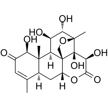
Bruceine D
Bruceine D は、抗癌活性を持つ Notch 阻害剤であり、いくつかのヒト癌細胞でアポトーシスを誘導します。
-
GC34070
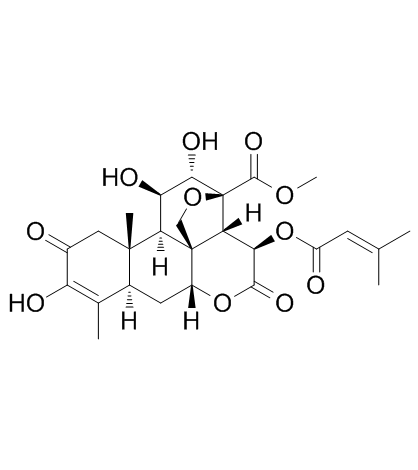
Brusatol (NSC 172924)
Brusatol (NSC 172924) (NSC172924) は、Nrf2 経路のユニークな阻害剤であり、広範囲のがん細胞をシスプラチンやその他の化学療法剤に感作させます。 Brusatol (NSC 172924) は、Nrf2 を介した防御メカニズムを阻害することにより、化学療法の有効性を高めます。 Brusatol (NSC 172924) は補助化学療法剤として開発することができます。 Brusatol (NSC 172924) は、細胞のアポトーシスを増加させます。
-
GC38014
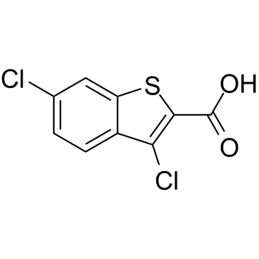
BT2
BT2 は、IC50 が 3.19 μM の BCKDC キナーゼ (BDK) 阻害剤です。
-
GC38467
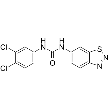
BTdCPU
BTdCPU は、強力なヘム調節 eIF2α キナーゼ (HRI) 活性化因子です。 BTdCPU は eIF2α リン酸化を促進し、耐性細胞のアポトーシスを誘導します。
-
GC34511
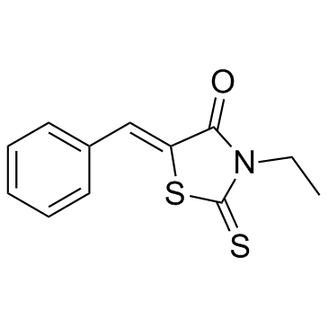
BTR-1
BTR-1 は活性抗がん剤であり、S 期停止を引き起こし、白血病細胞の DNA 複製に影響を与えます。 BTR-1 はアポトーシスを活性化し、細胞死を誘導します。
-
GC11141
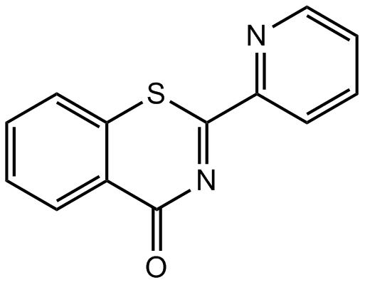
BTZO 1
BTZO 1 は 68.6 nM の Kd 値でマクロファージ遊走阻害因子 (MIF) に結合し、その結合には N 末端の Pro1 が必要です。
-
GC48376
Burnettramic Acid A
多様な生物学的活性を持つ真菌代謝産物

-
GC48409
Burnettramic Acid A aglycone
Burnettramic 酸 A アグリコンは、Aspergillus burnettii に見られる真菌代謝産物です 。

-
GC13671
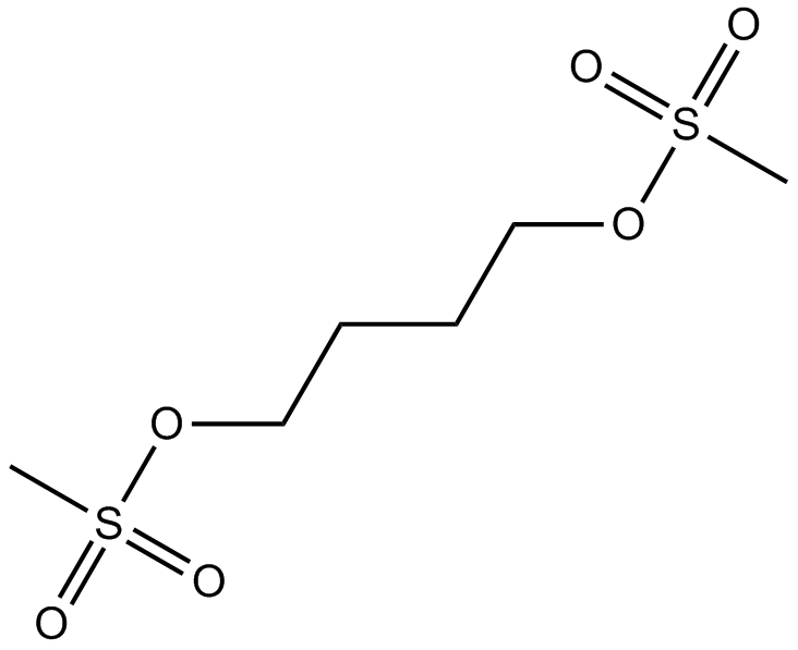
Busulfan
ブスルファンは、骨髄に対する選択的免疫抑制効果を持つ強力なアルキル化剤です。
-
GC46962
Busulfan-d8
Busulfan-D8 は、Busulfan と標識された重水素です。ブスルファンは、アルキル化抗腫瘍剤として作用するアルキルスルホン酸塩です。ブスルファンは、DNA の鎖内および鎖間架橋の両方を形成します。哺乳動物では、ブスルファンは、リンパ球レベルや体液性抗体反応に大きな影響を与えることなく、造血前駆細胞の生成を大幅かつ長期にわたって減少させます。

-
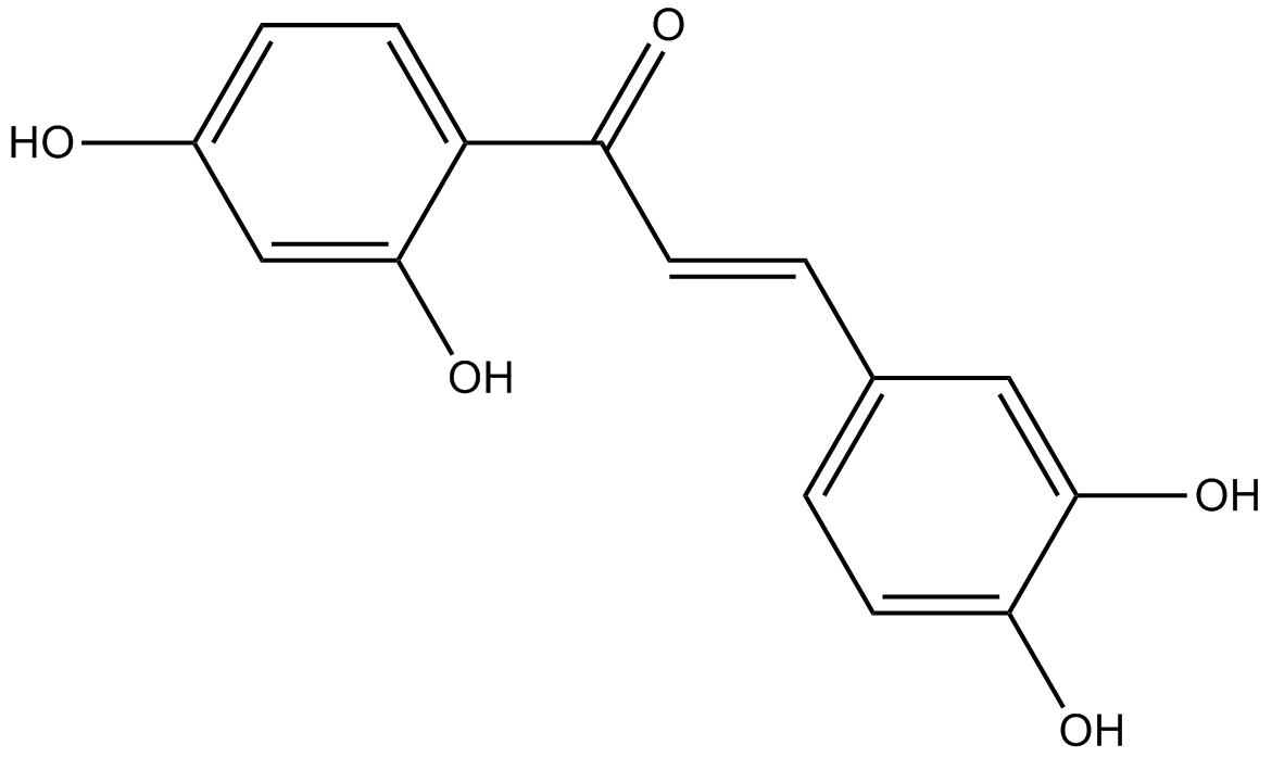
GC10944
Butein
ブテインは、PDE4 に対する IC50 が 10.4 μM の cAMP 特異的 PDE 阻害剤です。ブテインは、HepG2 細胞の EGFR および p60c-src に対して 16 および 65 μM の IC50 を持つ特定のタンパク質チロシンキナーゼ阻害剤です。ブテインは、FoxO3a を標的とすることにより、AKT および ERK/p38 MAPK 経路を介して HeLa 細胞をシスプラチンに感作します。ブテインは SIRT1 アクティベーター (STAC) です。
-
GC46104
Butyric Acid-d7
ナトリウムブチレートの定量化のための内部標準

-
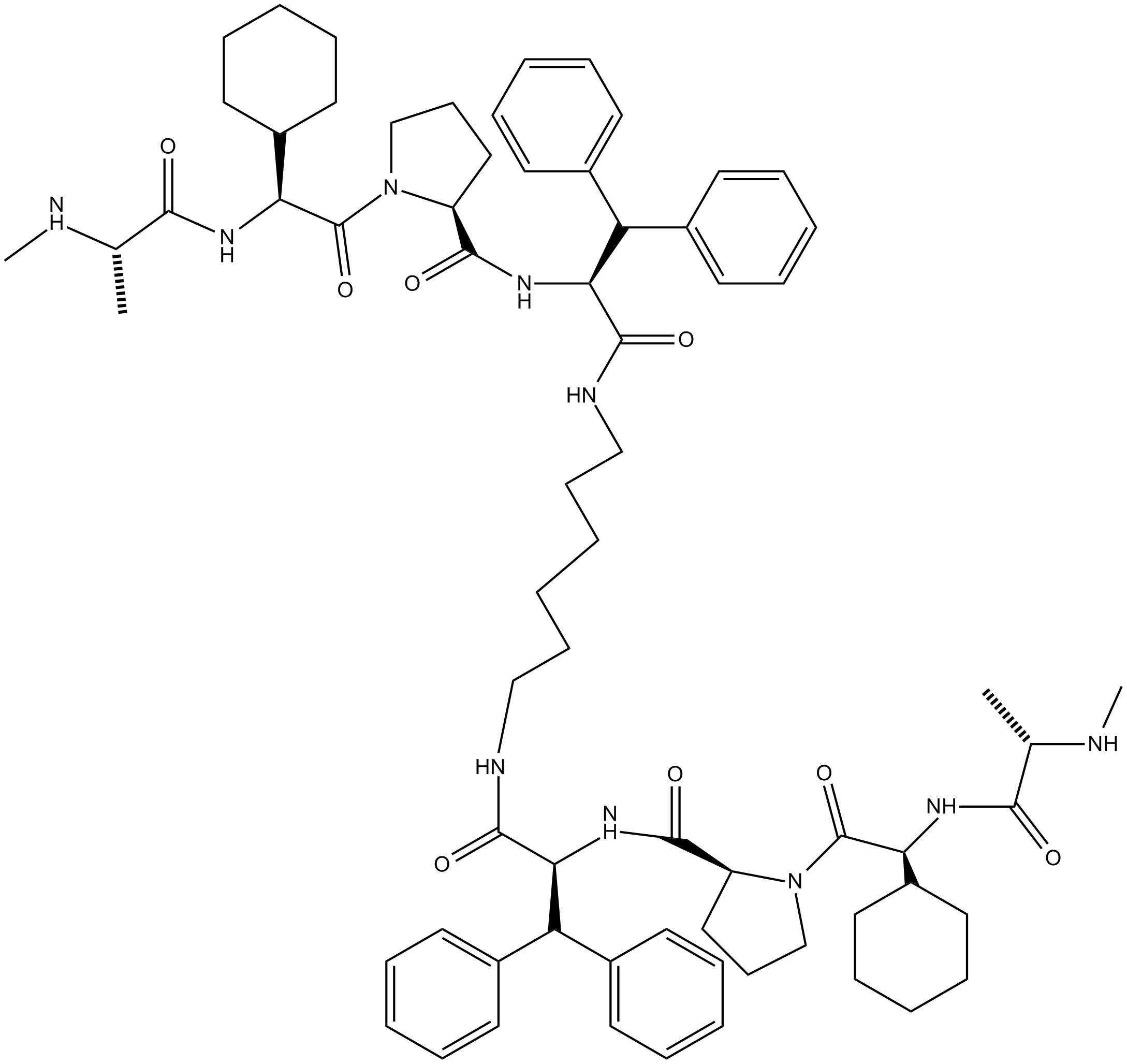
GC12333
BV6
BV6 は、アポトーシス阻害剤 (IAP) ファミリーのメンバーである cIAP1 および XIAP のアンタゴニストです。
-
GC48433
BX-320
BX-320 は、直接キナーゼアッセイ形式で 30 nM の IC50 を持つ、選択的、ATP 競合性、経口活性、および直接 PDK1 阻害剤です。 BX-320 はアポトーシスも誘導します。抗がん効果。

-
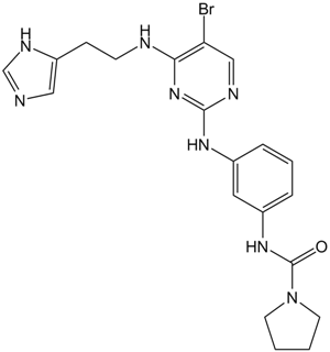
GC16818
BX-912
BX-912 は、直接、選択的、ATP 競合的な PDK1 阻害剤です (IC50=26 nM)。 BX-912 は、腫瘍細胞の PDK1/Akt シグナル伝達をブロックし、培養中のさまざまな腫瘍細胞株の足場依存性増殖を阻害するか、アポトーシスを誘導します。
-
GC31806
Bz 423 (BZ48)
Bz 423 (BZ48) は、自己反応性リンパ球に対する選択性を示す狼瘡のマウスモデルで治療特性を持つプロアポトーシス 1,4-ベンゾジアゼピンであり、Bax と Bak を活性化します。
-
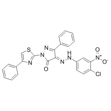
GC33826
C 87
C 87 は、新規の低分子 TNFα 阻害剤です。は、8.73 μM の IC50 で TNFα による細胞毒性を強力に阻害します。
-
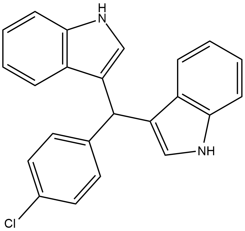
GC19095
C-DIM12
C-DIM12 は合成 Nurr1 活性剤であり、細胞株および初代ニューロンで Nurr1 および DA 遺伝子発現を誘導します。
-
GC43028
C16 Ceramide (d18:1/16:0)
セラミドは、スフィンゴミエリン酸化酵素の活性化によってまたはデノボ合成経路を介して生成されます。デノボ合成経路では、セリンパルミトイルトランスフェラーゼとセラミドシンターゼの協調作用が必要です。

-
GC46976
C16 Ceramide-d7 (d18:1-d7/16:0)
C-16セラミドの定量化のための内部標準

-
GC43052
C18 Phytoceramide (t18:0/18:0)
C18フィトセラミド(t18:0 / 18:0)(Cer(t18:0 / 18:0))は、S.に存在する生体活性スフィンゴ脂質です。

-
GC40141
C18 Phytoceramide-d3 (t18:0/18:0-d3)
C18フィトセラミド-d3(t18:0 / 18:0-d3)は、GC-またはLC-MSによるC18フィトセラミド(t18:0 / 18:0)の定量化のための内部標準として使用することを目的としています。

-
GC43065
C2 Phytoceramide (t18:0/2:0)
C2フィトセラミドは、懸濁した好中球細胞において、フォルミルペプチド誘導性酸化物放出を抑制する生体活性半合成スフィンゴ脂質であり、IC50値は0.38μMです。

-
GC43069
C22 Ceramide (d18:1/22:0)
C-22セラミドは内因性の生体活性スフィンゴ脂質です。

-
GC43075
C24 dihydro Ceramide (d18:0/24:0)
C24ジヒドロセラミドは、人間の皮膚の角質層に見つかったスフィンゴリピドです。

-
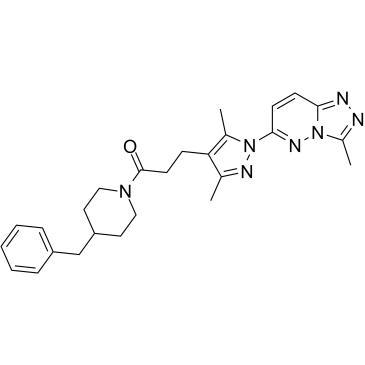
GC34513
C25-140
C25-140 は、クラス初の経口活性型でかなり選択的な TRAF6-Ubc13 阻害剤であり、TRAF6 に直接結合し、TRAF6 と Ubc13 との相互作用をブロックします。
-
GC43084
C4 Ceramide (d18:1/4:0)
C4セラミドは、生物活性スフィンゴ脂質であり、自然に存在するセラミドの細胞浸透性アナログです。

-
GC40688
C6 D-threo Ceramide (d18:1/6:0)
C6 D-threoセラミドは、生物活性スフィンゴ脂質であり、自然に存在するセラミドの細胞浸透性アナログです。C6 D-threoセラミドは、 vitro内のU937細胞に対して細胞毒性を示し(IC50 = 18μM)、作用します。

-
GC40689
C6 L-erythro Ceramide (d18:1/6:0)
C6 L-erythroセラミドは、生物活性スフィンゴ脂質であり、自然に存在するセラミドの細胞浸透性アナログです。

-
GC40690
C6 L-threo Ceramide (d18:1/6:0)
C6 L-threo セラミド (d18:1/6:0) は、生理活性スフィンゴ脂質であり、天然に存在するセラミドの細胞透過性類似体です。

-
GC45616
C6 Urea Ceramide
中性セラミダーゼの阻害剤

-
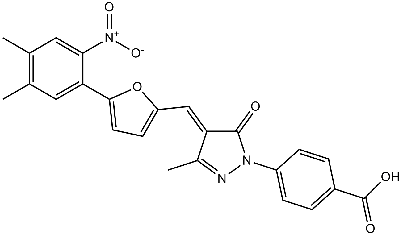
GC12733
C646
C646 は、Ki が 400 nM の選択的かつ競合的なヒストン アセチルトランスフェラーゼ p300 阻害剤であり、他のアセチルトランスフェラーゼに対してはあまり強力ではありません。
-
GC43105
C8 Ceramide (d18:1.8:0)
C8セラミド(d18:1.8:0)(N-オクタノイル-D-エリスロ-スフィンゴシン)は、自然界に存在するセラミドの細胞浸透性アナログです。

-
GC43109
C8 D-threo Ceramide (d18:1/8:0)
C8 D-スレオセラミドは、生物活性のあるスフィンゴ脂質であり、自然界に存在するセラミドの細胞浸透性アナログです。

-
GC43110
C8 Galactosylceramide (d18:1/8:0)
C8ガラクトシルセラミドは、膜マイクロドメイン形成スフィンゴリピッドの知られた合成C8短鎖誘導体です。

-
GC43111
C8 L-threo Ceramide (d18:1/8:0)
C8 L-スレオセラミドは、生物活性のあるスフィンゴ脂質であり、自然に存在するセラミドの細胞浸透性アナログです。

-
GC33218
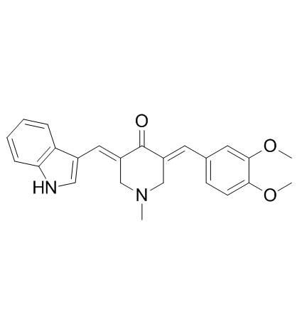
CA-5f
CA-5fは、オートファゴソームとリソソームの融合を阻害することにより、強力な後期マクロオートファジー/オートファジー阻害剤です。 CA-5f は LC3B-II (オートファジーを監視するマーカー) と SQSTM1 タンパク質を増加させ、ROS 産生も増加させます。抗腫瘍活性。
-
GC15779
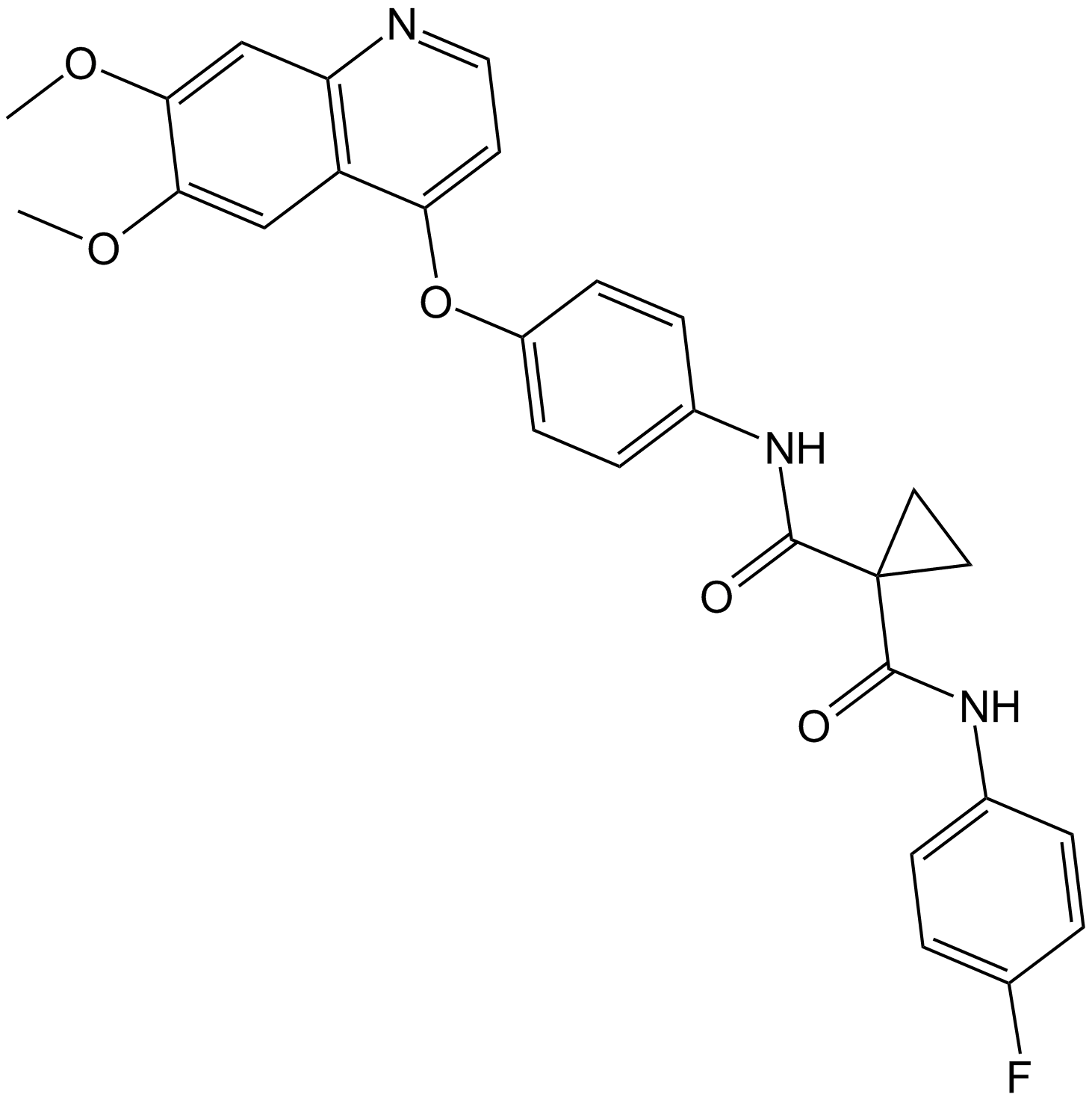
Cabozantinib (XL184, BMS-907351)
Cabozantinib (XL184,BMS-907351)は、新規なMETおよびVEGFR2阻害剤であり、同時に転移、血管新生、および腫瘍成長を抑制する。
-
GC12531
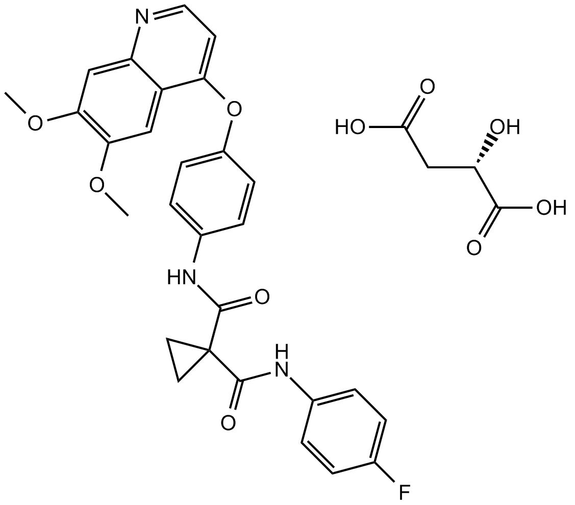
Cabozantinib malate (XL184)
カボザンチニブマレート(XL184)(XL184 S-マレート)は、VEGFR2、c-Met、Kit、AxlおよびFlt3をそれぞれ0.035、1.3、4.6、7および11.3 nMのIC50で阻害する強力な多数受容体チロシンキナーゼ阻害剤です。
-
GC10692
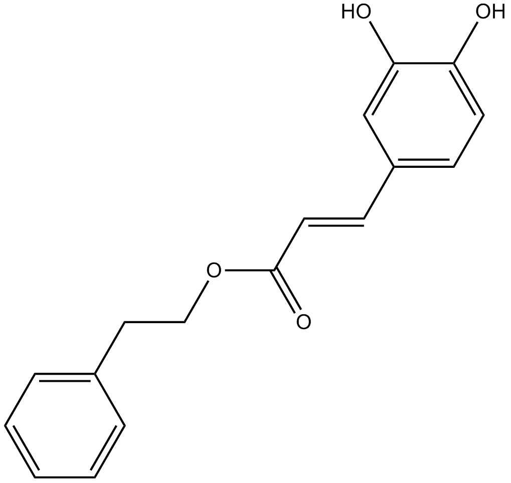
Caffeic Acid Phenethyl Ester
コーヒー酸フェネチルエステルはNF-κB阻害剤です。
-
GC18604
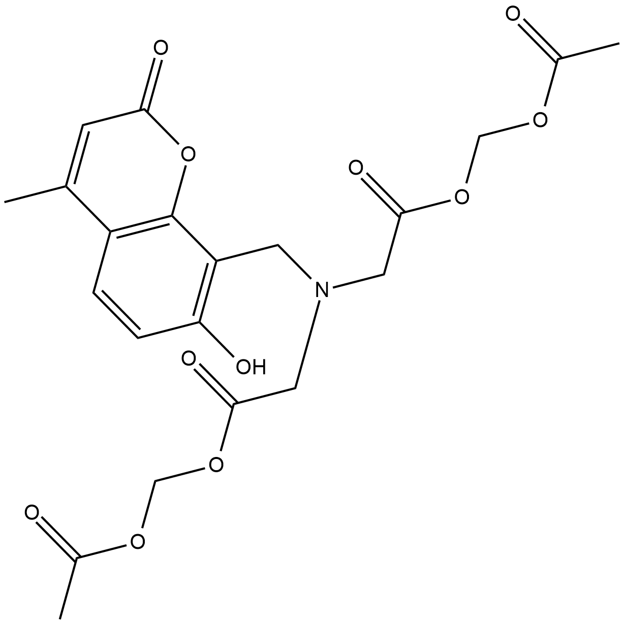
Calcein Blue AM
Calcein Blue AM は細胞染色剤です 。
-
GC43121
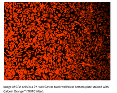
Calcein Orange™ Diacetate
カルセインオレンジジアセテートは、細胞の生存性を評価するために使用される蛍光色素です。
-
GC43123
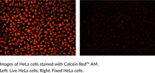
Calcein Red™ AM
カルセインレッド?AMは、細胞の生存性を評価するために使用される蛍光色素です。
-
GC60668
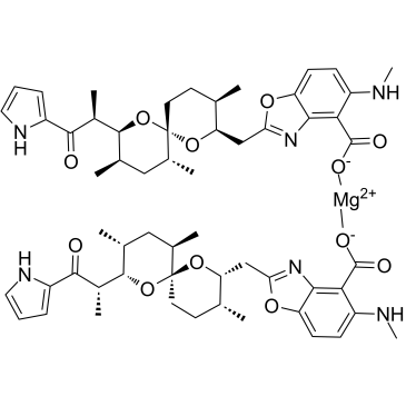
Calcimycin hemimagnesium
カルシマイシン (A-23187) ヘミマグネシウムは抗生物質であり、ユニークな二価カチオン イオノフォア (カルシウムやマグネシウムと同様) です。
-
GC15161
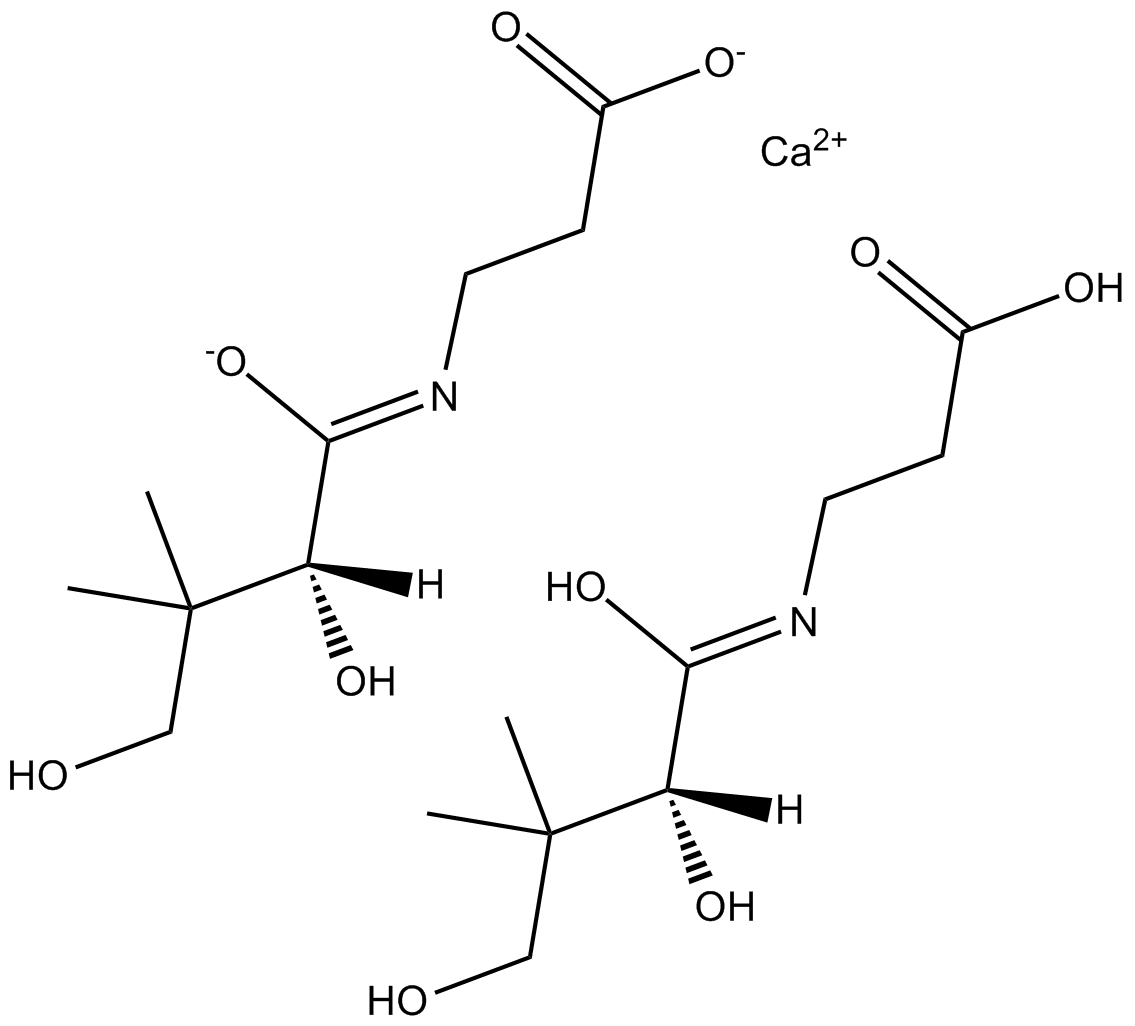
Calcium D-Panthotenate
ビタミンであるカルシウム D-パントテン酸 (ビタミン B5 カルシウム塩) は、リンゴ ジュースのパツリン含有量を減らすことができます。
-
GC30240
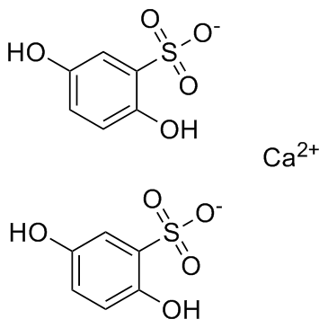
Calcium dobesilate
血管保護剤であるドベシル酸カルシウムは、多くの国で慢性静脈疾患、糖尿病性網膜症、および痔核発作の症状に広く使用されています。
-
GC19086
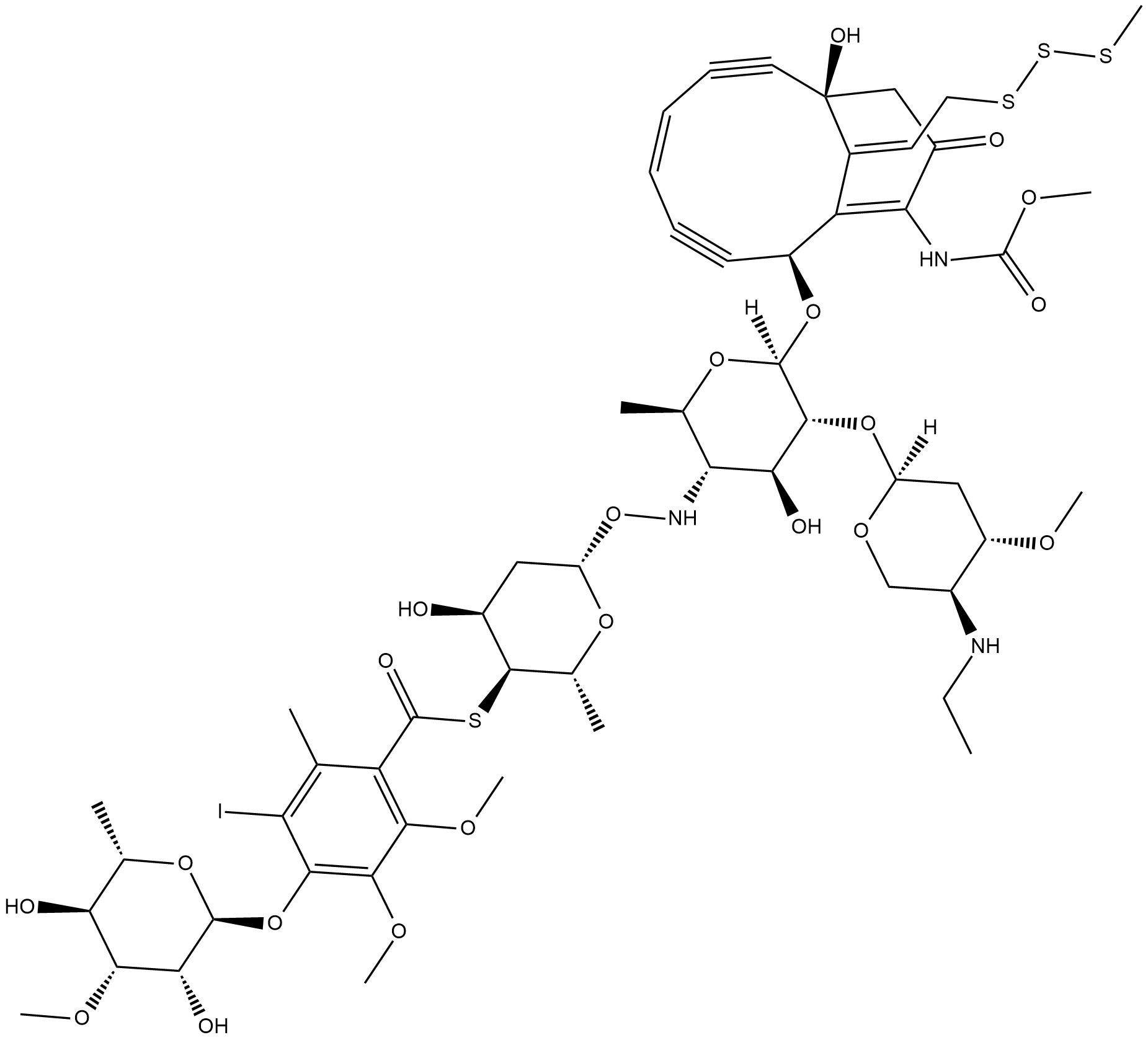
Calicheamicin
抗腫瘍抗生物質であるカリケアマイシンは、二本鎖 DNA 切断を引き起こす細胞傷害剤です。
-
GC65081
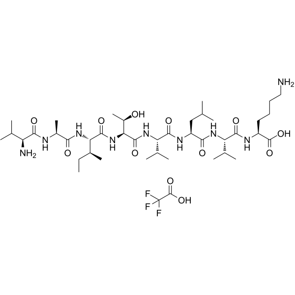
CALP1 TFA
CALP1 TFA は、CaM EF-hand/Ca2+-結合部位に結合するカルモジュリン (CaM) アゴニスト (Kd 88 μM) です。
-
GC18315
Calpain Inhibitor VI
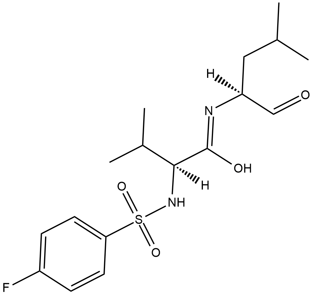
カルペイン阻害剤VIは、カルシウム依存性のシステインプロテアーゼであるu-カルペイン(カルペイン1;IC50 = 7.5 nM)およびm-カルペイン(カルペイン2;IC50 = 78 nM)の阻害剤です。
-
GC10342
Calpeptin
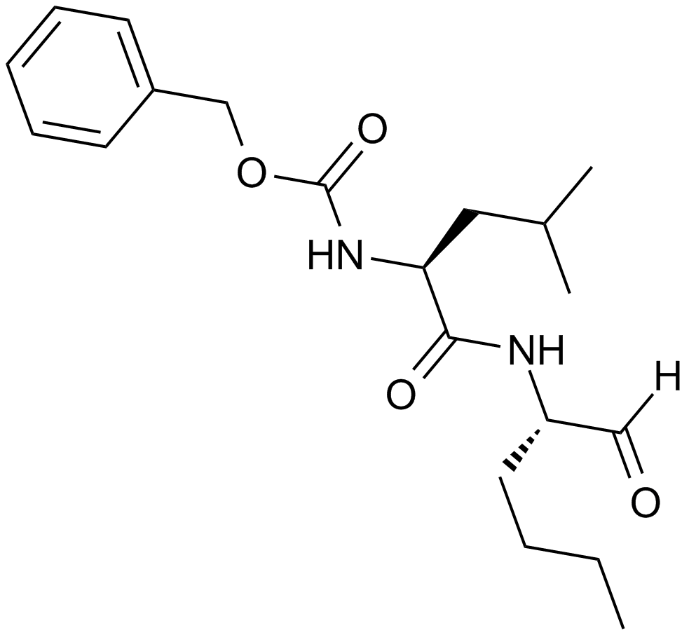
カルパイン阻害剤
-
GN10667
Calycosin
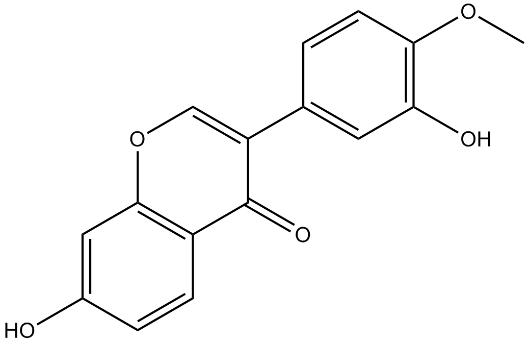
Calycosin (CA, 7, 3-dihydroxy-4-methoxy isoflavone, C16H12O5) is one of the flavonoids extracted from astragalus root, also known as the typical phytoestrogens.
-
GC35598
Camellianin A
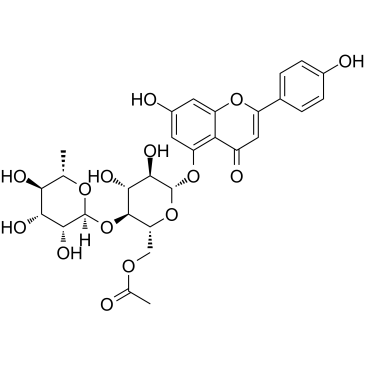
A. ニチダの葉の主要なフラボノイドであるカメリアニン A は、抗癌活性とアンギオテンシン変換酵素 (ACE) 阻害活性を示します。 Camellianin A は、ヒト Hep G2 および MCF-7 細胞株の増殖を阻害し、G0/G1 細胞集団の有意な増加を誘導します。
-
GC62253
Camrelizumab
カムレリズマブ (SHR-1210) は、PD-1 に対する強力なヒト化高親和性 IgG4-κ モノクローナル抗体 (mAb) です。カムレリズマブ は 3 nM の高親和性で PD-1 に結合し、PD-1 と PD-L1 の結合相互作用を 0.70 nM の IC50 で阻害します。カムレリズマブは抗 PD-1/PD-L1 剤として作用し、NSCLC、ESCC、ホジキンリンパ腫、進行性 HCC などのがん研究に使用できます。

-
GC12318
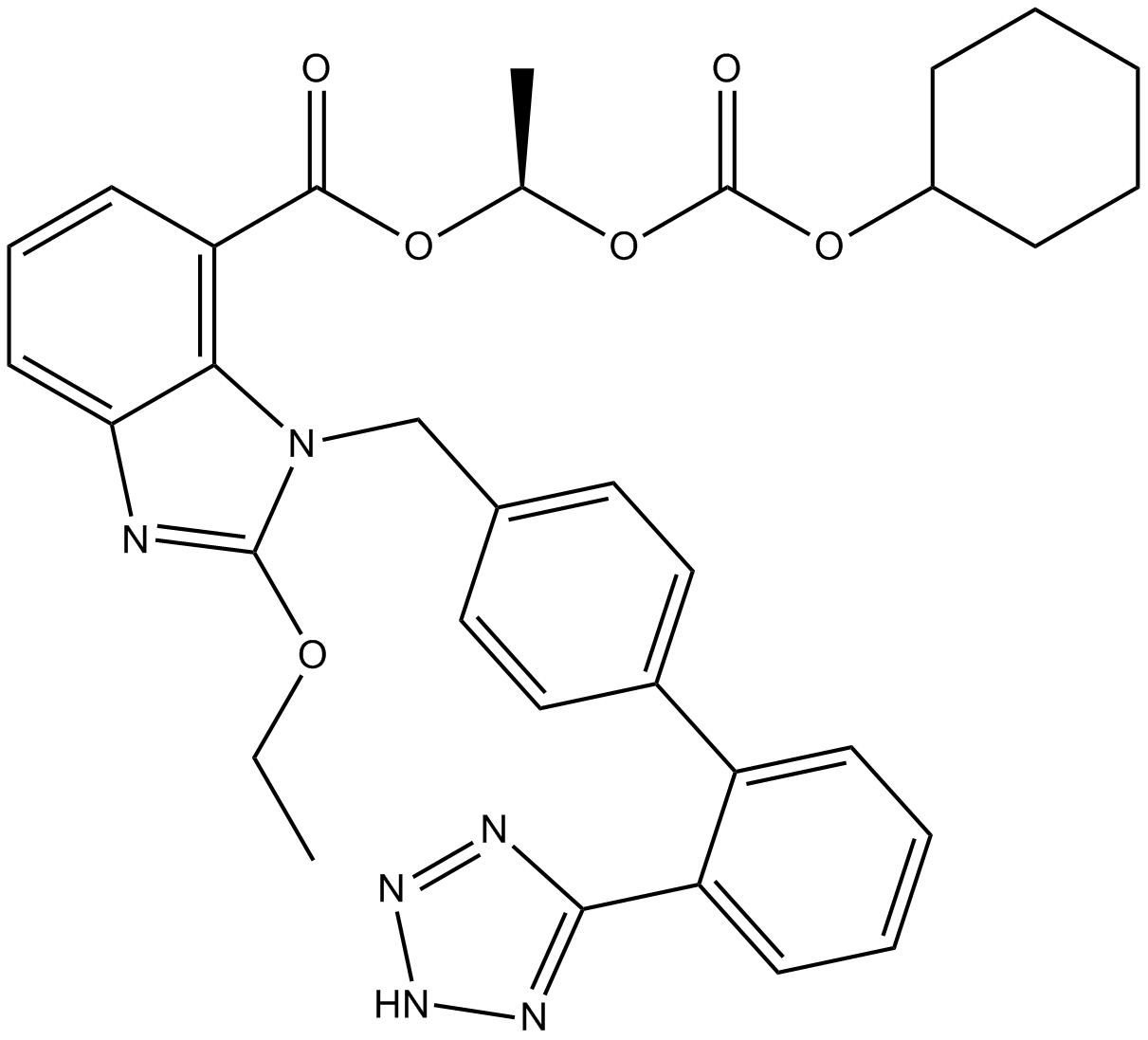
Candesartan Cilexetil
カンデサルタン シレキセチル (TCV-116) は、主に高血圧症の治療に使用されるアンギオテンシン II 受容体拮抗薬です。
-
GC15866
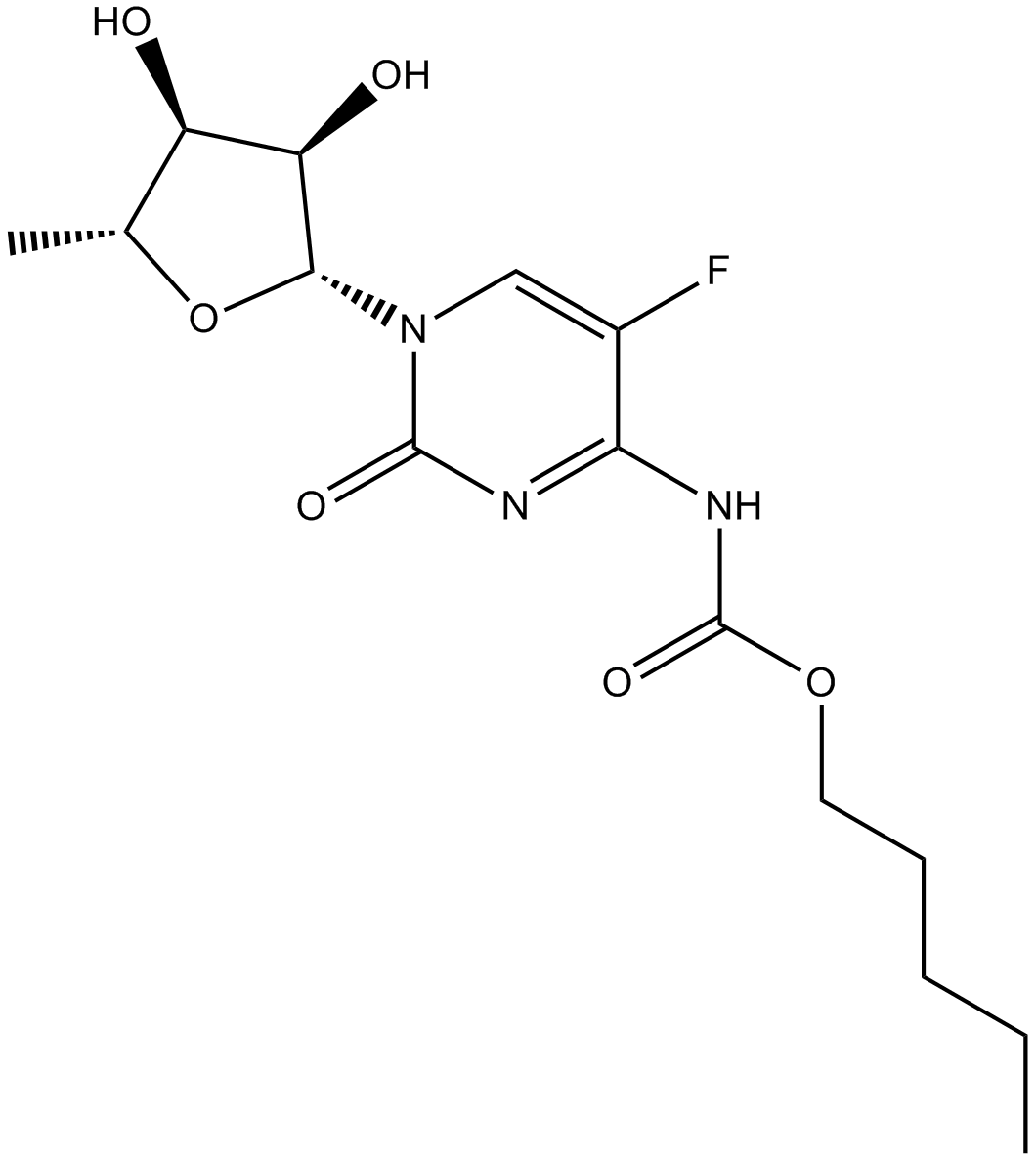
Capecitabine
カペシタビンは、チミジンホスホリラーゼによって活性代謝物である 5-FU に変換される経口プロドラッグです。
-
GC14065
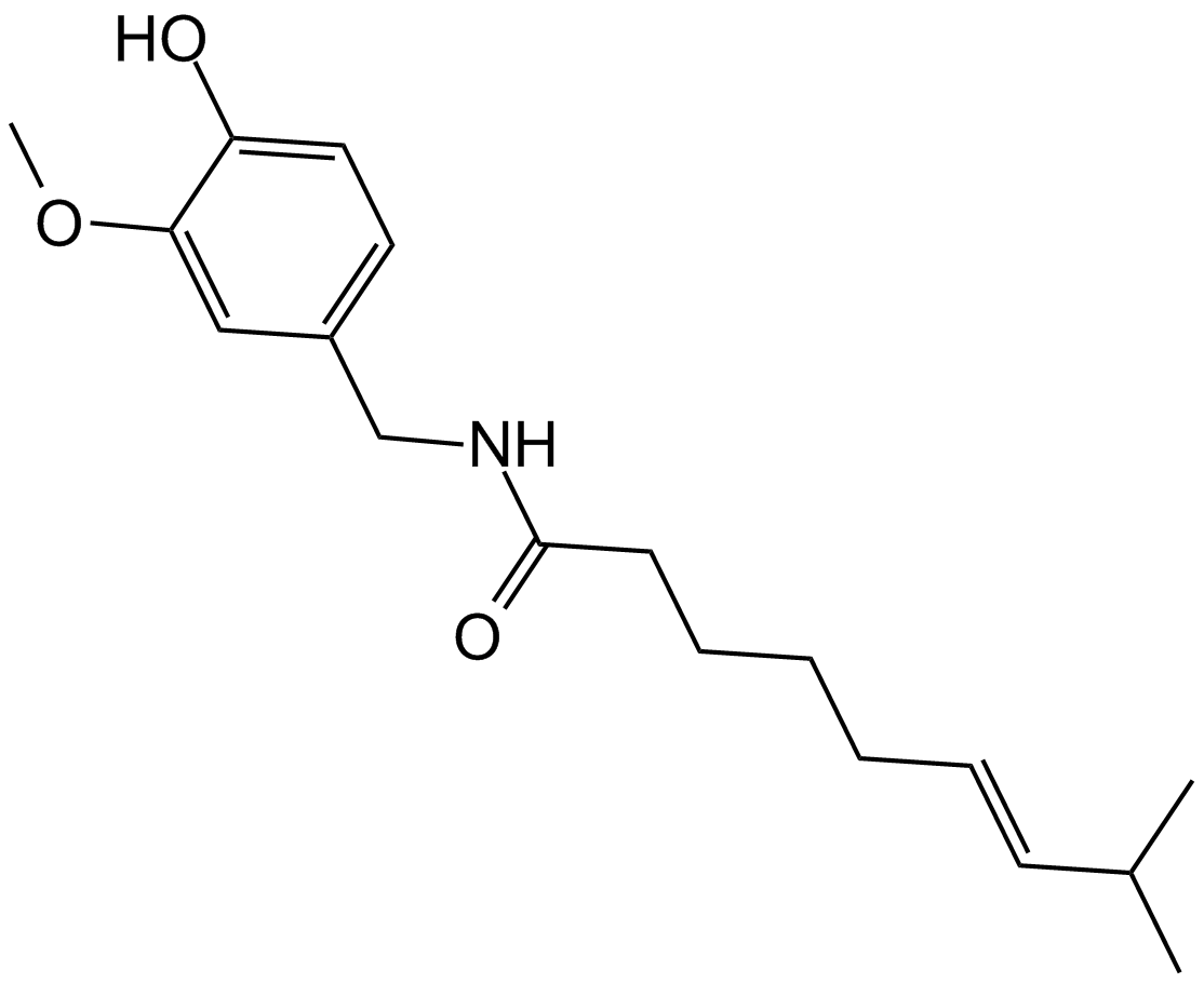
Capsaicin
多様な生物学的活性を持つテルペンアルカロイド
-
GC17918
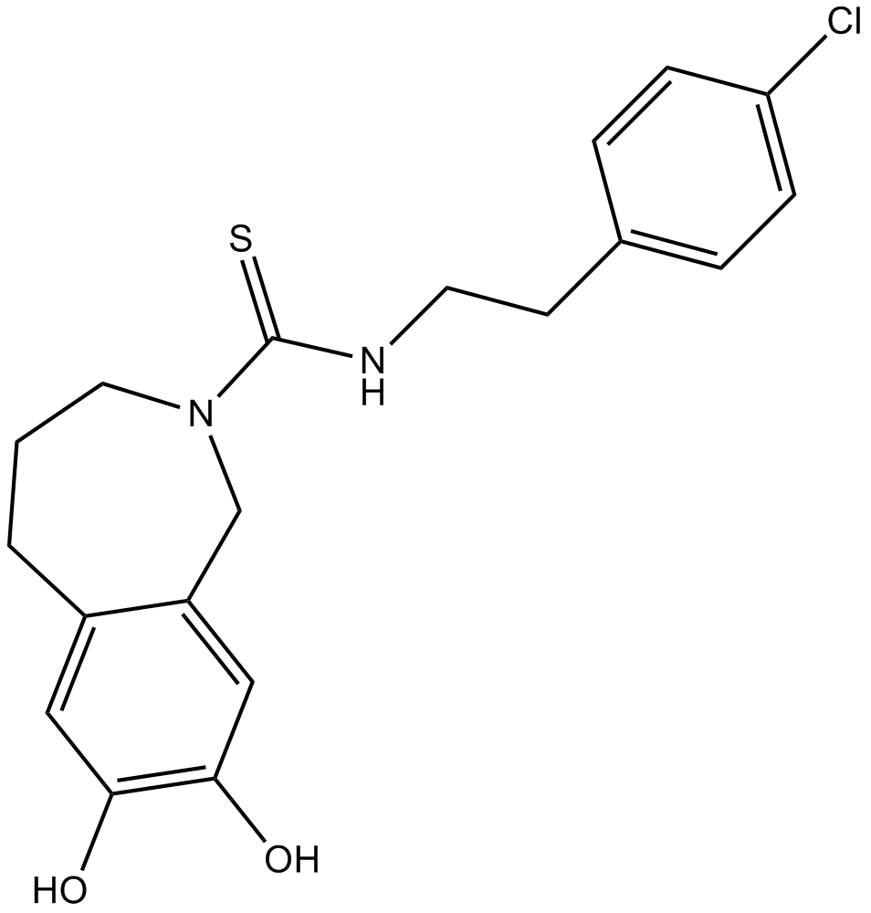
Capsazepine
TRPV1拮抗剤
-
GC49415
Capsorubin
多様な生物学的活性を持つカロテノイド

-
GC48878
Carbazomycin A
多様な生物学的活性を持つ細菌代謝産物

-
GC48893
Carbazomycin B
多様な生物学的活性を持つ細菌代謝産物

-
GC48850
Carbazomycin C
多様な生物学的活性を持つ細菌代謝産物

-
GC48826
Carbazomycin D
多様な生物学的活性を持つ細菌代謝産物

-
GC49147
Carboxyphosphamide
シクロホスファミドの不活性代謝物

-
GC18069
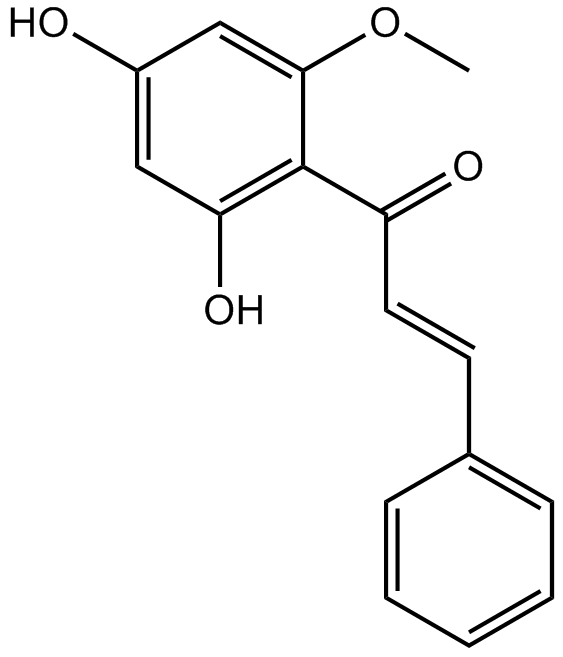
Cardamonin
カルダモニン ((E)-カルダモミン) は、454 nM の IC50 を持つ hTRPA1 カチオン チャネルの新規アンタゴニストです。
-
GC18449
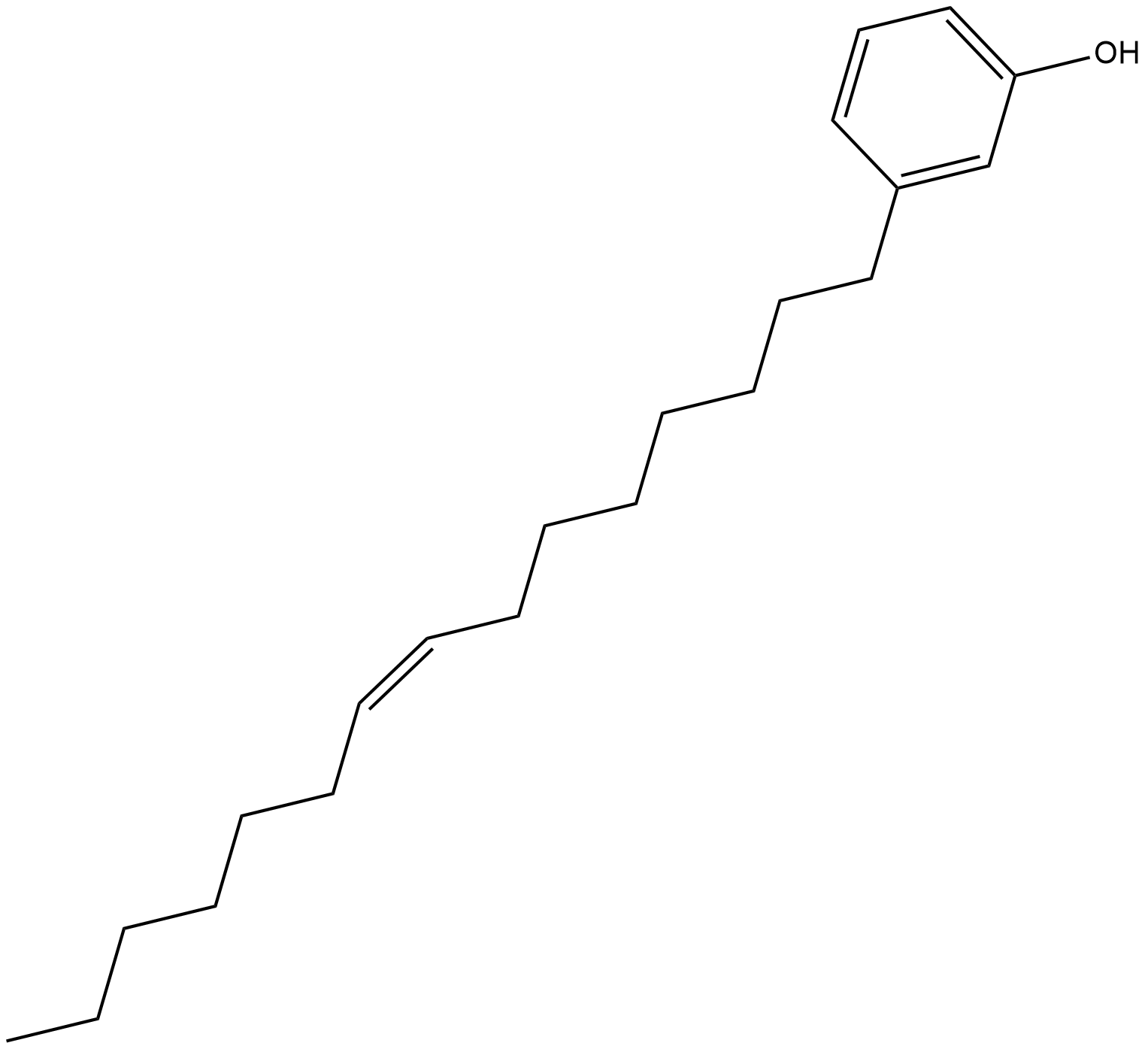
Cardanol monoene
カルダノール モノエン (カルダノール C15:1) は、カシュー ナッツの殻の液体に含まれるフェノール化合物です。カルダノールモノエンは、ヒト黒色腫細胞でミトコンドリア関連アポトーシスを誘導できます。
-
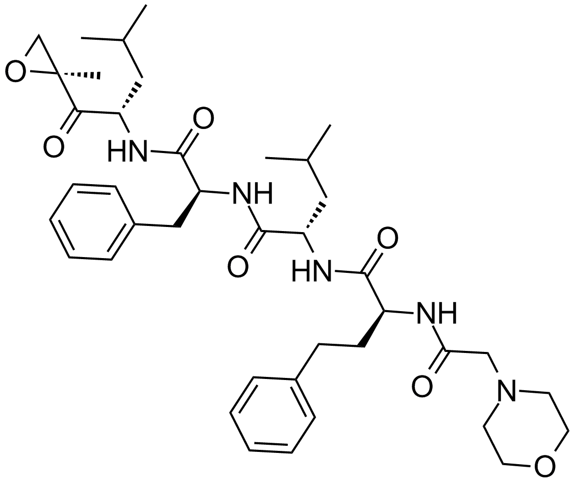
GC15089
Carfilzomib (PR-171)
プロテアソーム阻害剤
