Apoptosis
As one of the cellular death mechanisms, apoptosis, also known as programmed cell death, can be defined as the process of a proper death of any cell under certain or necessary conditions. Apoptosis is controlled by the interactions between several molecules and responsible for the elimination of unwanted cells from the body.
Many biochemical events and a series of morphological changes occur at the early stage and increasingly continue till the end of apoptosis process. Morphological event cascade including cytoplasmic filament aggregation, nuclear condensation, cellular fragmentation, and plasma membrane blebbing finally results in the formation of apoptotic bodies. Several biochemical changes such as protein modifications/degradations, DNA and chromatin deteriorations, and synthesis of cell surface markers form morphological process during apoptosis.
Apoptosis can be stimulated by two different pathways: (1) intrinsic pathway (or mitochondria pathway) that mainly occurs via release of cytochrome c from the mitochondria and (2) extrinsic pathway when Fas death receptor is activated by a signal coming from the outside of the cell.
Different gene families such as caspases, inhibitor of apoptosis proteins, B cell lymphoma (Bcl)-2 family, tumor necrosis factor (TNF) receptor gene superfamily, or p53 gene are involved and/or collaborate in the process of apoptosis.
Caspase family comprises conserved cysteine aspartic-specific proteases, and members of caspase family are considerably crucial in the regulation of apoptosis. There are 14 different caspases in mammals, and they are basically classified as the initiators including caspase-2, -8, -9, and -10; and the effectors including caspase-3, -6, -7, and -14; and also the cytokine activators including caspase-1, -4, -5, -11, -12, and -13. In vertebrates, caspase-dependent apoptosis occurs through two main interconnected pathways which are intrinsic and extrinsic pathways. The intrinsic or mitochondrial apoptosis pathway can be activated through various cellular stresses that lead to cytochrome c release from the mitochondria and the formation of the apoptosome, comprised of APAF1, cytochrome c, ATP, and caspase-9, resulting in the activation of caspase-9. Active caspase-9 then initiates apoptosis by cleaving and thereby activating executioner caspases. The extrinsic apoptosis pathway is activated through the binding of a ligand to a death receptor, which in turn leads, with the help of the adapter proteins (FADD/TRADD), to recruitment, dimerization, and activation of caspase-8 (or 10). Active caspase-8 (or 10) then either initiates apoptosis directly by cleaving and thereby activating executioner caspase (-3, -6, -7), or activates the intrinsic apoptotic pathway through cleavage of BID to induce efficient cell death. In a heat shock-induced death, caspase-2 induces apoptosis via cleavage of Bid.
Bcl-2 family members are divided into three subfamilies including (i) pro-survival subfamily members (Bcl-2, Bcl-xl, Bcl-W, MCL1, and BFL1/A1), (ii) BH3-only subfamily members (Bad, Bim, Noxa, and Puma9), and (iii) pro-apoptotic mediator subfamily members (Bax and Bak). Following activation of the intrinsic pathway by cellular stress, pro‑apoptotic BCL‑2 homology 3 (BH3)‑only proteins inhibit the anti‑apoptotic proteins Bcl‑2, Bcl-xl, Bcl‑W and MCL1. The subsequent activation and oligomerization of the Bak and Bax result in mitochondrial outer membrane permeabilization (MOMP). This results in the release of cytochrome c and SMAC from the mitochondria. Cytochrome c forms a complex with caspase-9 and APAF1, which leads to the activation of caspase-9. Caspase-9 then activates caspase-3 and caspase-7, resulting in cell death. Inhibition of this process by anti‑apoptotic Bcl‑2 proteins occurs via sequestration of pro‑apoptotic proteins through binding to their BH3 motifs.
One of the most important ways of triggering apoptosis is mediated through death receptors (DRs), which are classified in TNF superfamily. There exist six DRs: DR1 (also called TNFR1); DR2 (also called Fas); DR3, to which VEGI binds; DR4 and DR5, to which TRAIL binds; and DR6, no ligand has yet been identified that binds to DR6. The induction of apoptosis by TNF ligands is initiated by binding to their specific DRs, such as TNFα/TNFR1, FasL /Fas (CD95, DR2), TRAIL (Apo2L)/DR4 (TRAIL-R1) or DR5 (TRAIL-R2). When TNF-α binds to TNFR1, it recruits a protein called TNFR-associated death domain (TRADD) through its death domain (DD). TRADD then recruits a protein called Fas-associated protein with death domain (FADD), which then sequentially activates caspase-8 and caspase-3, and thus apoptosis. Alternatively, TNF-α can activate mitochondria to sequentially release ROS, cytochrome c, and Bax, leading to activation of caspase-9 and caspase-3 and thus apoptosis. Some of the miRNAs can inhibit apoptosis by targeting the death-receptor pathway including miR-21, miR-24, and miR-200c.
p53 has the ability to activate intrinsic and extrinsic pathways of apoptosis by inducing transcription of several proteins like Puma, Bid, Bax, TRAIL-R2, and CD95.
Some inhibitors of apoptosis proteins (IAPs) can inhibit apoptosis indirectly (such as cIAP1/BIRC2, cIAP2/BIRC3) or inhibit caspase directly, such as XIAP/BIRC4 (inhibits caspase-3, -7, -9), and Bruce/BIRC6 (inhibits caspase-3, -6, -7, -8, -9).
Any alterations or abnormalities occurring in apoptotic processes contribute to development of human diseases and malignancies especially cancer.
References:
1.Yağmur Kiraz, Aysun Adan, Melis Kartal Yandim, et al. Major apoptotic mechanisms and genes involved in apoptosis[J]. Tumor Biology, 2016, 37(7):8471.
2.Aggarwal B B, Gupta S C, Kim J H. Historical perspectives on tumor necrosis factor and its superfamily: 25 years later, a golden journey.[J]. Blood, 2012, 119(3):651.
3.Ashkenazi A, Fairbrother W J, Leverson J D, et al. From basic apoptosis discoveries to advanced selective BCL-2 family inhibitors[J]. Nature Reviews Drug Discovery, 2017.
4.McIlwain D R, Berger T, Mak T W. Caspase functions in cell death and disease[J]. Cold Spring Harbor perspectives in biology, 2013, 5(4): a008656.
5.Ola M S, Nawaz M, Ahsan H. Role of Bcl-2 family proteins and caspases in the regulation of apoptosis[J]. Molecular and cellular biochemistry, 2011, 351(1-2): 41-58.
What is Apoptosis? The Apoptotic Pathways and the Caspase Cascade
Targets for Apoptosis
- Pyroptosis(15)
- Caspase(77)
- 14.3.3 Proteins(3)
- Apoptosis Inducers(71)
- Bax(15)
- Bcl-2 Family(136)
- Bcl-xL(13)
- c-RET(15)
- IAP(32)
- KEAP1-Nrf2(73)
- MDM2(21)
- p53(137)
- PC-PLC(6)
- PKD(8)
- RasGAP (Ras- P21)(2)
- Survivin(8)
- Thymidylate Synthase(12)
- TNF-α(141)
- Other Apoptosis(1145)
- Apoptosis Detection(0)
- Caspase Substrate(0)
- APC(6)
- PD-1/PD-L1 interaction(60)
- ASK1(4)
- PAR4(2)
- RIP kinase(47)
- FKBP(22)
Products for Apoptosis
- Cat.No. Product Name Information
-
GC47042
Carfilzomib-d8
An internal standard for the quantification of carfilzomib
-
GN10733
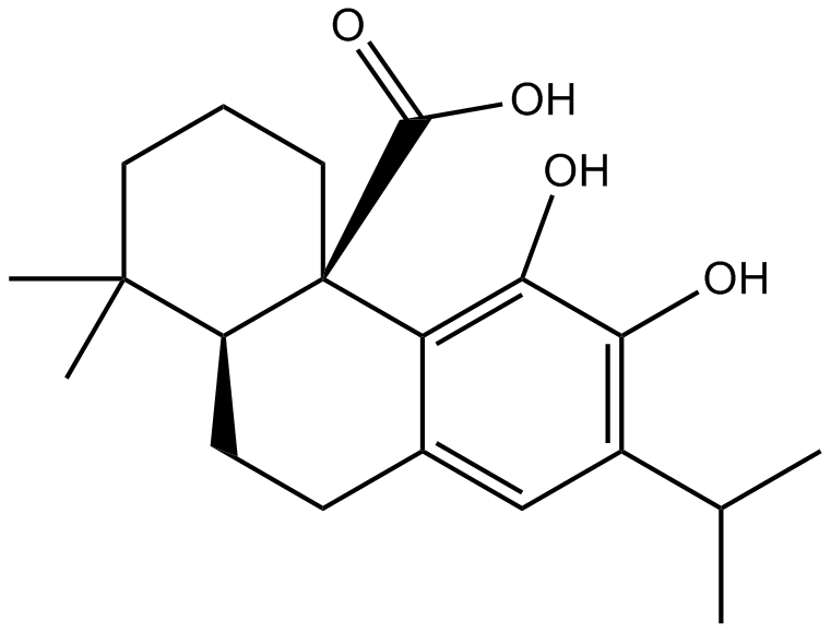
Carnosic acid
-
GC45679
Carubicin
An anthracycline with anticancer activity
-
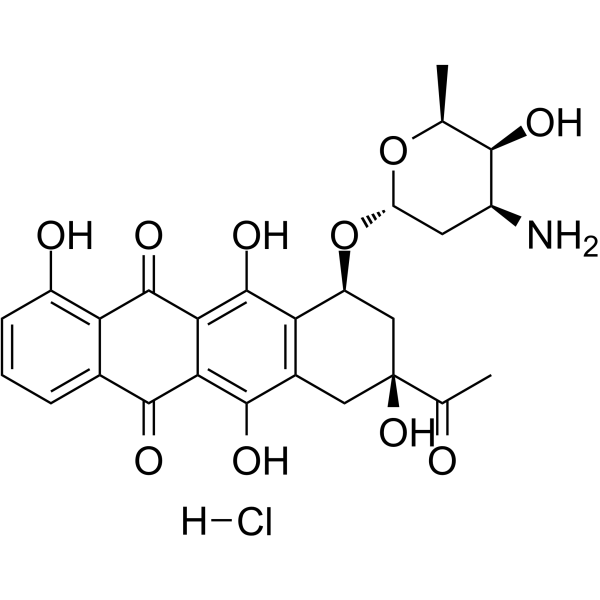
GC64110
Carubicin hydrochloride
Carubicin hydrochloride is a microbially-derived compound.
-
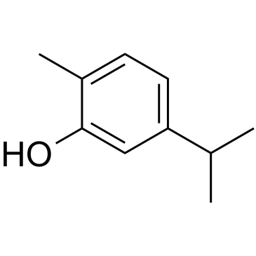
GC35612
Carvacrol
Carvacrol is a monoterpenoid phenol isolated from Thymus mongolicus Ronn.
-
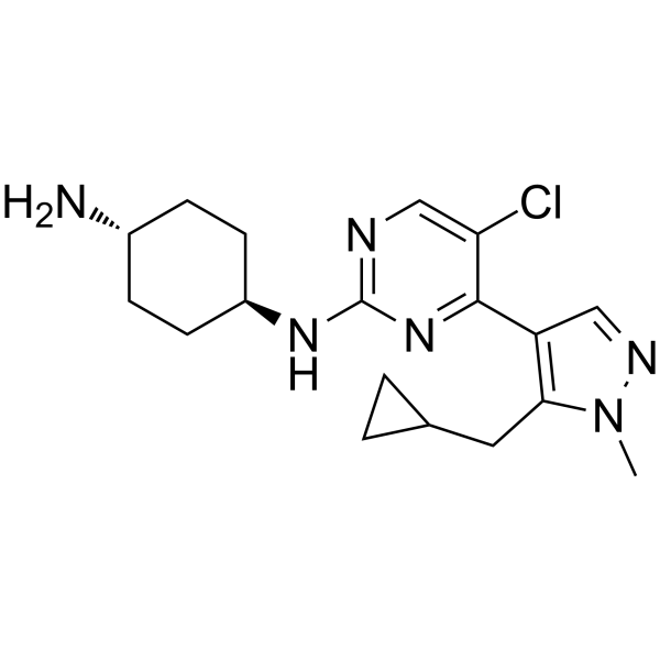
GC62442
Casein Kinase inhibitor A51
Casein Kinase inhibitor A51 is a potent and orally active casein kinase 1α (CK1α) inhibitor. Casein Kinase inhibitor A51 induces leukemia cell apoptosis, and has potent anti-leukemic activities.
-
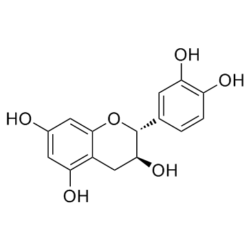
GC32841
Catechin ((+)-Catechin)
Catechin ((+)-Catechin) ((+)-Catechin ((+)-Catechin)) inhibits cyclooxygenase-1 (COX-1) with an IC50 of 1.4 μM.
-
GN10543
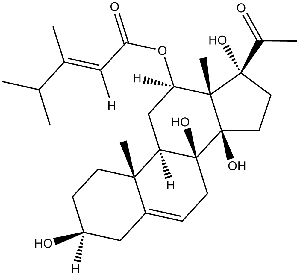
caudatin
-
GC43149
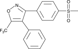
CAY10404
Many non-steroidal anti-inflammatory drugs (NSAIDs) are potent but non-selective inhibitors of both COX-1 and COX-2 in humans.
-
GC43150
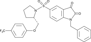
CAY10406
CAY10406 is a trifluoromethyl analog of an isatin sulfonamide compound that selectively inhibits caspases 3 and 7.
-
GC43154
CAY10443
Mitochondrial release of cytochrome c triggers apoptosis via the assembly of a multimeric complex including caspase-9, Apaf-1, and other components, sometimes called the apoptosome.

-
GC43176
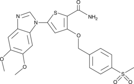
CAY10575
CAY10575 (Compound 8) is an IKK2 inhibitor with an IC50 of 0.075 μM.
-
GC18530
CAY10616
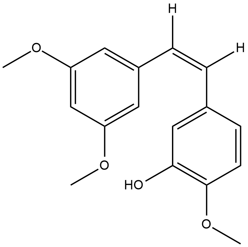
Resveratrol is a natural polyphenolic antioxidant that has anti-cancer properties.
-
GC41317
CAY10625
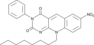
Survivin is a cellular protein implicated in cell survival by interacting with and inhibiting the apoptotic function of several proteins including Smac/DIABLO, caspase-3, and caspase-7.
-
GC43189
CAY10681
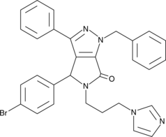
Inactivation of the tumor suppressor p53 commonly coincides with increased signaling through NF-κB in cancer.
-
GC43190
CAY10682
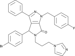
(±)-Nutlin-3 blocks the interaction of p53 with its negative regulator Mdm2 (IC50 = 90 nM), inducing the expression of p53-regulated genes and blocking the growth of tumor xenografts in vivo.
-
GC40650
CAY10706
CAY10706 is a ligustrazine-curcumin hybrid that promotes intracellular reactive oxygen species accumulation preferentially in lung cancer cells.

-
GC43198
CAY10717
CAY10717 is a multi-targeted kinase inhibitor that exhibits greater than 40% inhibition of 34 of 104 kinases in an enzymatic assay at a concentration of 100 nM.

-
GC43203
CAY10726
CAY10726 is an arylurea fatty acid.

-
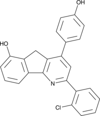
GC46113
CAY10744
A topoisomerase II-α poison
-
GC47053
CAY10746
A ROCK1 and ROCK2 inhibitor

-
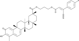
GC48392
CAY10747
An inhibitor of the Hsp90-Cdc37 protein-protein interaction
-
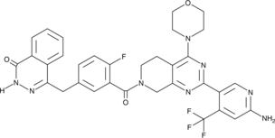
GC47055
CAY10749
CAY10749 (compound 15) is a potent PARP/PI3K inhibitor with pIC50 values of 8.22, 8.44, 8.25, 6.54, 8.13, 6.08 for PARP-1, PARP-2, PI3Kα, PI3Kβ, PI3Kδ, and PI3Kγ, respectively. CAY10749 is a highly effective anticancer compound targeted against a wide range of oncologic diseases.
-
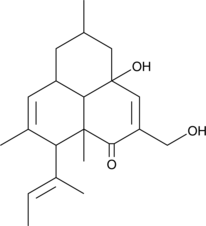
GC47057
CAY10755
A fungal metabolite with anticancer activity
-
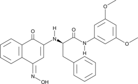
GC47061
CAY10763
A dual inhibitor of IDO1 and STAT3 activation
-
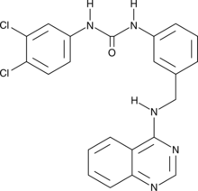
GC47065
CAY10773
A derivative of sorafenib
-
GC49080
CAY10786
CAY10786 (Compound 43) is a GPR52 antagonist with an IC50 of 0.63 μM.

-
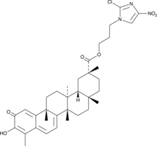
GC52245
CAY10792
An anticancer agent
-
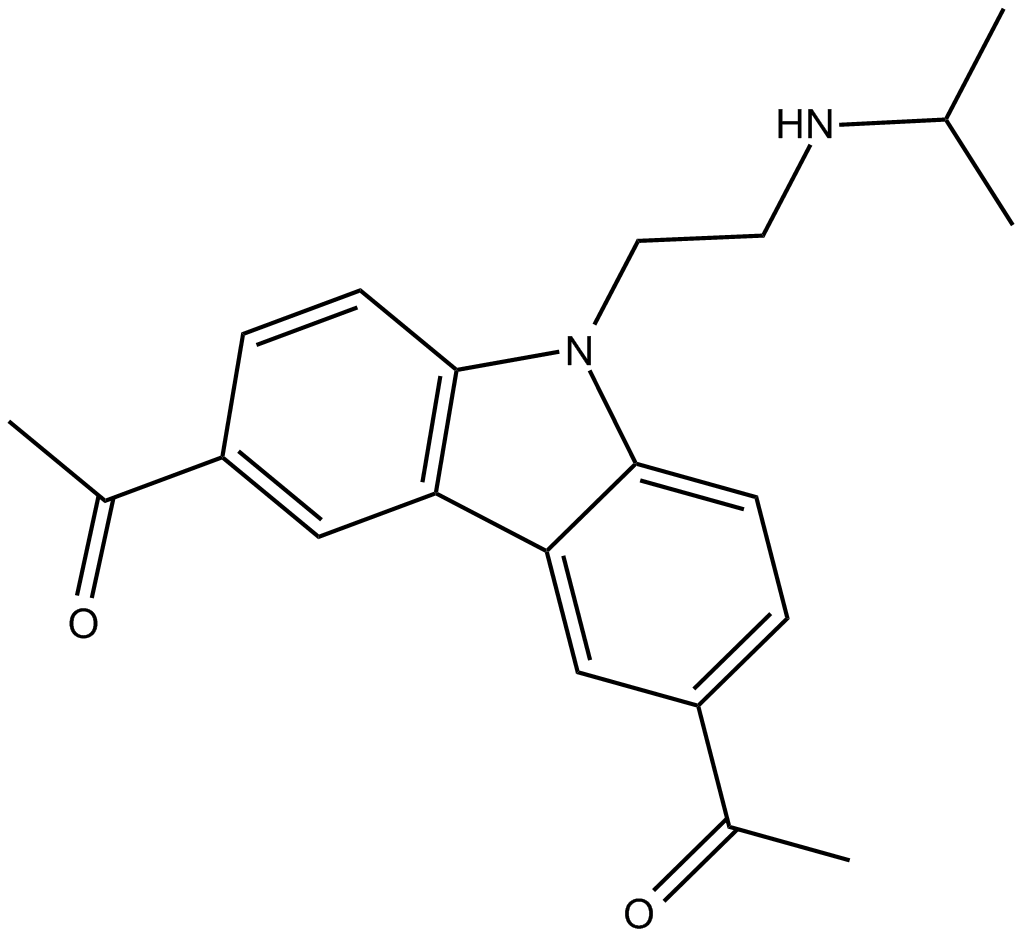
GC14634
CBL0137
curaxin that activates p53 and inhibits NF-κB
-
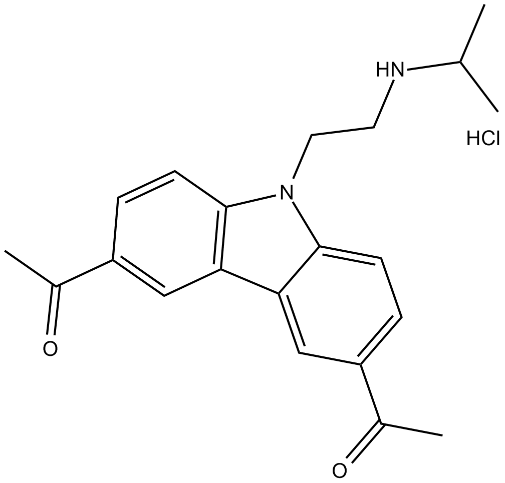
GC15394
CBL0137 (hydrochloride)
curaxin that activates p53 and inhibits NF-κB
-
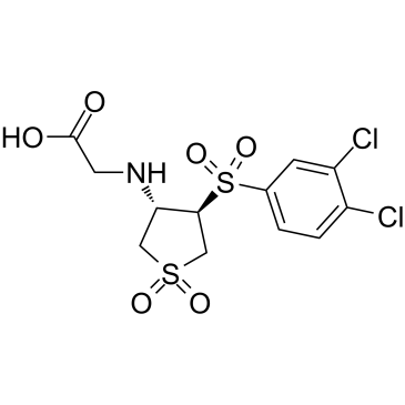
GC61636
CBR-470-2
CBR-470-2, a glycine-substituted analog, can activate NRF2 signaling.
-
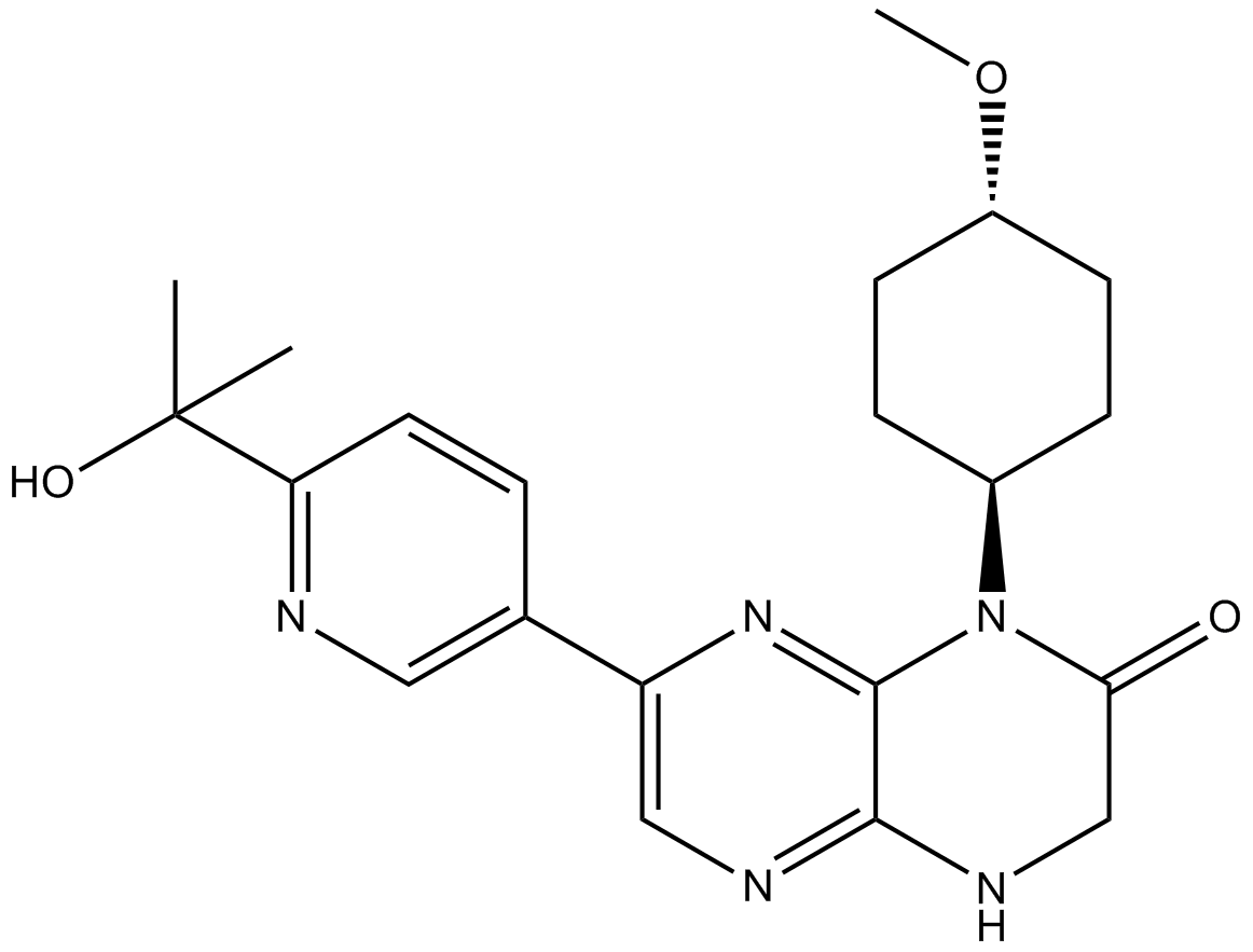
GC13648
CC-223
CC-223 (CC-223) is a potent, selective, and orally bioavailable inhibitor of mTOR kinase, with an IC50 value for mTOR kinase of 16 nM. CC-223 inhibits both mTORC1 and mTORC2.
-
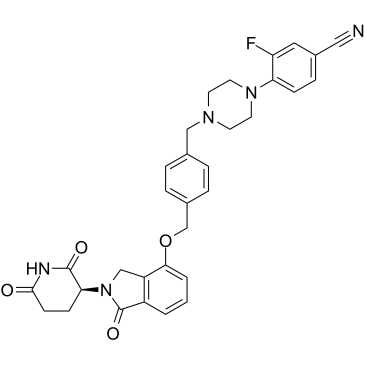
GC39169
CC-92480
CC-92480 (CC-92480), a cereblon E3 ubiquitin ligase modulating drug (CELMoD), acts as a molecular glue. CC-92480 shows high affinity to cereblon, resulting in potent antimyeloma activity.
-
GC19088
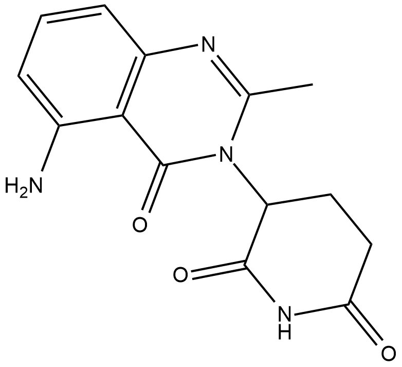
CC122
CC122 (CC 122) is an orally active cereblon modulator.
-
GC61532
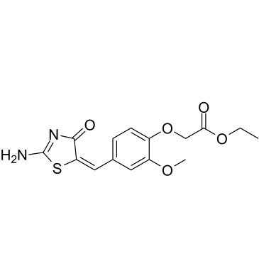
CCI-007
CCI-007 is a small molecule with cytotoxic activity against infant leukemia with MLL rearrangements, with IC50 values of 2.5-6.2 μM in sensitive cells.
-
GC12891
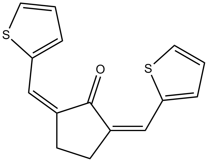
CCT007093
PPM1D inhibitor
-
GC14566
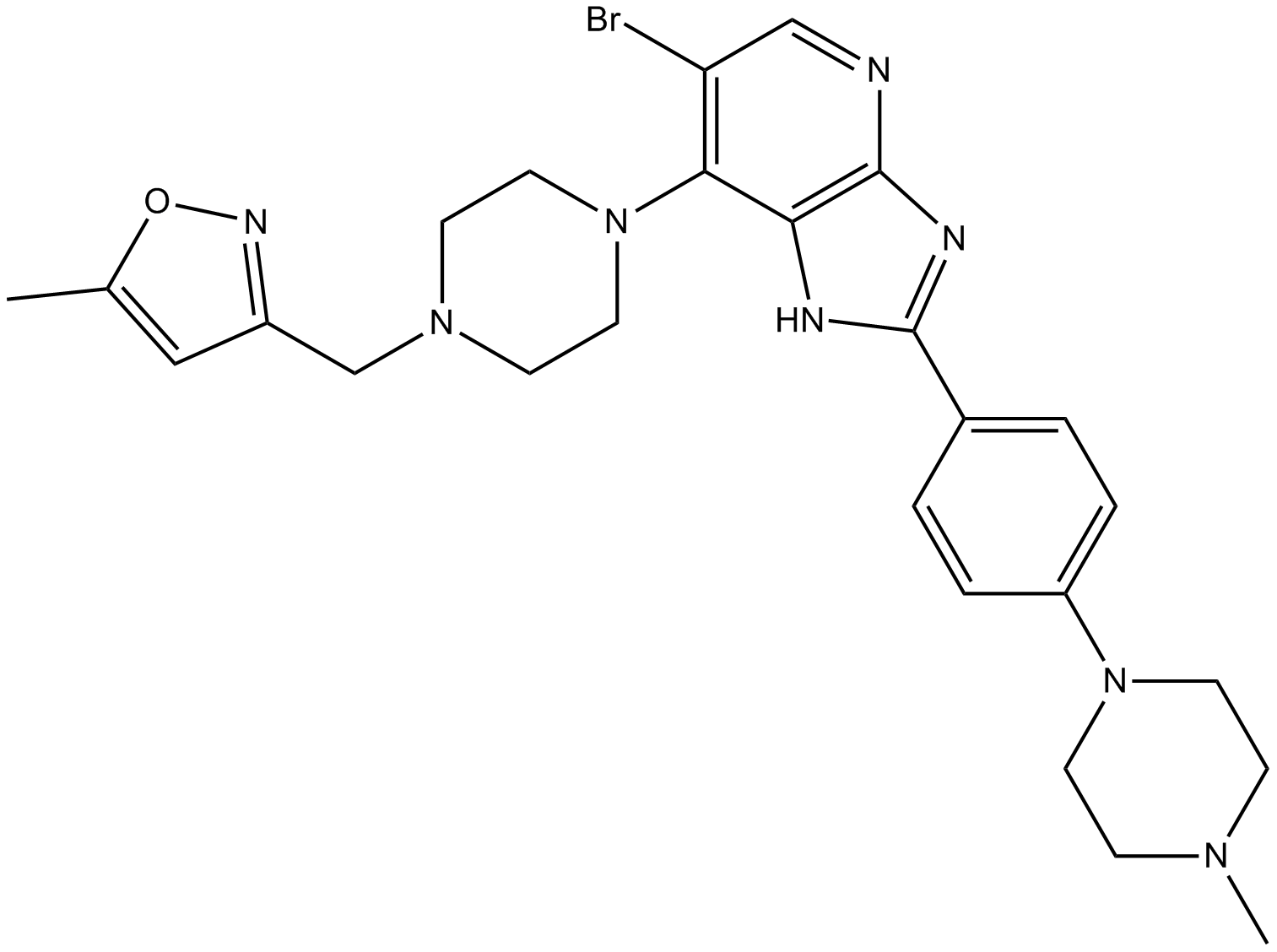
CCT137690
An inhibitor of Aurora kinases and FLT3
-
GC62561
CCT369260
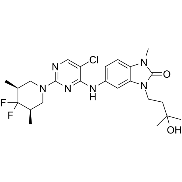
CCT369260 (compound 1) is an orally avtive B-cell lymphoma 6 (BCL6) inhibitor with anti-tumor activity. CCT369260 (compound 1) exhibits an IC50 of 520 nM.
-
GC33337
CDC801
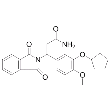
CDC801 is a potent and orally active phosphodiesterase 4 (PDE4) and tumor necrosis factor-α (TNF-α) inhibitor with IC50 of 1.1 μM and 2.5 μM, respectively.
-
GC39555
CDDO-2P-Im
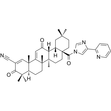
CDDO-2P-Im is an analogue of CDDO-Imidazolide with chemopreventive effect. CDDO-2P-Im can reduce the size and the severity of the lung tumors in mouse lung cancer model.
-
GC39556
CDDO-3P-Im
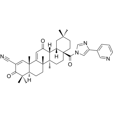
CDDO-3P-Im is an analogue of CDDO-Imidazolide with chemopreventive effect. CDDO-3P-Im can reduce the size and the severity of the lung tumors in mouse lung cancer model. CDDO-3P-Im is a orally active necroptosis inhibitor that can be used for the research of ischemia/reperfusion (I/R).
-
GC35629
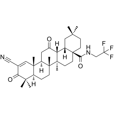
CDDO-dhTFEA
CDDO-dhTFEA (RTA dh404) is a synthetic oleanane triterpenoid compound which potently activates Nrf2 and inhibits the pro-inflammatory transcription factor NF-κB.
-
GC35630
CDDO-EA
CDDO-EA is an NF-E2 related factor 2/antioxidant response element (Nrf2/ARE) activator.

-
GC32723
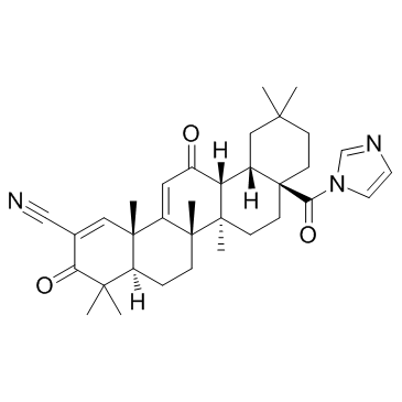
CDDO-Im (RTA-403)
CDDO-Im (RTA-403) (RTA-403) is an activator of Nrf2 and PPAR, with Kis of 232 and 344 nM for PPARα and PPARγ.
-
GC16625
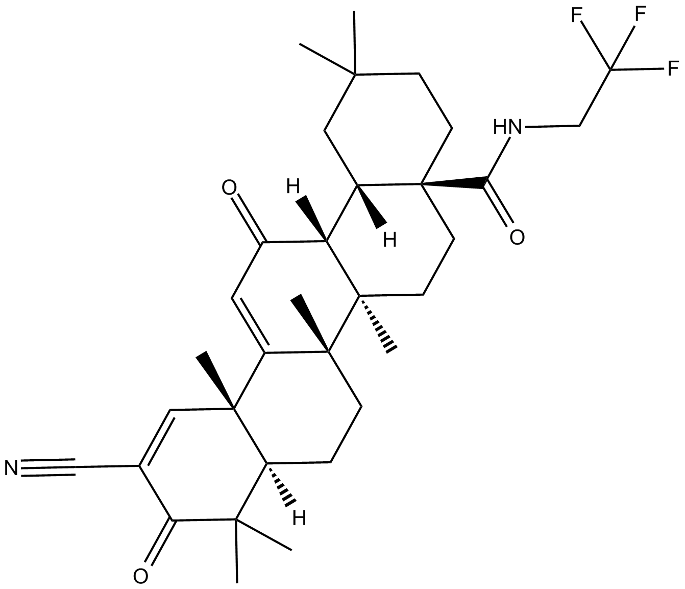
CDDO-TFEA
Nrf2 activator
-
GC43217
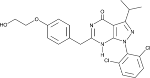
CDK/CRK Inhibitor
CDK/CRK inhibitor is an inhibitor of cyclin-dependent kinases (CDK) and CDK-related kinases (CRK) with IC50 values ranging from 9-839 nM in vitro.
-
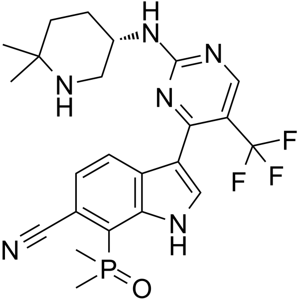
GC62596
CDK7-IN-3
CDK7-IN-3 (CDK7-IN-3) is an orally active, highly selective, noncovalent CDK7 inhibitor with a KD of 0.065 nM. CDK7-IN-3 shows poor inhibition on CDK2 (Ki=2600 nM), CDK9 (Ki=960 nM), CDK12 (Ki=870 nM). CDK7-IN-3 induces apoptosis in tumor cells and has antitumor activity.
-
GC35636
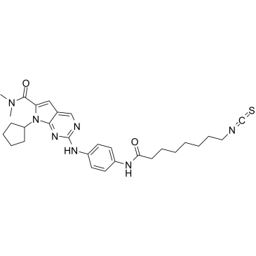
CDK9-IN-7
CDK9-IN-7 (compound 21e) is a selective, highly potent, and orally active CDK9/cyclin T inhibitor (IC50=11 nM), which exhibits more potent over other CDKs (CDK4/cyclinD=148 nM; CDK6/cyclinD=145 nM). CDK9-IN-7 shows antitumor activity without obvious toxicity. CDK9-IN-7 induces NSCLC cell apoptosis, arrests the cell cycle in the G2 phase, and suppresses the stemness properties of NSCLC.
-
GC19096
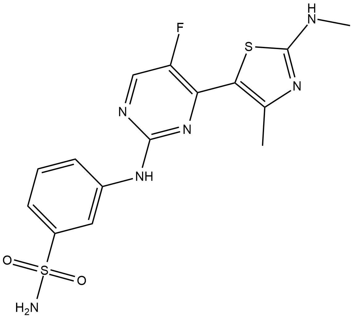
CDKI-73
CDKI-73 is a potent CDK9 inhibitor with Ki of 4 nM; shows selective toxicity to CLL cells(LD50=80 nM) versus normal B cell and normal CD34+ cell(LD50>20 uM).
-
GC61865
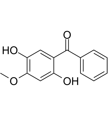
Cearoin
Cearoin increases autophagy and apoptosis through the production of ROS and the activation of ERK.
-
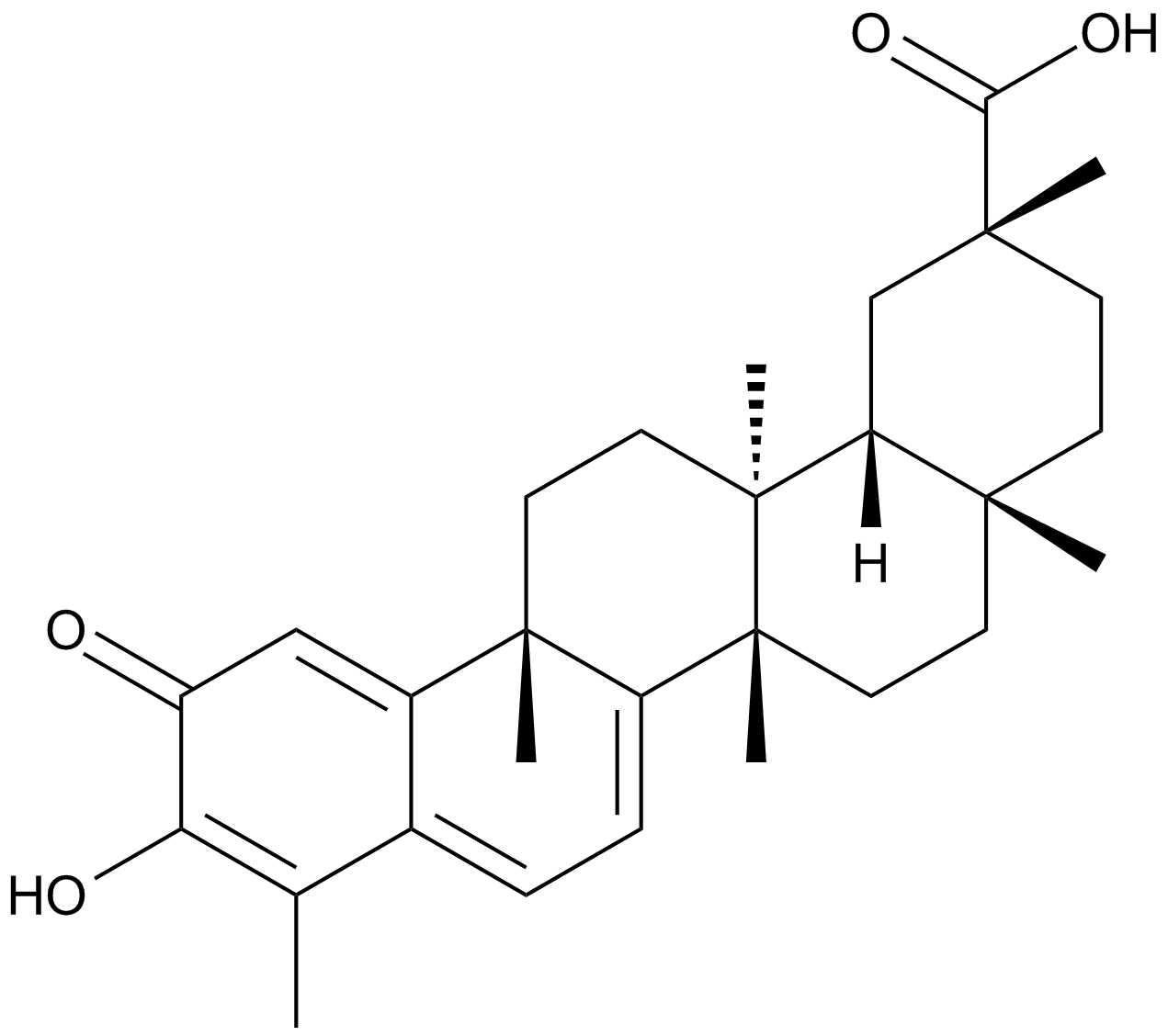
GC15083
Celastrol
A triterpenoid antioxidant
-
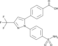
GC49152
Celecoxib Carboxylic Acid
An inactive metabolite of celecoxib
-
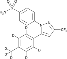
GC47070
Celecoxib-d7
An internal standard for the quantification of celecoxib
-
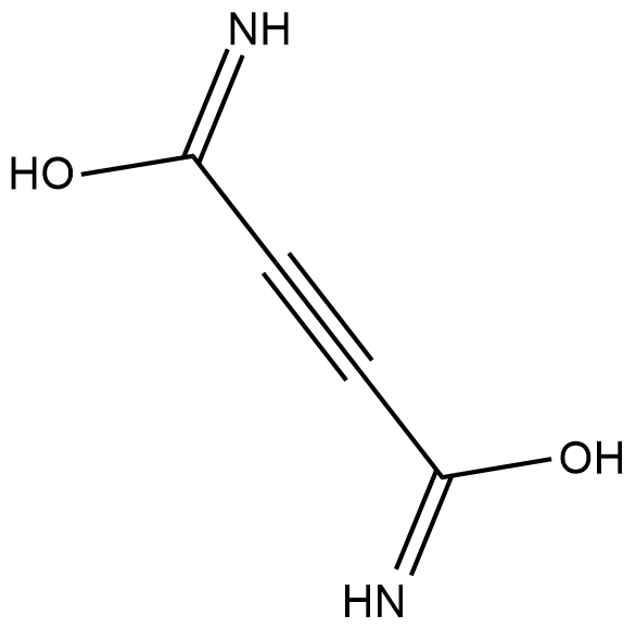
GC18392
Cellocidin
Cellocidin is an antibiotic originally isolated from S.
-
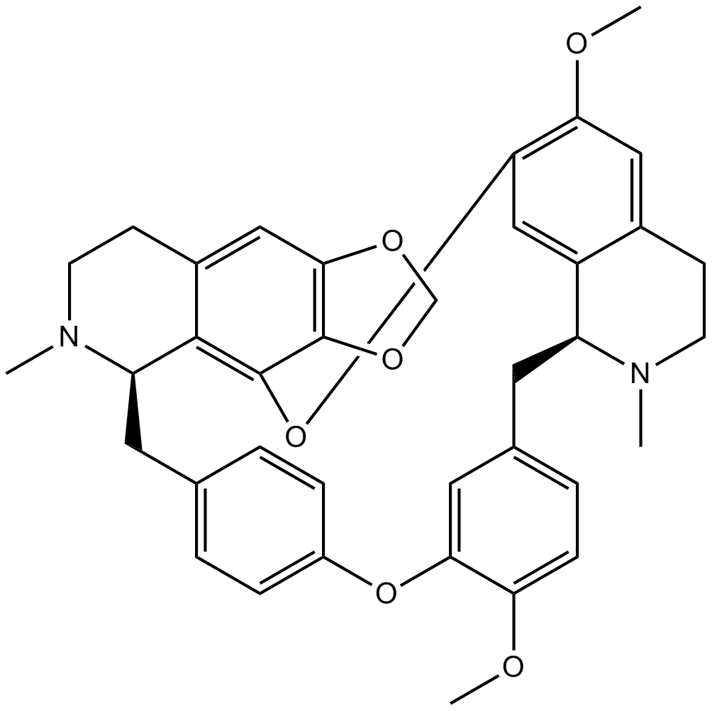
GN10113
Cepharanthine
-
GC52489
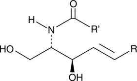
Ceramide (hydroxy) (bovine spinal cord)
A sphingolipid

-
GC52485
Ceramide (non-hydroxy) (bovine spinal cord)
A sphingolipid
-
GC52486
Ceramide Phosphoethanolamine (bovine)
A sphingolipid

-
GC43229
Ceramide Phosphoethanolamines (bovine)
Ceramide phosphoethanolamine (CPE) is an analog of sphingomyelin that contains ethanolamine rather than choline as the head group.

-
GC47073
Ceramides (hydroxy)
A mixture of hydroxy fatty acid-containing ceramides

-
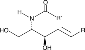
GC43230
Ceramides (non-hydroxy)
Ceramides are generated from sphingomyelin through activation of sphingomyelinases or through the de novo synthesis pathway, which requires the coordinated action of serine palmitoyl transferase and ceramide synthase.
-
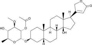
GC49706
Cerberin
A cardiac glycoside with cytotoxic and cardiac modulatory activities
-
GC60688
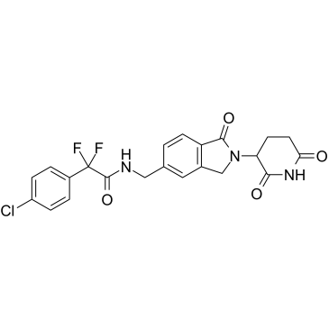
Cereblon modulator 1
Cereblon modulator 1 (CC-90009) is a first-in-class GSPT1-selective cereblon (CRBN) E3 ligase modulator, acts as a molecular glue.
-
GC65487
Certolizumab pegol
Certolizumab pegol (Certolizumab) is a recombinant, polyethylene glycolylated, antigen-binding fragment of a humanized monoclonal antibody that selectively targets and neutralizes tumour necrosis factor-α (TNF-α).
-
GC11543
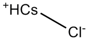
Cesium chloride
-
GC11710
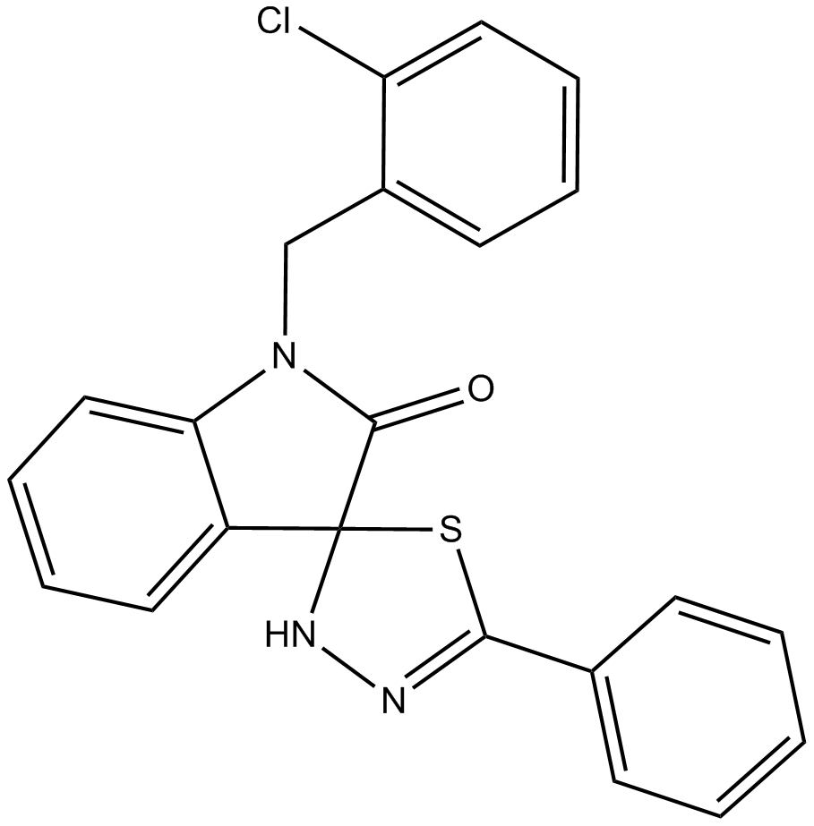
CFM 4
CFM 4 is a potent small molecular antagonist of CARP-1/APC-2 binding. CFM 4 prevents CARP-1 binding with APC-2, causes G2M cell cycle arrest, and induces apoptosis with an IC50 range of 10-15 μM. CFM 4 also suppresses growth of drug-resistant human breast cancer cells.
-
GC35668
CG-200745
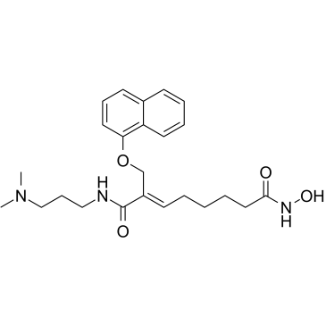
CG-200745 (CG-200745) is an orally active, potent pan-HDAC inhibitor which has the hydroxamic acid moiety to bind zinc at the bottom of catalytic pocket. CG-200745 inhibits deacetylation of histone H3 and tubulin. CG-200745 induces the accumulation of p53, promotes p53-dependent transactivation, and enhances the expression of MDM2 and p21 (Waf1/Cip1) proteins. CG-200745 enhances the sensitivity of Gemcitabine-resistant cells to Gemcitabine and 5-Fluorouracil (5-FU; ). CG-200745 induces apoptosis and has anti-tumour effects.
-
GC10666
CGP 57380
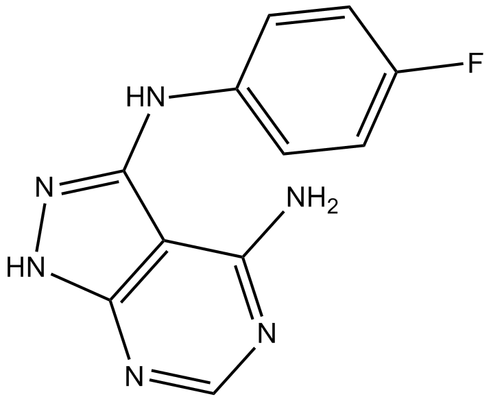
MNK1 inhibitor, specific and selective
-
GC43234
Chaetoglobosin A
Chaetoglobosin A is a mycotoxic cytochalasin that was first isolated from the marine-derived endophytic fungus C.
-
GC18536
Chartreusin
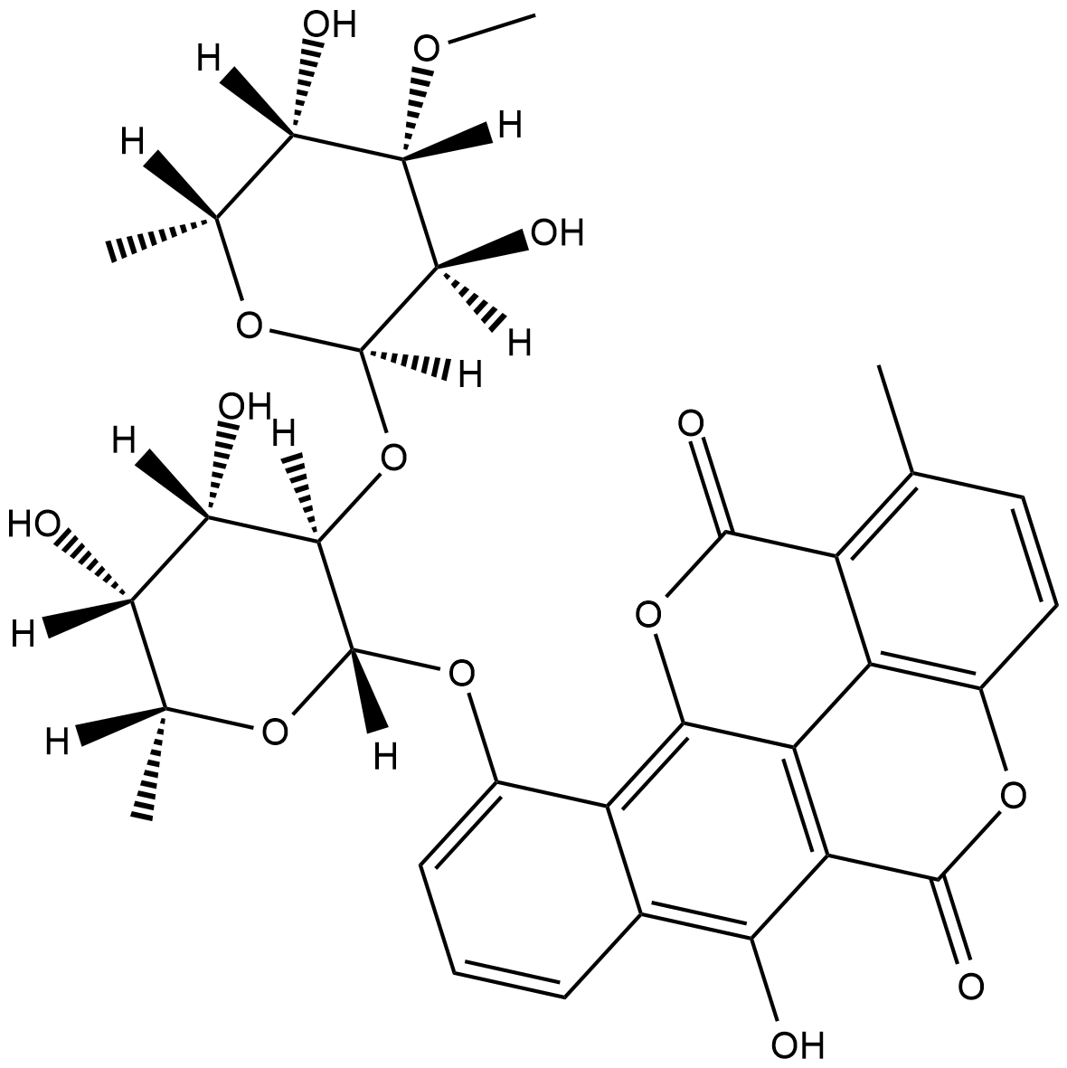
Chartreusin is an antibiotic originally isolated from S.
-
GN10463
Chelerythrine
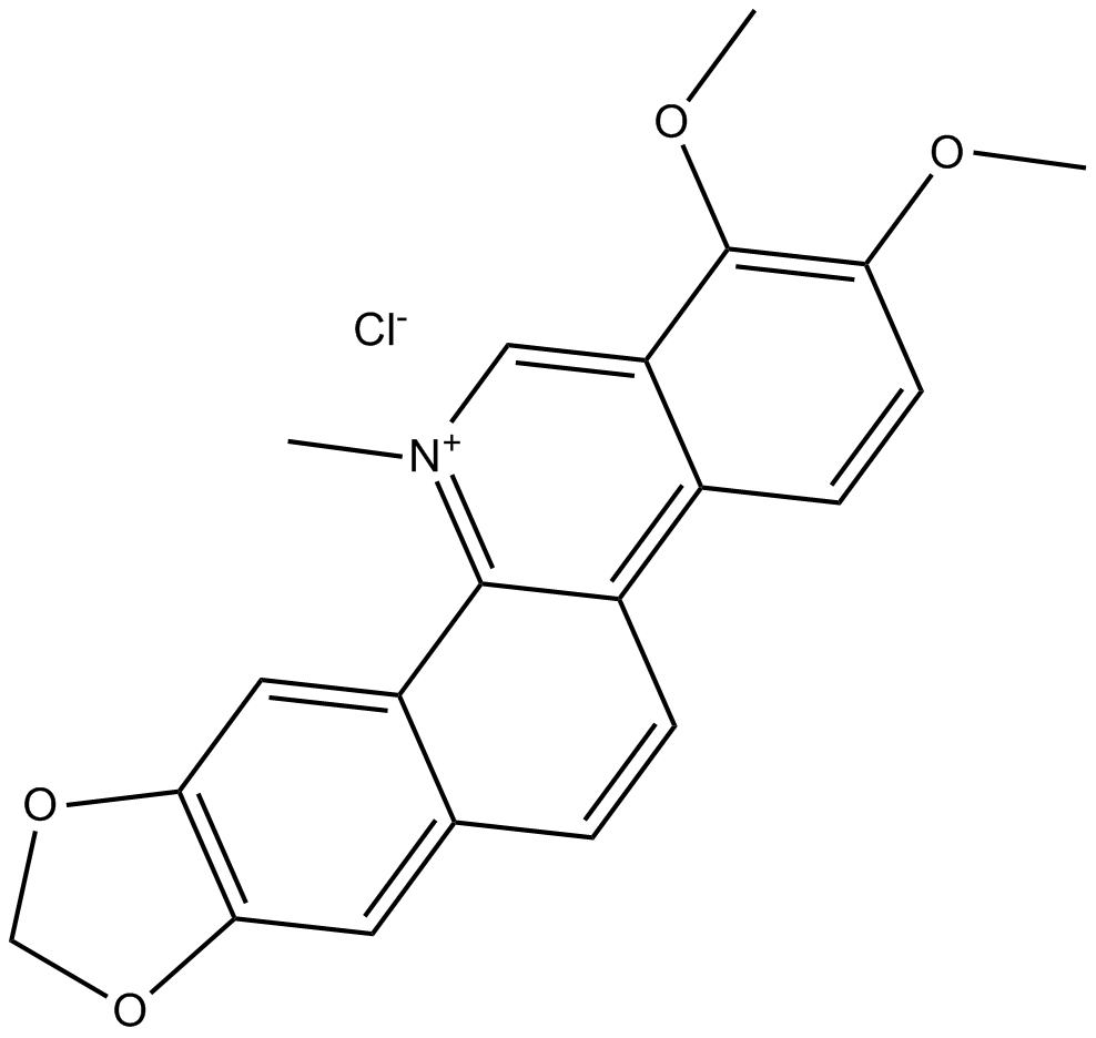
-
GC13065
Chelerythrine Chloride
Potent inhibitor of PKC and Bcl-xL
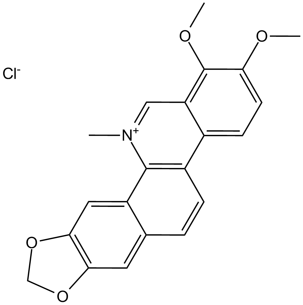
-
GC31886
Chelidonic acid
A pyran with diverse biological activities
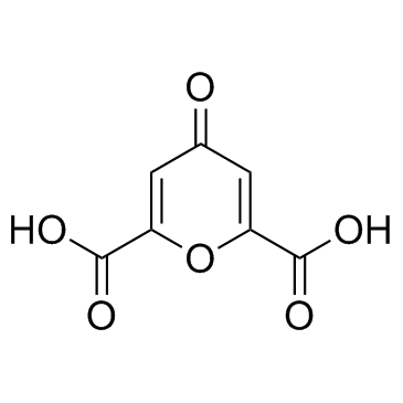
-
GC40878
Chelidonine
Chelidonine is a benzophenanthridine alkaloid that has been isolated from C.
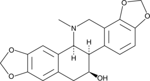
-
GC43236
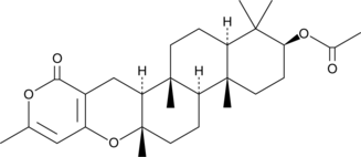
Chevalone B
Chevalone B is a meroterpenoid originally isolated from the fungus E.
-
GC43237
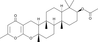
Chevalone C
Chevalone C is a meroterpenoid fungal metabolite originally isolated from E.
-
GC64993
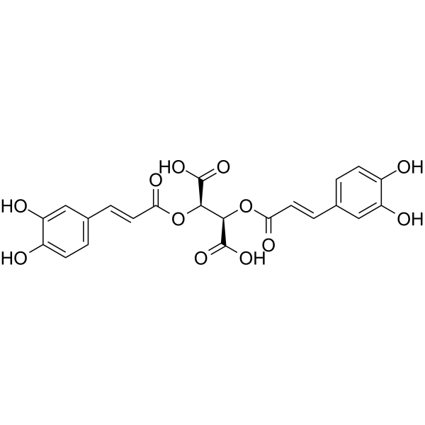
Chicoric acid
Chicoric acid (Cichoric acid), an orally active dicaffeyltartaric acid, induces reactive oxygen species (ROS) generation.
-
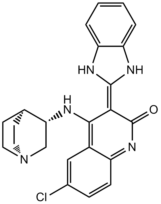
GC15739
CHIR-124
Chk1 inhibitor,novel and potent
-
GC43239
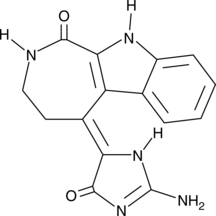
Chk2 Inhibitor
Chk2 Inhibitor (compound 1) is a potent and selective inhibitor of checkpoint kinase 2 (Chk2), with IC50s of 13.5 nM and 220.4 nM for Chk2 and Chk1, respectively. Chk2 Inhibitor can elicit a strong ataxia telangiectasia mutated (ATM)-dependent Chk2-mediated radioprotection effect.
-
GC45717
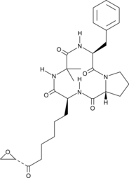
Chlamydocin
An HDAC inhibitor
-
GC17969
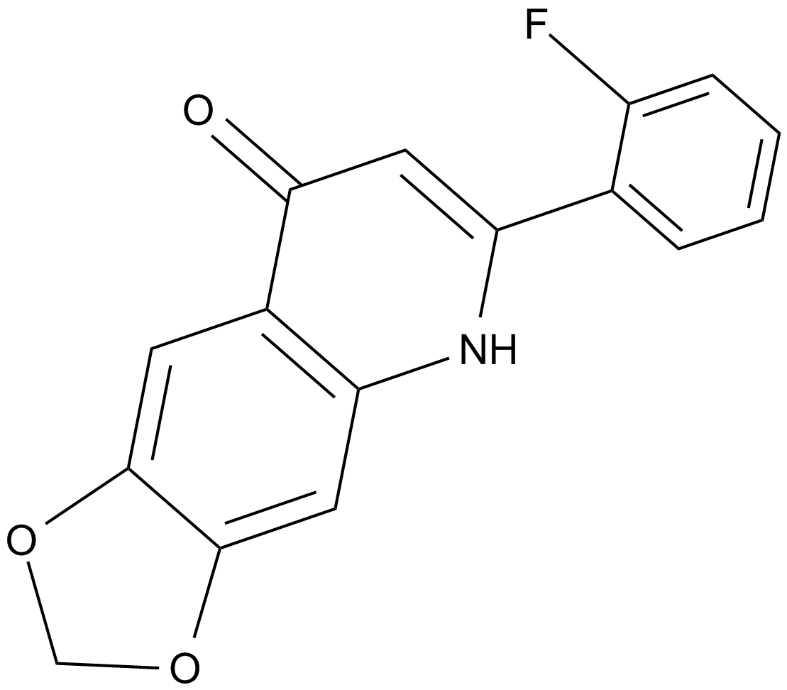
CHM 1
An inhibitor of tubulin polymerization
-
GC35682
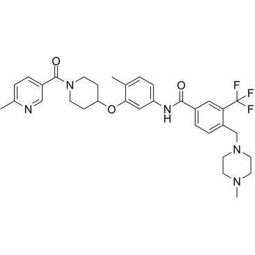
CHMFL-ABL/KIT-155
CHMFL-ABL/KIT-155 (CHMFL-ABL-KIT-155; compound 34) is a highly potent and orally active type II ABL/c-KIT dual kinase inhibitor (IC50s of 46 nM and 75 nM, respectively), and it also presents significant inhibitory activities to BLK (IC50=81 nM), CSF1R (IC50=227 nM), DDR1 (IC50=116 nM), DDR2 (IC50=325 nM), LCK (IC50=12 nM) and PDGFRβ (IC50=80 nM) kinases. CHMFL-ABL/KIT-155 (CHMFL-ABL-KIT-155) arrests cell cycle progression and induces apoptosis.
-
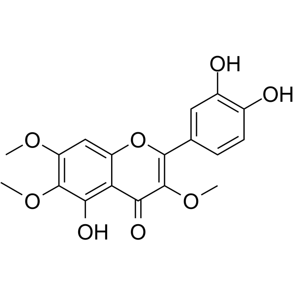
GC64028
Chrysosplenol D
Chrysosplenol D is a methoxy flavonoid that induces ERK1/2-mediated apoptosis in triple negative human breast cancer cells.
-
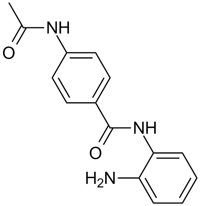
GC13408
CI994 (Tacedinaline)
An inhibitor of HDAC1, -2, and -3
-
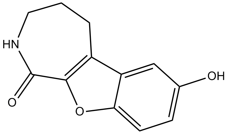
GC13589
CID 755673
PKD inhibitor
-
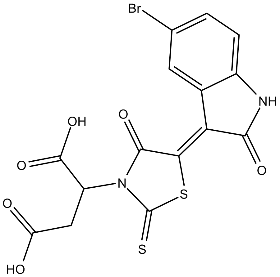
GC19436
CID-5721353
CID5721353 is an inhibitor of BCL6 with an IC50 value of 212 μM, which corresponds to a Ki of 147 μM.
-
GC32997
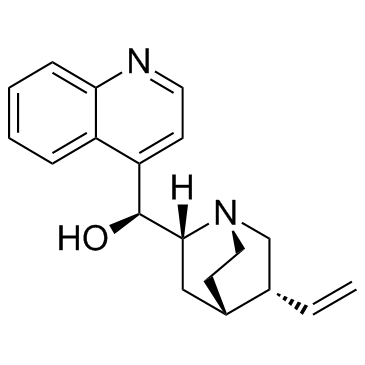
Cinchonine ((8R,9S)-Cinchonine)
Cinchonine ((8R,9S)-Cinchonine) is a natural compound present in Cinchona bark. Cinchonine ((8R,9S)-Cinchonine) activates endoplasmic reticulum stress-induced apoptosis in human liver cancer cells.
-
GC60708
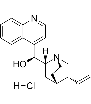
Cinchonine hydrochloride
Cinchonine hydrochloride ((8R,9S)-Cinchonine hydrochloride) is a natural alkaloid present in Cinchona bark, with antimalarial activity. Cinchonine hydrochloride activates endoplasmic reticulum (ER) stress-induced apoptosis in human liver cancer cells.
-
GC52269
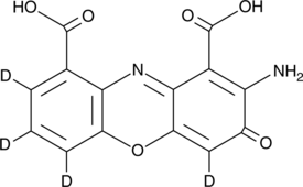
Cinnabarinic Acid-d4
An internal standard for the quantification of cinnabarinic acid
-
GC40986
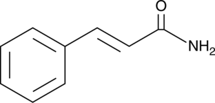
Cinnamamide
Cinnamamide is an amide form of of trans-cinnamic acid and a metabolite of Streptomyces.
-
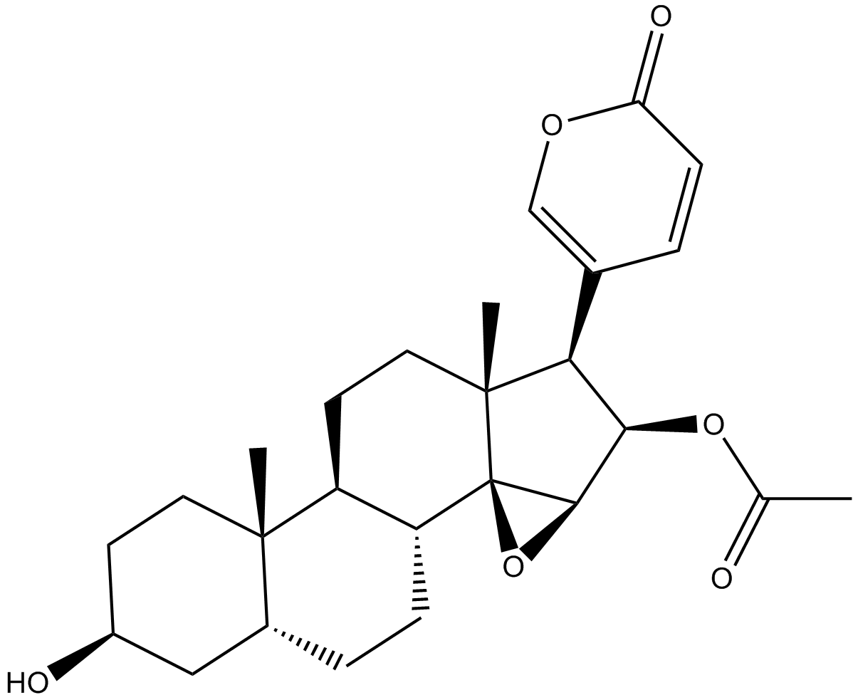
GN10189
Cinobufagin
-
GC11908
Cisplatin
Cisplatin is one of the best and first metal-based chemotherapeutic drugs, which is used for wide range of solid cancers such as testicular, ovarian, bladder, lung, cervical, head and neck cancer, gastric cancer and some other cancers.

-
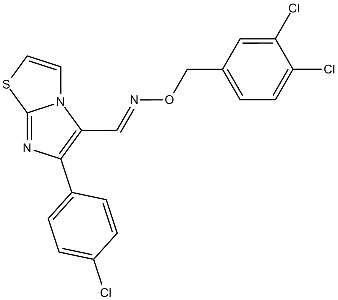
GC17491
CITCO
Constitutive androstane receptor (CAR) agonist
-
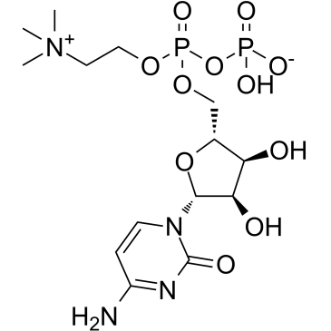
GC35703
Citicoline
Citicoline (Cytidine diphosphate-choline) is an intermediate in the synthesis of phosphatidylcholine, a component of cell membranes.
-
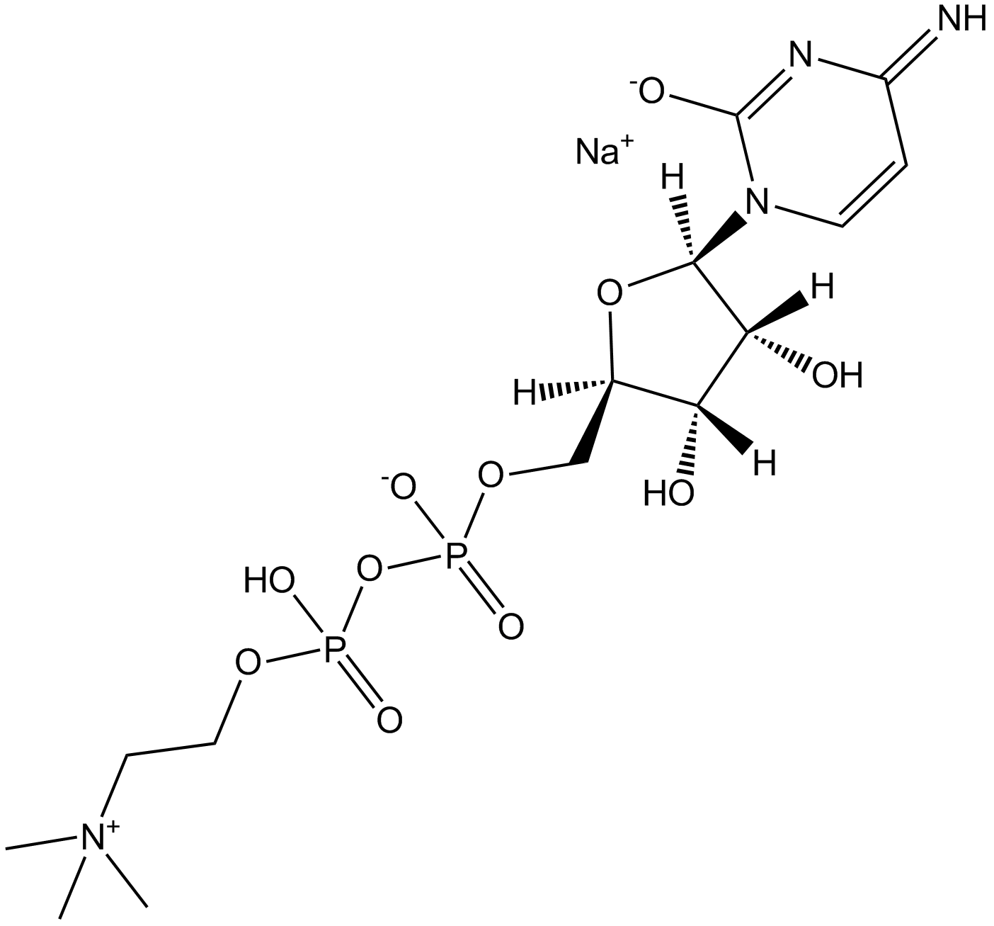
GC31186
Citicoline sodium salt
Citicoline sodium salt salt is an intermediate in the synthesis of phosphatidylcholine which is a component of cell membranes and also exerts neuroprotective effects.
-
GC43273
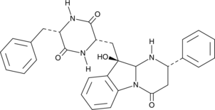
Citreoindole
Citreoindole is a diketopiperazine metabolite isolated from a hybrid cell fusion of two strains of P.
-
GC41514
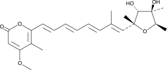
Citreoviridin
Citreoviridin is a mycotoxin isolated from several Penicillium species that has been shown to inhibit the mitochondrial ATP synthetase system.
-
GC14203
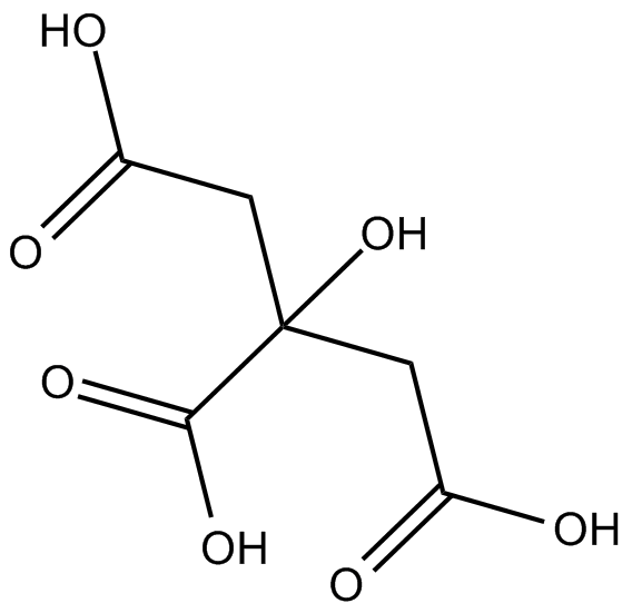
Citric acid
Commonly used laboratory reagent
-
GC68051
Citric acid-d4

-
GC16661
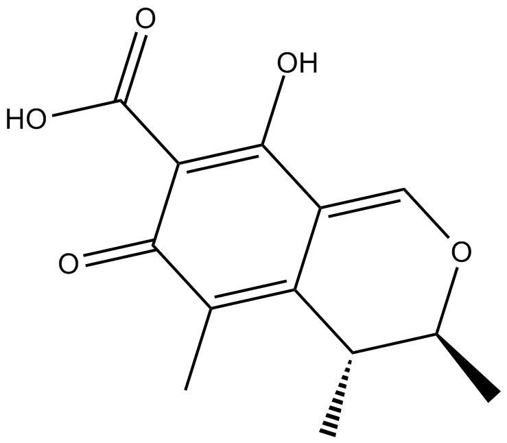
Citrinin
A mycotoxin inducing apoptosis
